Material specially prepared for Radozhiva by Rodion Eshmakov.
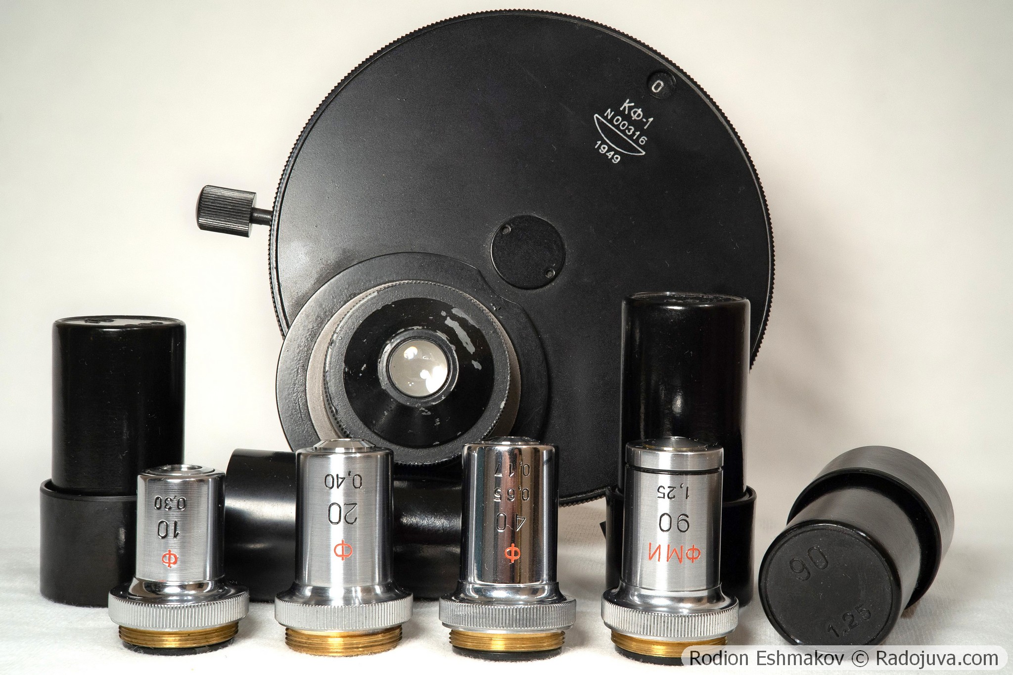
Condenser KF-1 and complete lenses.
Content
- Briefly about the KF-1 device
- Phase contrast in brief
- Technical characteristics of the KF-1 condenser
- Design and execution of the condenser
- Optical properties of the condenser
- Lenses for the KF-1: General Information
- Lenses for KF-1: 10×0.3 F OM-5
- Lenses for KF-1: 20×0.4 F OM-27
- Lenses for KF-1: 40×0.65 F MS
- Lenses for the KF-1: 90×1.25 F MI OM-41
- Lenses for the KF-1: 40×0.75 F VI OM-23
- Lenses for the KF-1: 70×1.25 F APO VI OM-25
- Conclusions
More micro-optics reviews here.
Briefly about the KF-1 device
The KF-1 device is designed for microscopic observation using phase contrast in transmitted light. In short, this kit significantly improves the experience of using simple biological microscopes, as it allows for working with unstained specimens, including live ones, without prior sample preparation.
This is the earliest version of the Soviet phase-contrast device, which is an analogue of the set produced by Carl Zeiss Jena - the condenser presented in this review was made in 1949. In addition to the Zernike condenser with annular diaphragms (the KF-1 itself), the KF-1 set also includes an auxiliary microscope Mir-4 for combining the rings and four achromatic objectives of the RMS standard - 10×0.3 F OM-5 (review of the regular analog here ), 20×0.4 F OM-27 (review of the regular version here ), 40×0.65 F MS (review of the regular version here ), 90×1.25 F MI OM-41 oil immersion, equipped with special phase rings etched in the lenses or on plates installed in the interlens space. A distinctive feature of the objectives in the KF-1 kit is the red letter "F" on the barrel, although all objectives except the 90×1.25 are also compatible with the later Zernike condensers, the KF-4 and KF-4M. In addition to the objectives mentioned, this review also presents additional water immersion objectives that are not initially included in the KF-1 kit but can also be used with it: the 40×0.75 F VI OM-23 achromat (review of the standard version here ) and the 70×1.25 F APO VI OM-25 fluorite apochromat.
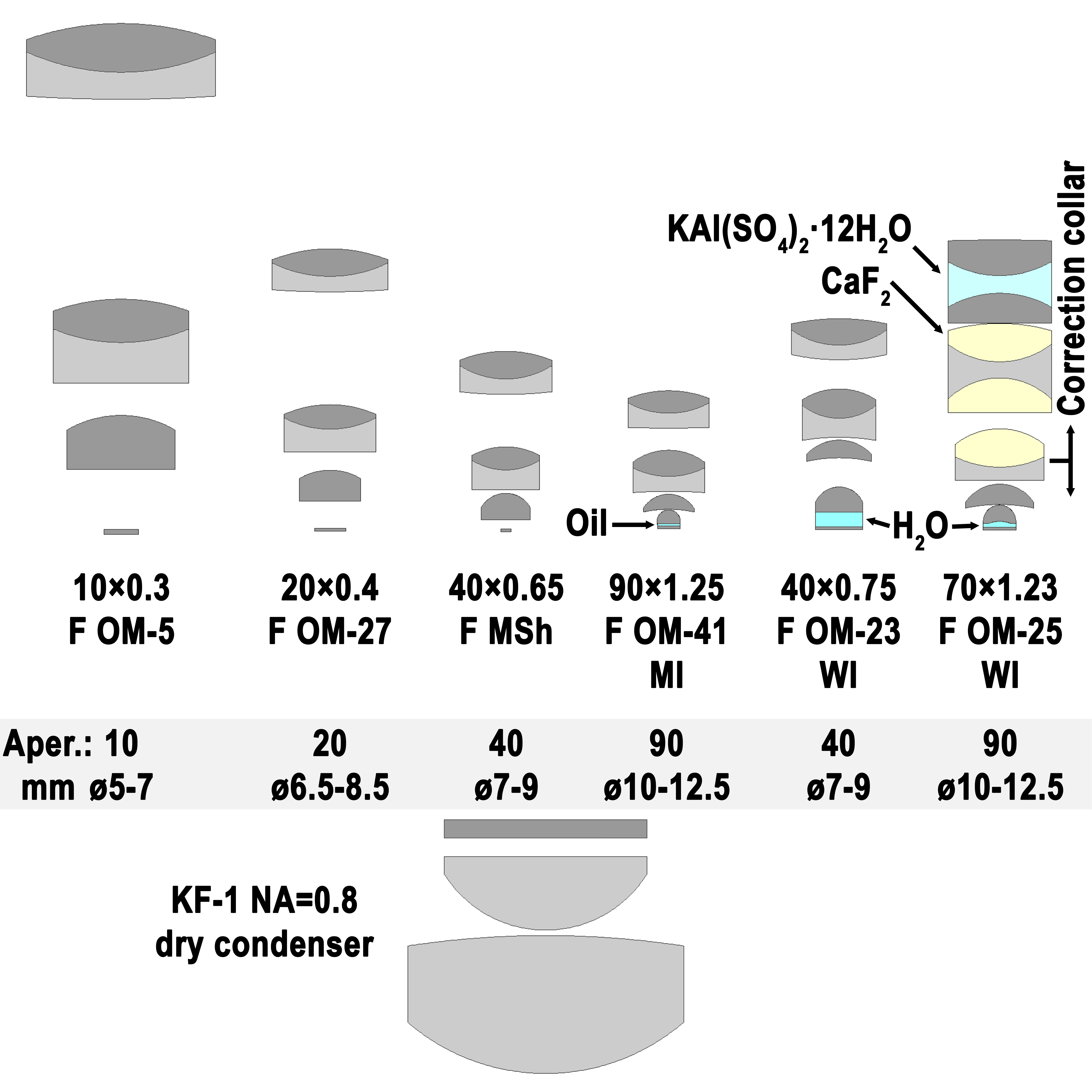
Drawing of optical diagrams of the KF-1 condenser and phase-contrast lenses for it.
Phase contrast in brief
Detailed information about the principle of the phase contrast method is given on the Nikon company website here , you can also read the translated version in Russian here.
In principle, the phase contrast method is a very advanced development of dark-field microscopy, which you can learn about in this article . In the dark-field method, a high-contrast, bright image of an object against a black background is created by selecting an illumination angle such that direct beams of light from the illuminator do not enter the objective, but instead receive beams of radiation scattered by the object.
In the phase contrast method, an object is illuminated using a condenser with an annular diaphragm whose numerical aperture is smaller than the objective aperture. The object is represented as a diffraction grating-like structure, through which some light passes without phase change, while others do so. The light beams from the condenser may change direction, or they may remain constant.
After interacting with the object, light enters the objective lens, which has a special ring in the plane of the entrance pupil with a diameter corresponding to the numerical aperture of the condenser ring. The objective's phase ring is etched into the glass and coated with aluminum or silver. Due to the high effective refractive index of the ring material, the phase of the transmitted light is rotated by 90°. The phase ring primarily captures light that has passed through the object without changing direction, while the remaining aperture of the objective captures light scattered by the object. In the image plane, these light beams interfere, resulting in the addition of the beams that passed through the object with a slight phase shift (~90°) and the subtraction of the scattered radiation.
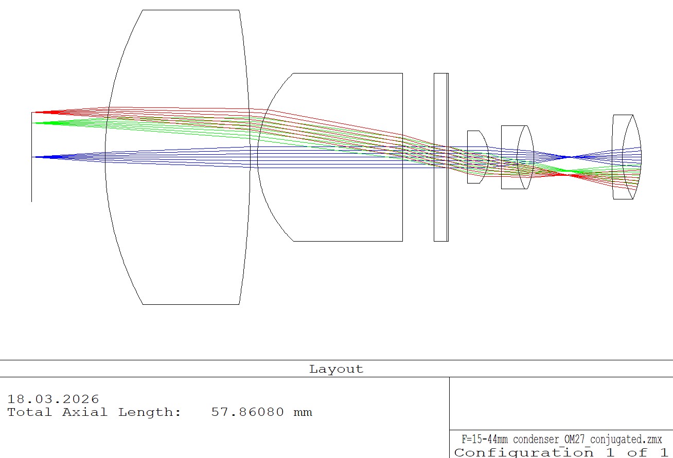
Ray path in a condenser-lens system, where the condenser's aperture diaphragm is imaged and aligned with the entrance pupil of the microscope objective, where a plate with a phase annulus is mounted. The blue beam is on-axis, while the red and green beams correspond to the annular diaphragm.
Thus, the classical method of positive phase contrast allows visualization of structural features of unstained preparations associated with very small fluctuations in the refractive index, such as living cells in water, cracks and inclusions in crystals, cell organelles, etc.
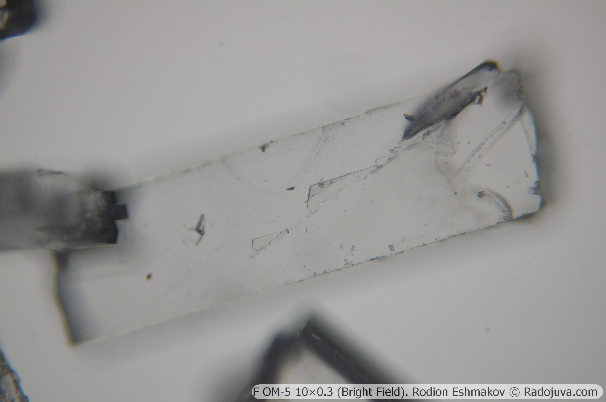
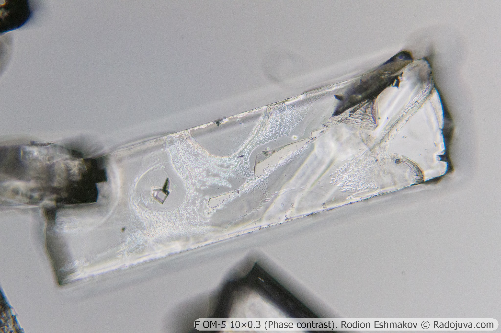
Below are paired photographs of a tetraphenylgermanium crystal taken with a Sony NEX-6 camera and an F OM-5 10×0.3 lens in bright field and using the phase contrast method with a KF-1 condenser.
Below are photographs of tulip pollen taken with a Progress O6AM-60 60×1.0-0.7 MI immersion lens under bright and dark field illumination, as well as with an F MI OM-41 90×1.25 lens with a KF-1 condenser under phase contrast.
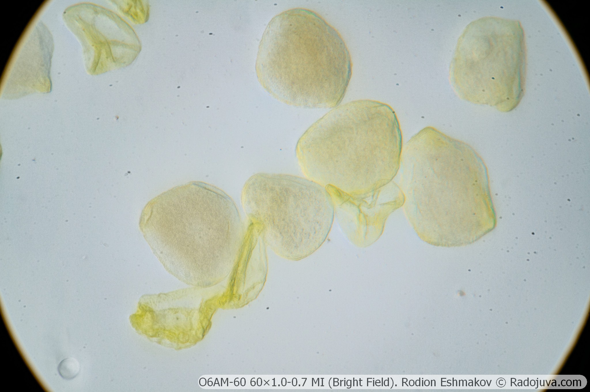
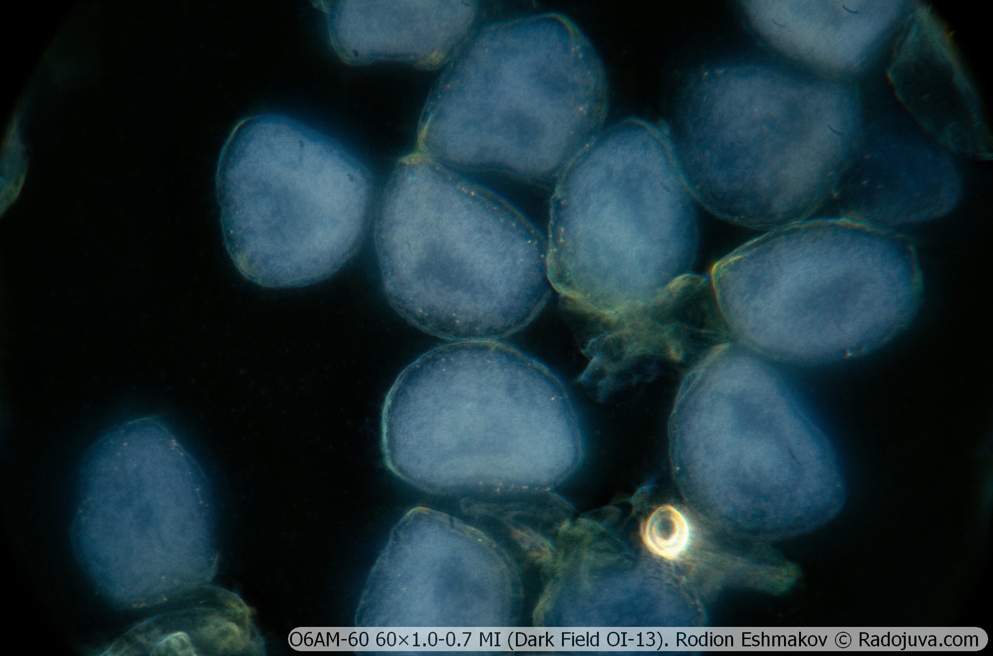
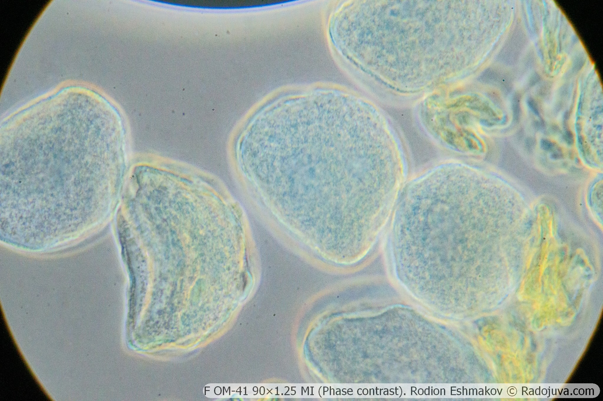
It's easy to see that phase contrast allows one to highlight structural features of the specimen that are invisible under normal brightfield illumination. Specifically, phase contrast perfectly conveys surface relief and hidden structural defects, increasing overall image contrast. However, it can also significantly distort the object's color and introduce characteristic halos.
Technical characteristics of the KF-1 condenser
Optical design – 2 lenses in 2 groups, high-aperture anachromat (Abbe condenser);
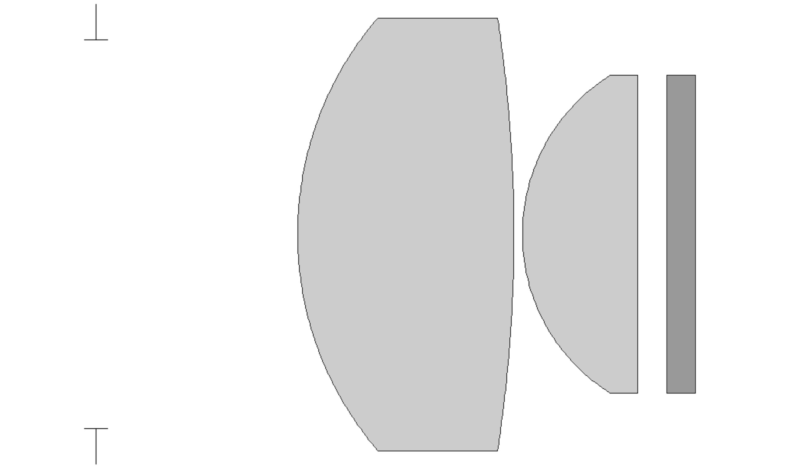
Optical diagram of the KF-1 condenser.
Focal length - 15.44 mm;
Numerical aperture – 0.8;
The size of the illuminated object is ~8 mm (with a numerical aperture of 0.1, including in dark field mode with an aperture of 90);
Immersion required - no;
Working distance – ~2-4 mm;
Aperture diameters – ø27 mm (NA=0.8, slot 0), ø15 mm (NA=0.44, intermediate empty slot), ø5-7 mm (NA=0.2, slot 10), ø6.5-8.5 mm (NA=0.25, slot 20), ø7-9 mm (NA=0.26, slot 40), ø10-12.5 mm (NA=0.37, slot 90);
Iris aperture diaphragm – yes;
Frame for light filters – yes;
Landing diameter - 37 mm;
Compatible lenses: F OM-5 10×0.3, LOMO CX 10/0.25 160/0.17 F, F OM-27 20×0.4, LOMO 20/0.4 160/0.17 F (DIN), F MS 40×0.65, F VI OM-23 40×0.75, LOMO CX 40/0.65 160/0.17 F, F APO VI OM-25 70×1.25, F MI OM-41 90×1.25 (red F only).
Design and execution of the condenser
The KF-1 condenser is made entirely of glass and metal and consists of a large flat plate with a diaphragm drum. The lens block is located at the top of the KF-1, the shiny side surface of which is used for mounting in the microscope ring under the stage. Unlike the OI-10 , the KF-1 condenser does not interfere with the rotating stages of the M-10, MBI-1, MBR-1, and Biolam-R microscopes. The KF-1 lens block is compatible with later KF-4 and KF-4M condensers, but is not compatible with standard brightfield condensers: a different mounting thread, different lenses, and different parameters. The caps with the upper lenses of the KF-1/KF-4 condensers are often confused with the lenses from a standard KON-3 condenser – however, the KF lens is easily distinguished by its much lesser surface curvature compared to the hemispherical lens of the KON-3.
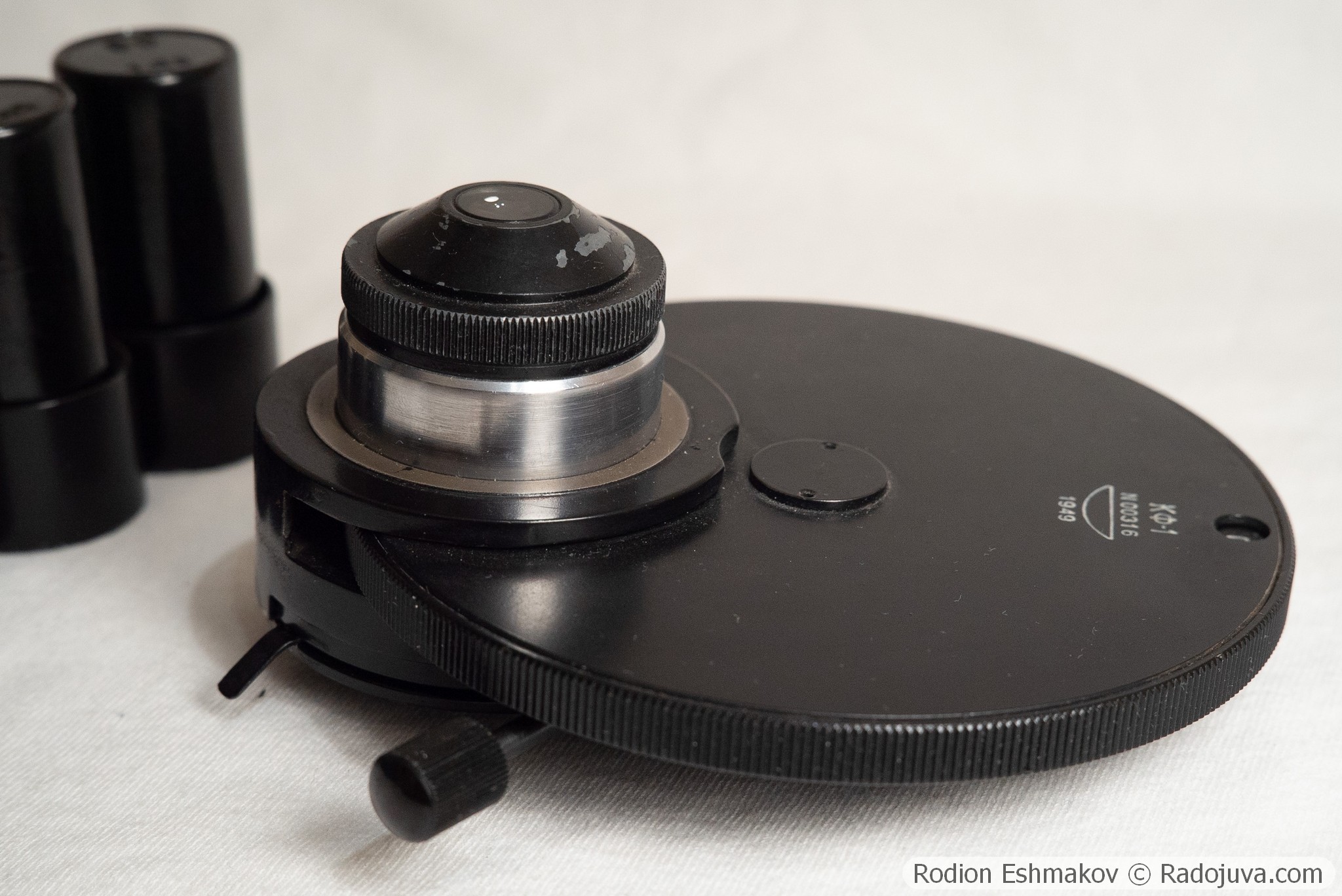
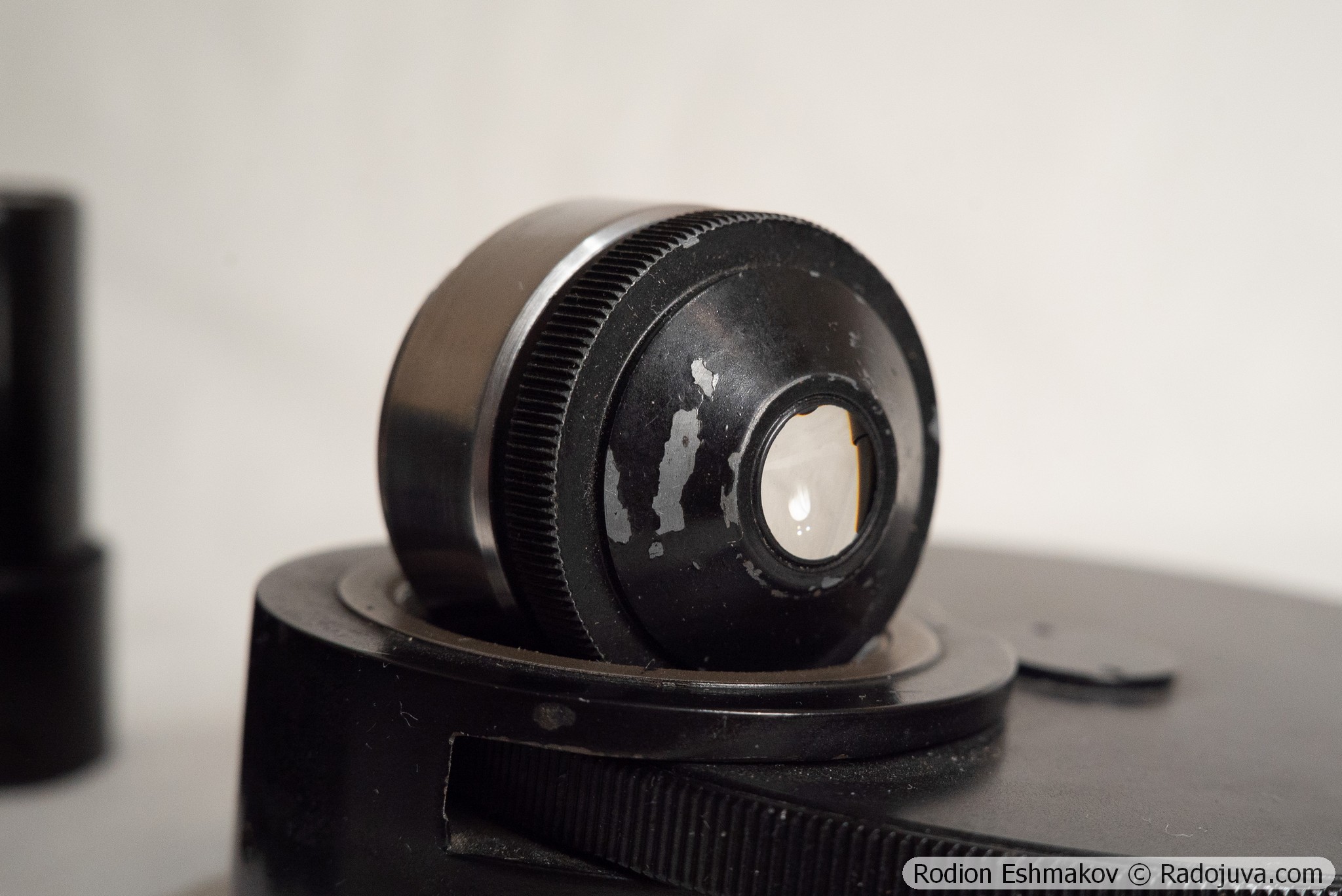
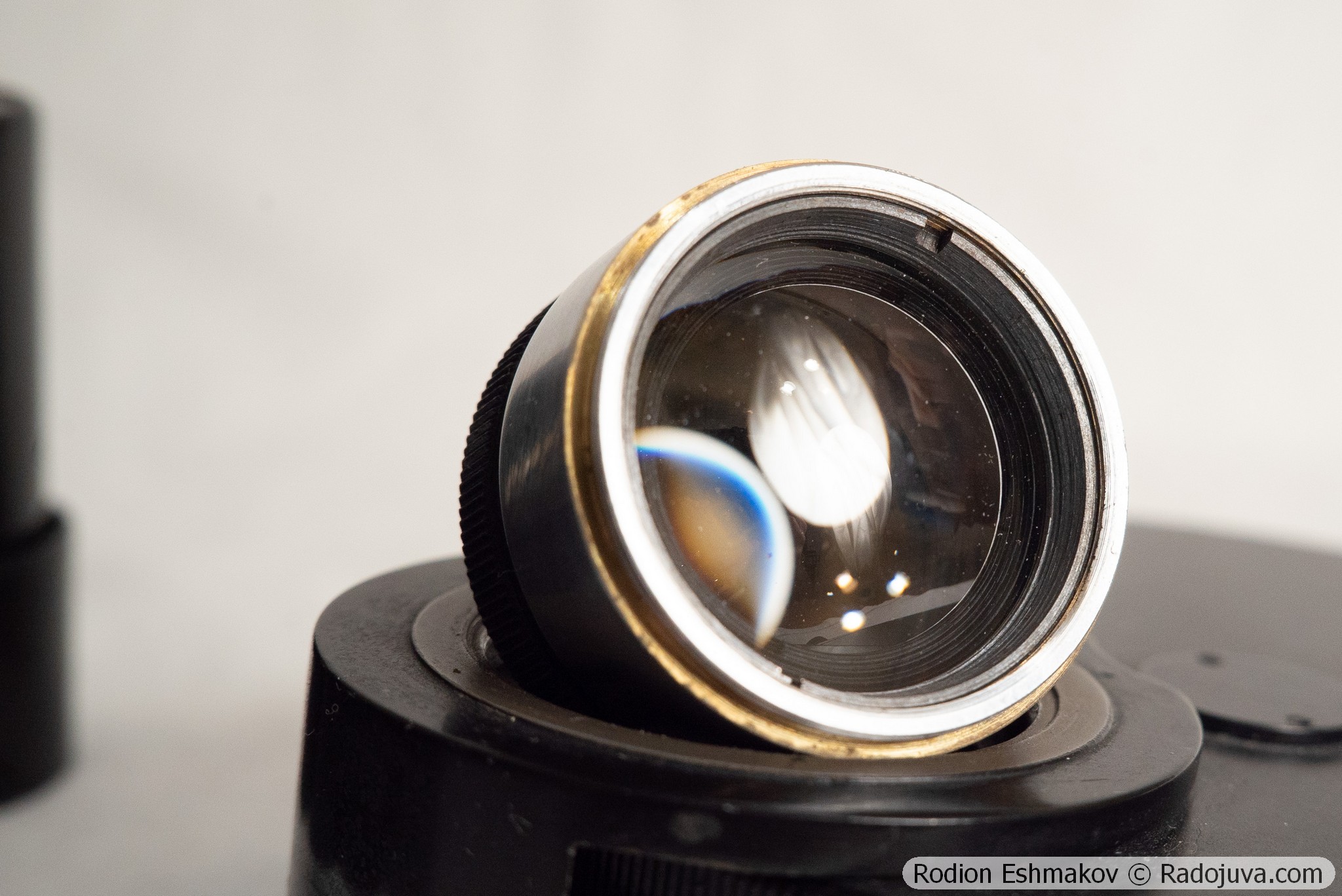
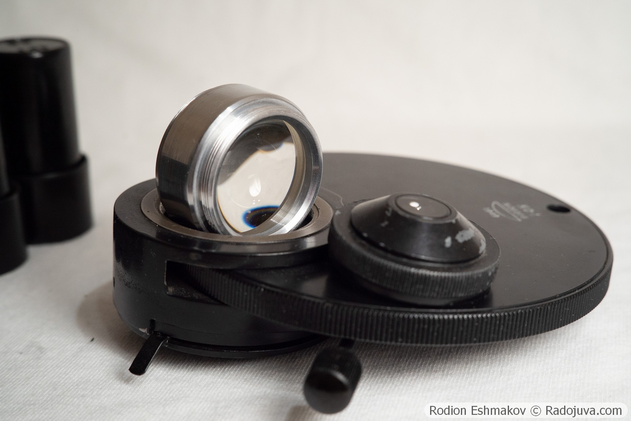
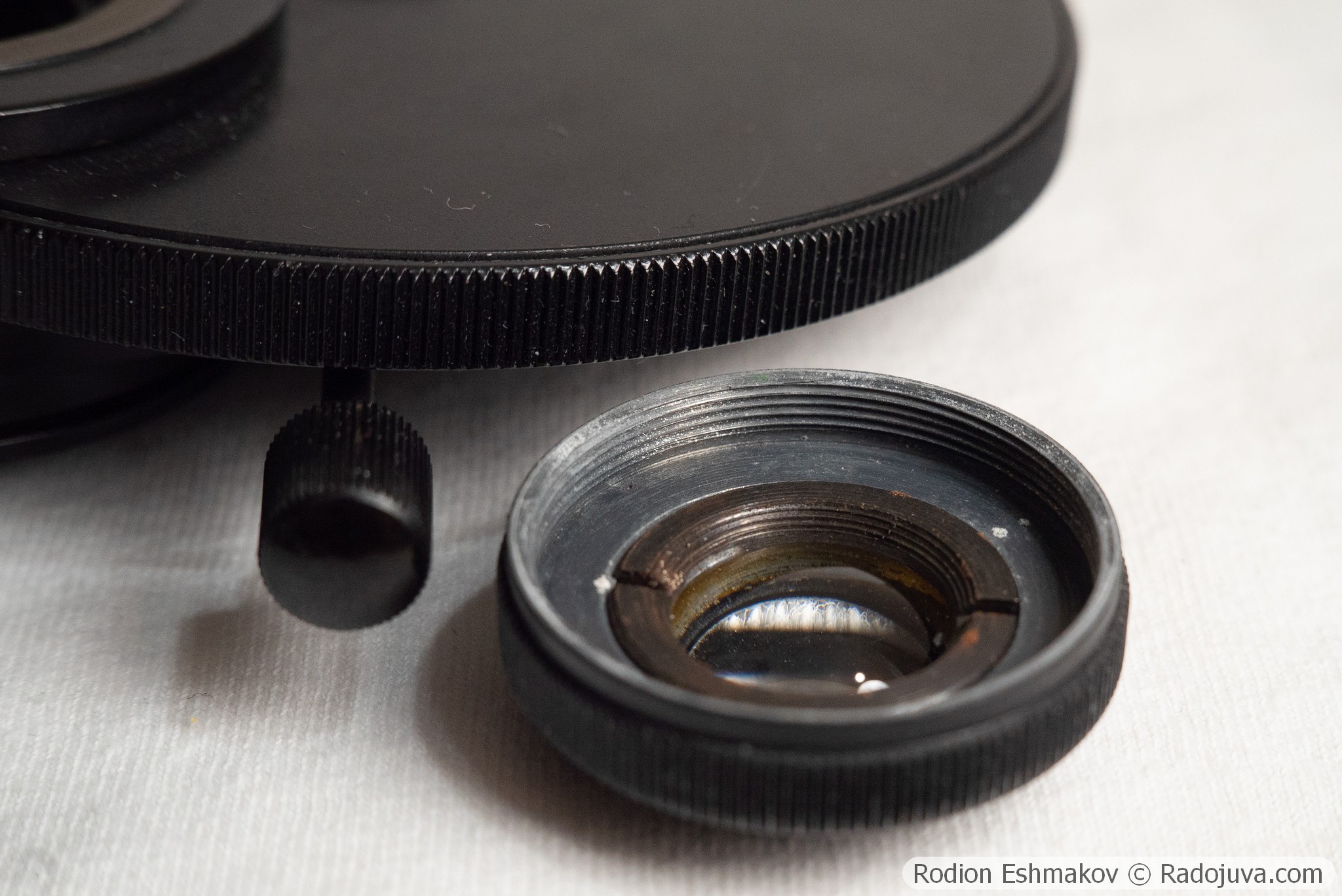
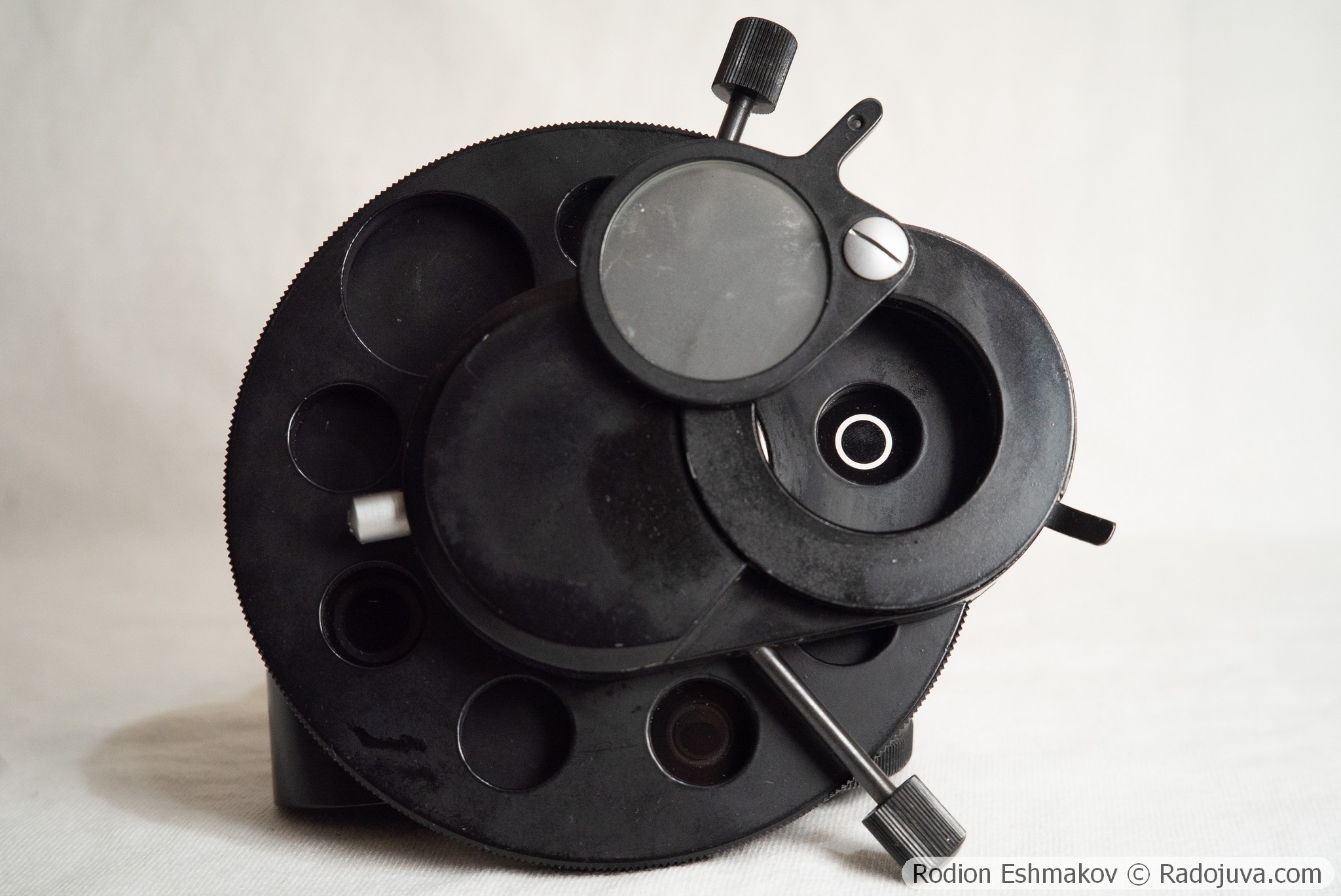
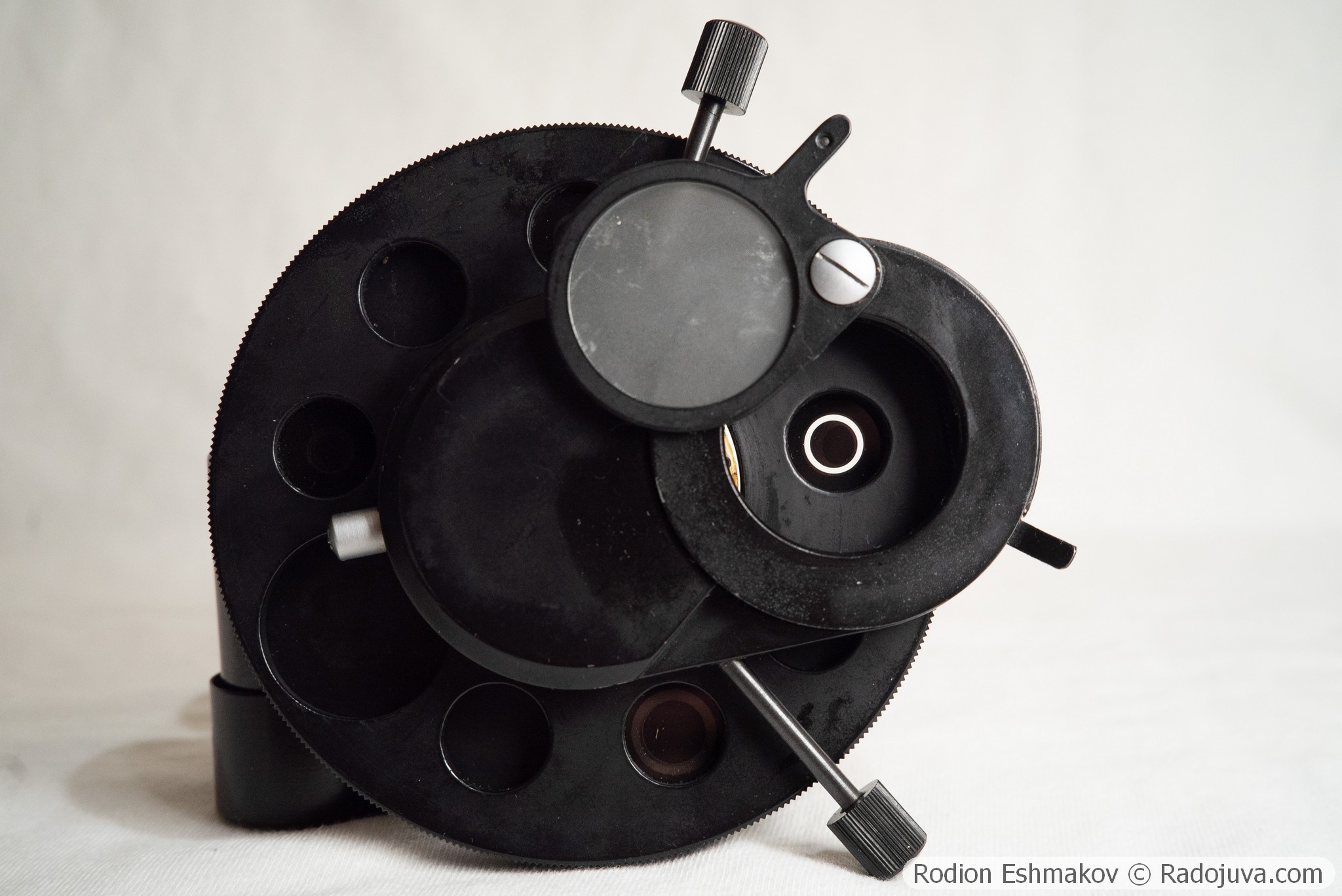
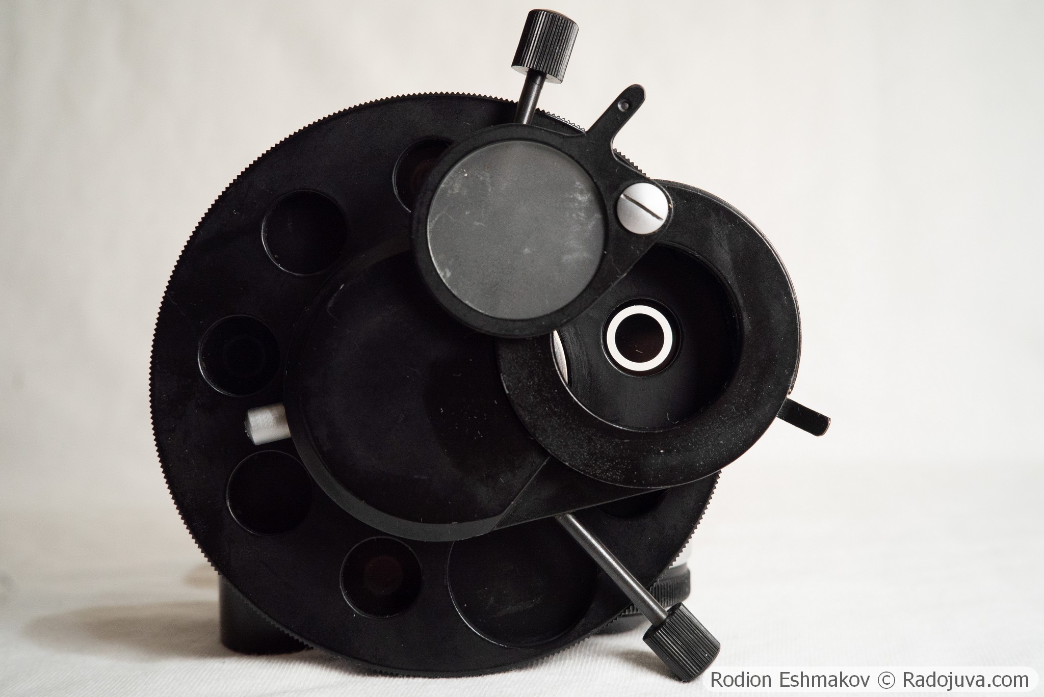
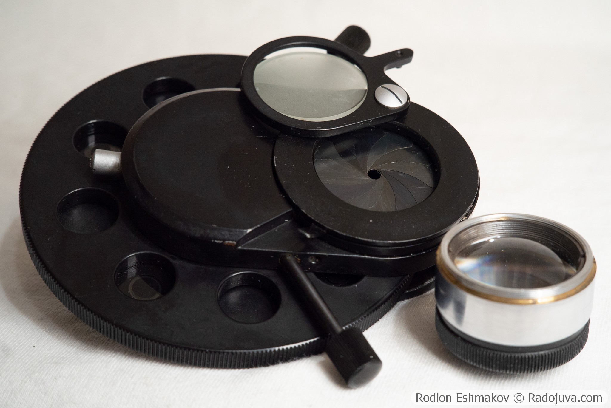
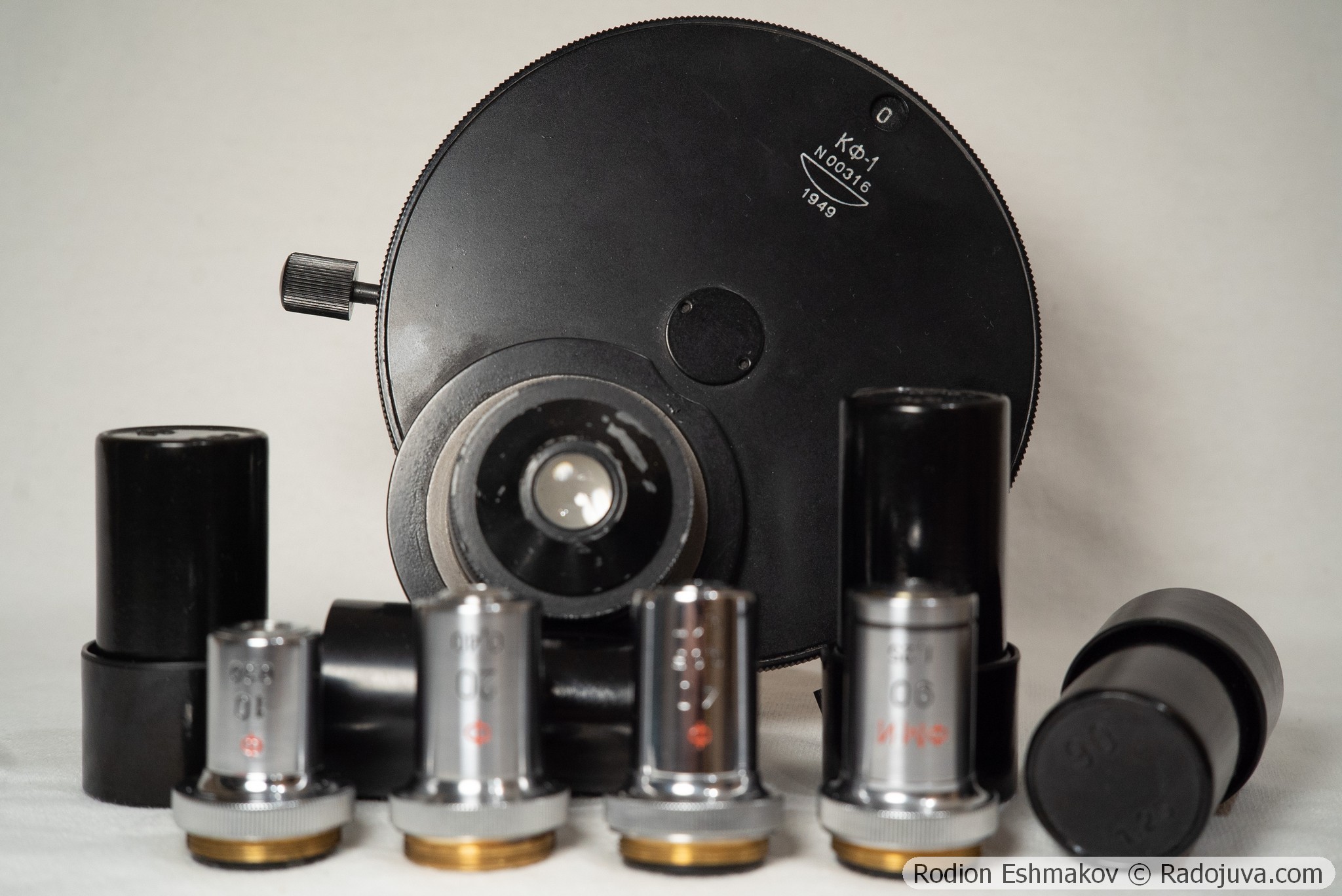
Also, unlike the KON-3, the KF-1's larger lens is very thick. Both KF-1 lenses are made of F-2 flint glass or TF-1 heavy flint glass, as indicated by the detection of lead in the X-ray fluorescence spectra.
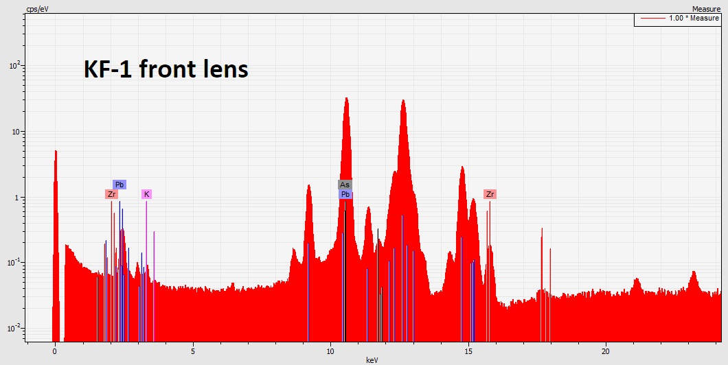
X-ray fluorescence spectrum of the front lens of the KF-1 condenser.
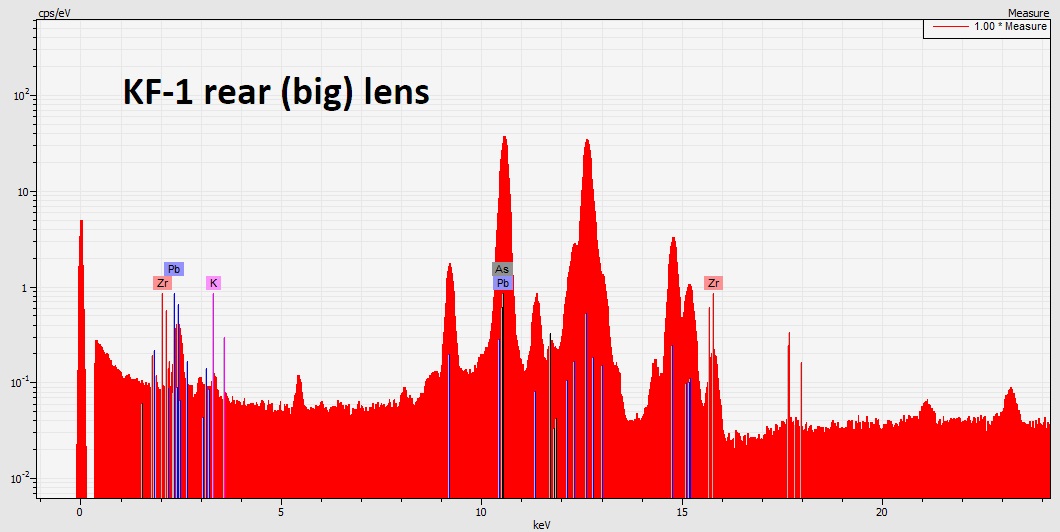
X-ray fluorescence spectrum of the rear lens of the KF-1 condenser.
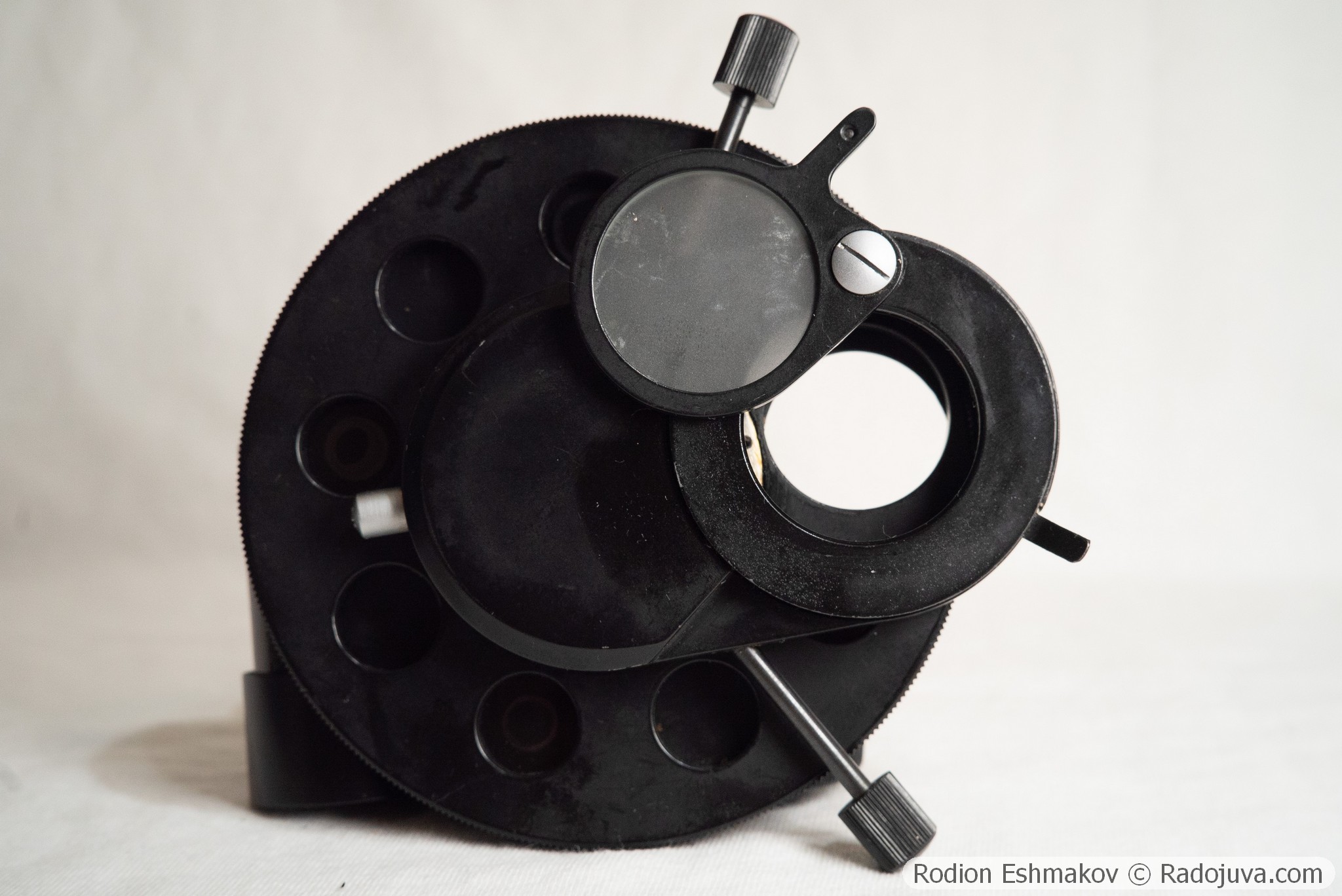
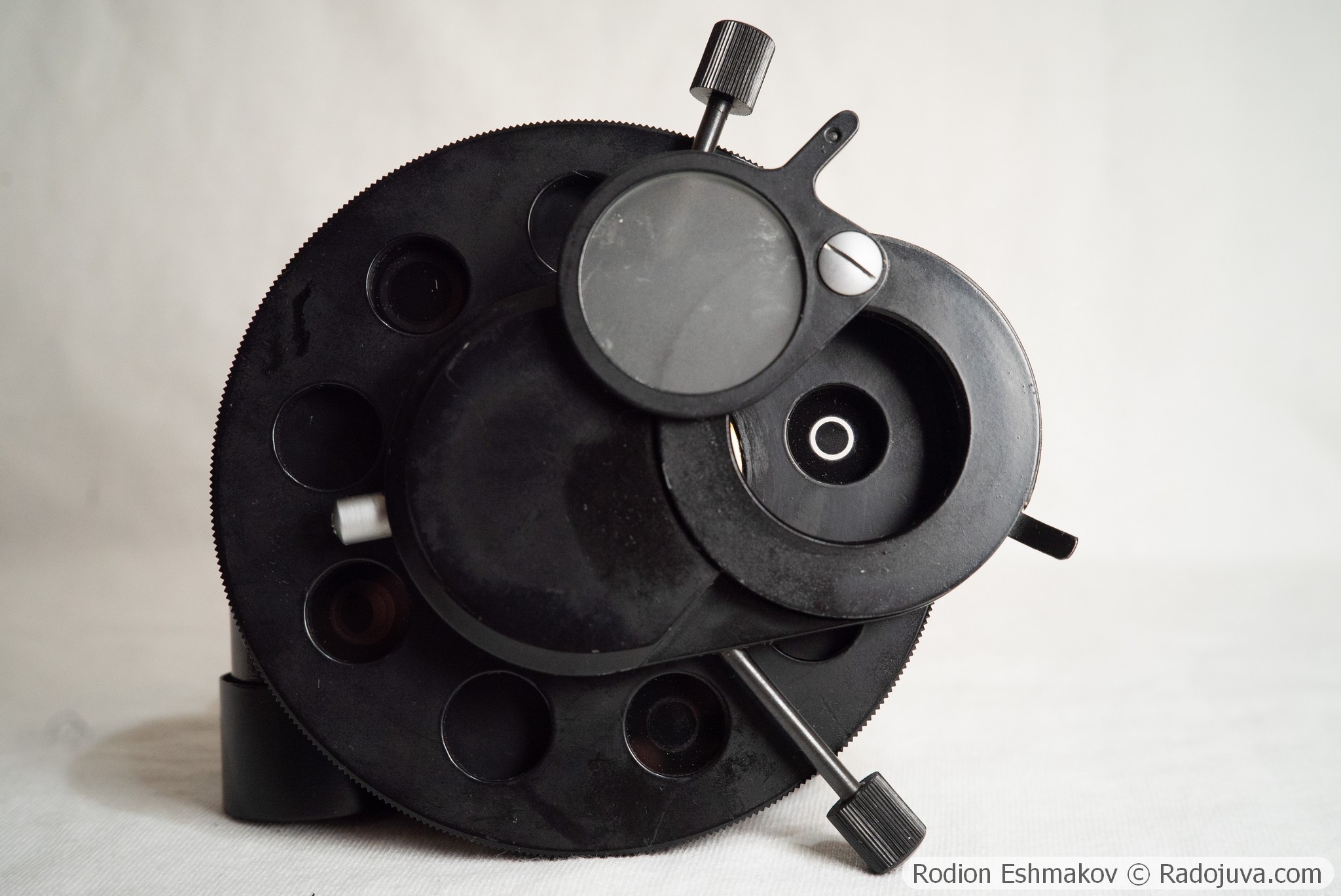
The ribbed side surface of the KF-1 condenser plate serves to switch the apertures located in a rotating drum. The condenser has a total of nine slots, one of which is empty, 27 mm in diameter, for brightfield operation. Four contain glass inserts with painted annular diaphragms for phase-contrast lenses. Another four are empty, 15 mm in diameter, located between the annular diaphragms. These empty slots are presumably used for subject retrieval in brightfield mode after switching lenses. They can also be used to install custom inserts (for example, for third-party lenses), making the KF-1 a good platform for modification.
The lower part of the KF-1 houses an iris diaphragm for brightfield operation, as well as two screw-type knobs for centering the movable annular diaphragm drum. Using these knobs and adjusting the condenser's height relative to the stage, the objective's phase ring is aligned with the condenser's annular diaphragm. This simple operation can be performed using a Mir-4 auxiliary microscope, installed in place of the eyepiece, or using a very nearsighted eye, also installed in place of the eyepiece (my method).
Typical problems with the KF-1 and newer KF-4/KF-4M condensers include loosening of the clamps in the central part of the plate, which leads to the rotation of individual housing parts along with the diaphragm block; hardening of the lubricant in the movable drum centering block, particularly in the spring supporting the block, located opposite the adjustment knobs; hardening of the oil on the iris diaphragm blades; and peeling of paint from the annular diaphragms. Generally, servicing the KF-1/KF-4/KF-4M does not present any major problems. However, the rather flimsy design and the inability to independently center the diaphragms are the main drawbacks of the KF-1.
Optical properties of the condenser
The KF-1 is a simple two-lens Abbe condenser with no correction for spherical and chromatic aberrations . The KF-1 (and later KF-4 and KF-4M) has a subtle advantage over most standard Abbe microscope condensers: it is designed for use without immersion and boasts a relatively high numerical aperture of 0.8. This is sufficient for use with most dry objectives, as their maximum numerical aperture never exceeds 0.95.
Furthermore, the KF-1 features a large working field, sufficient for use with 4-5x wide-angle lenses with an image field of up to 30mm. To enable use with 2-3.5x wide-angle lenses, the KF-1's top lens can be removed, leaving only the large lower lens, which has a numerical aperture of approximately 0.3. The standard Abbe condensers with NA=1.25 often produce very strong vignetting with wide-angle lenses.
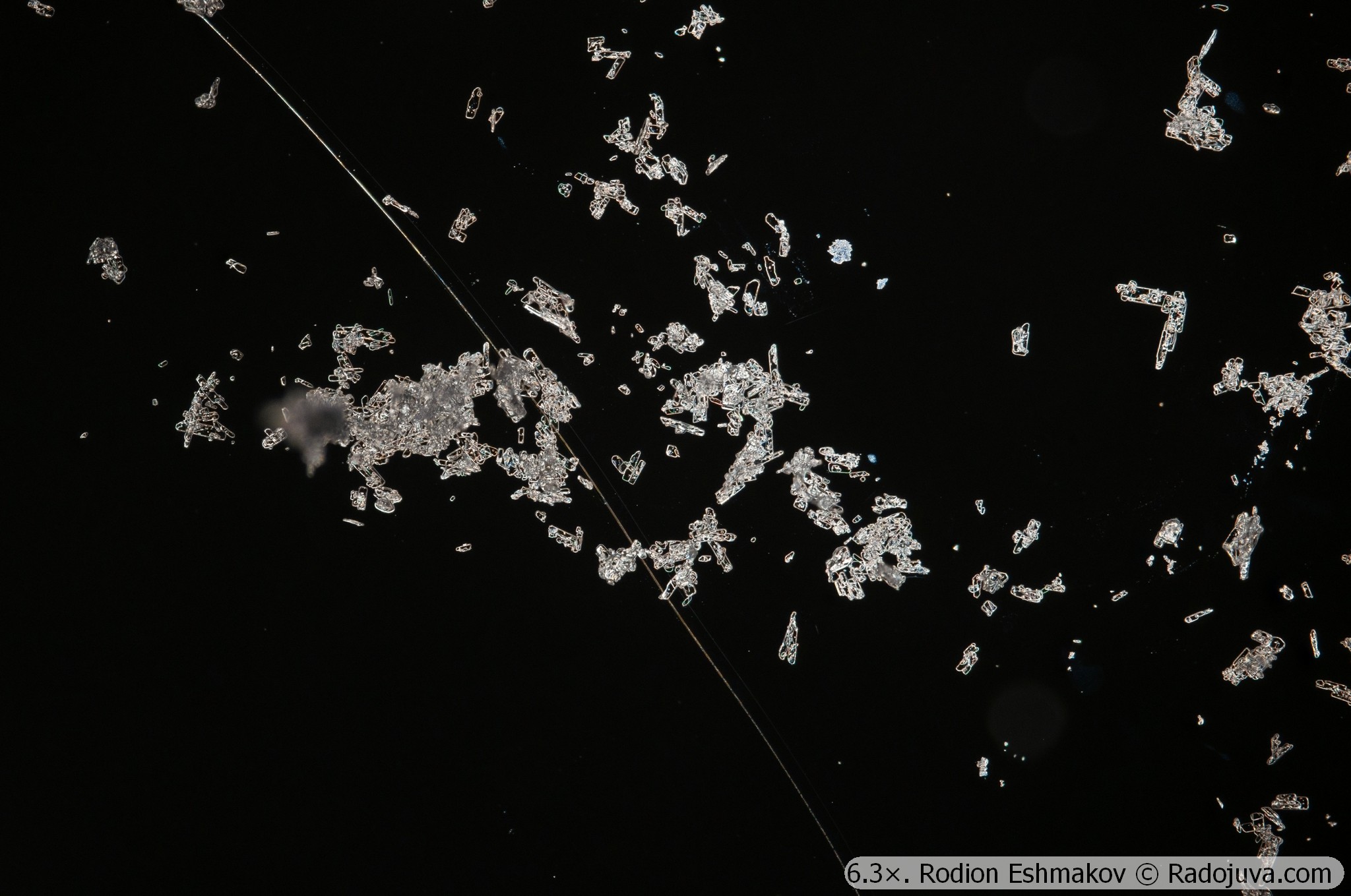
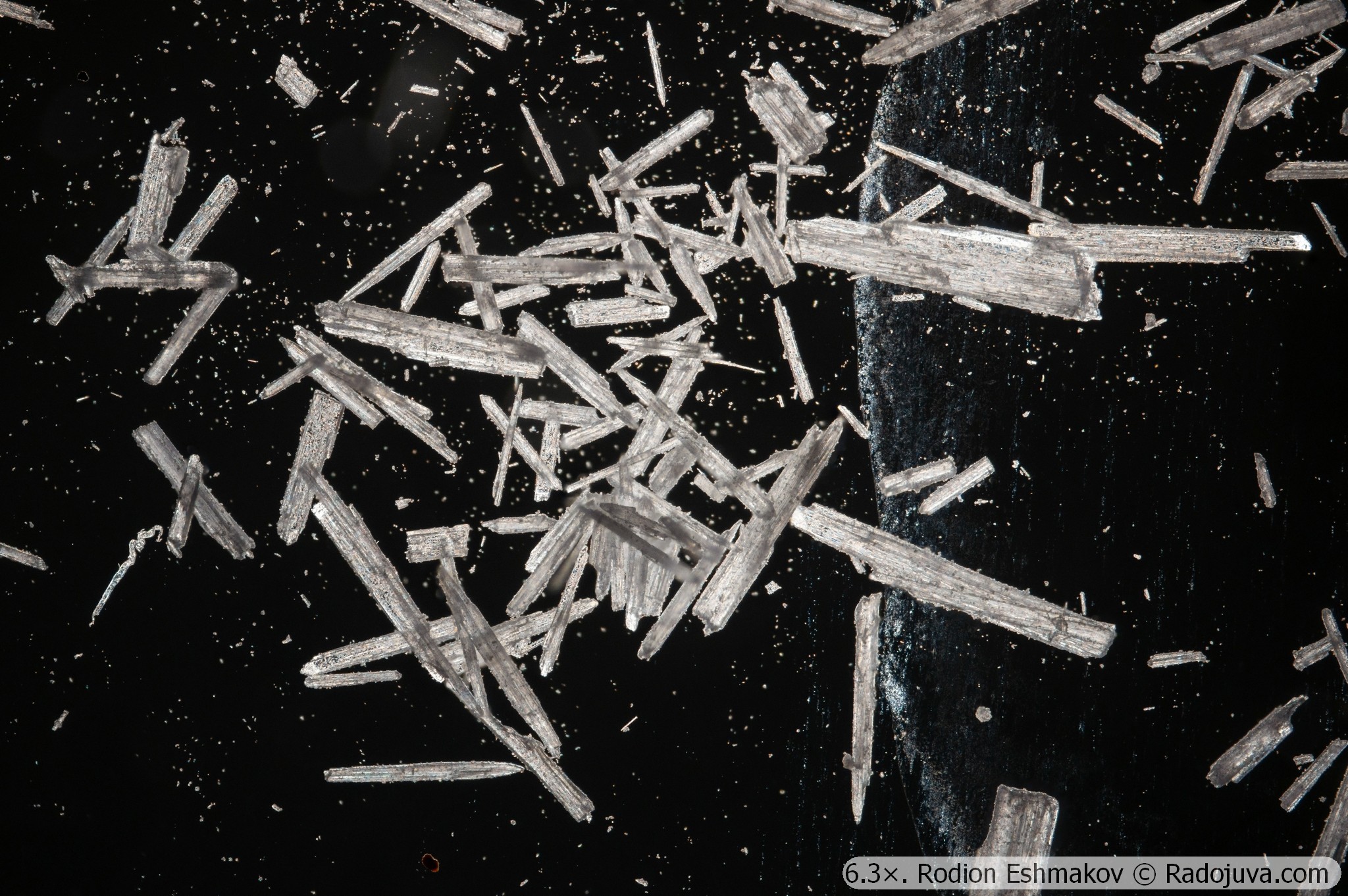
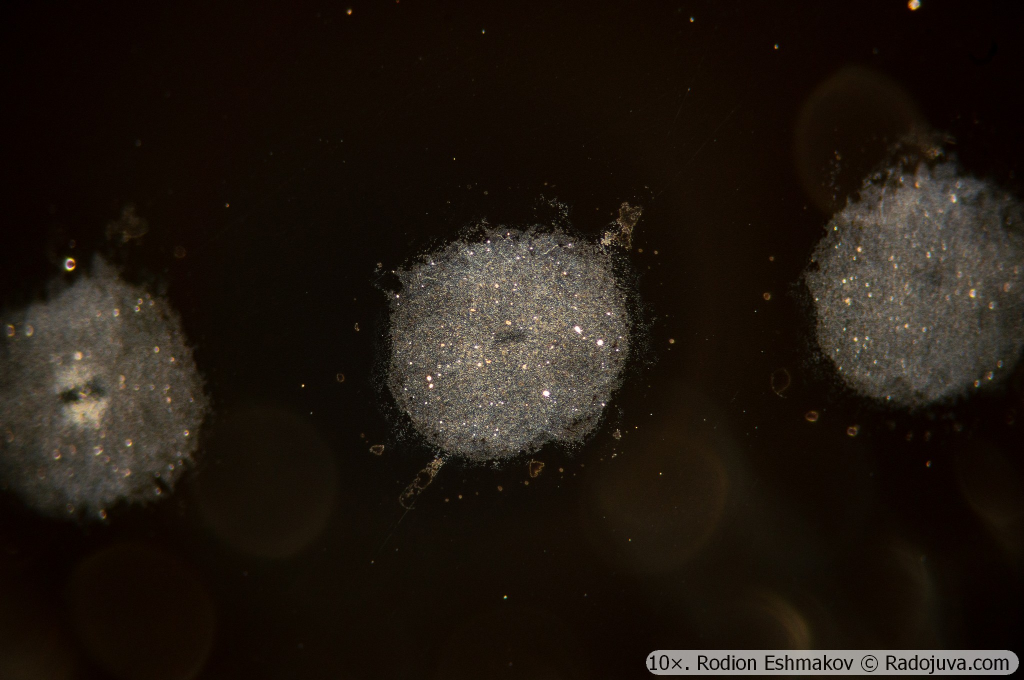
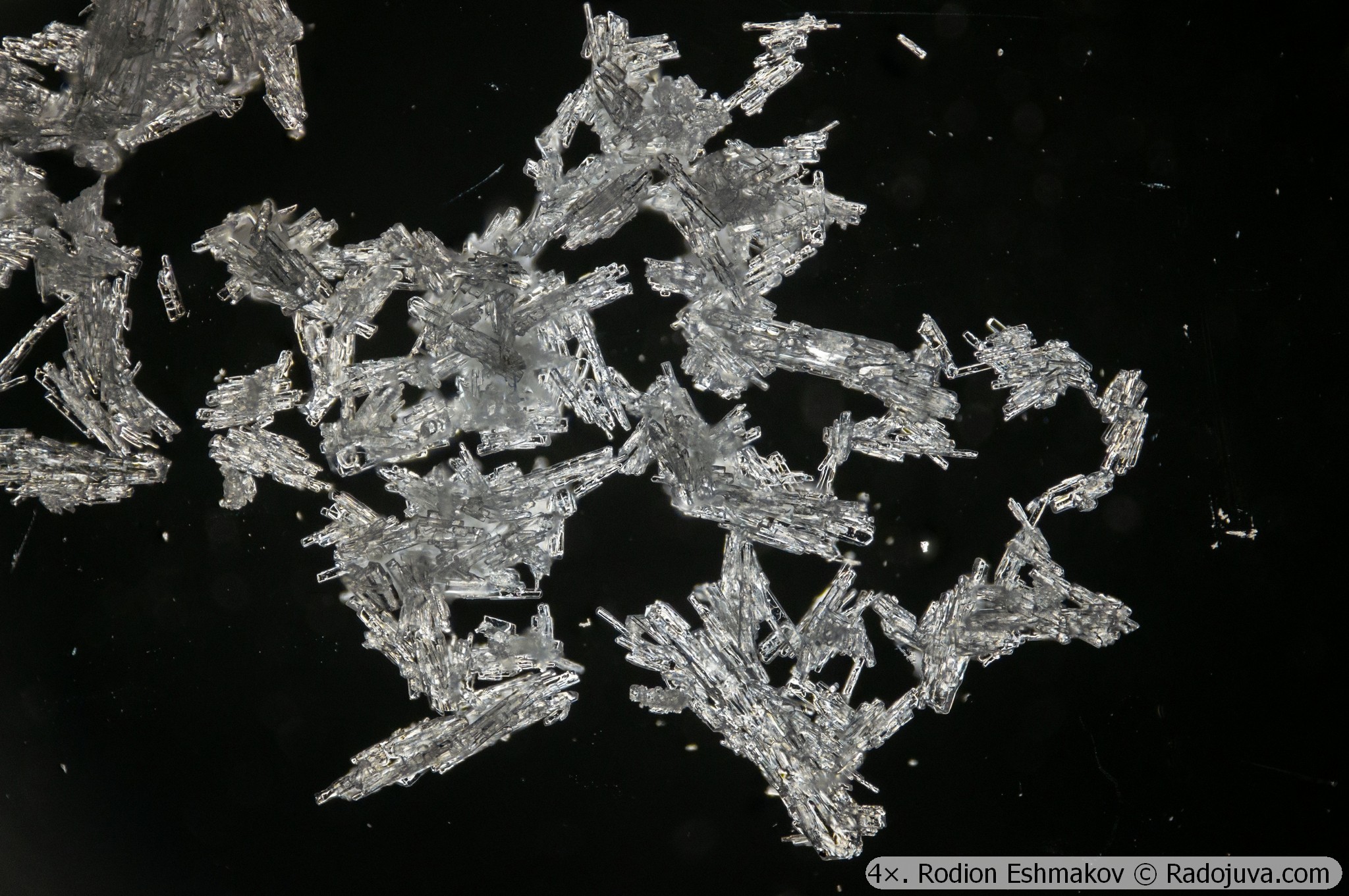
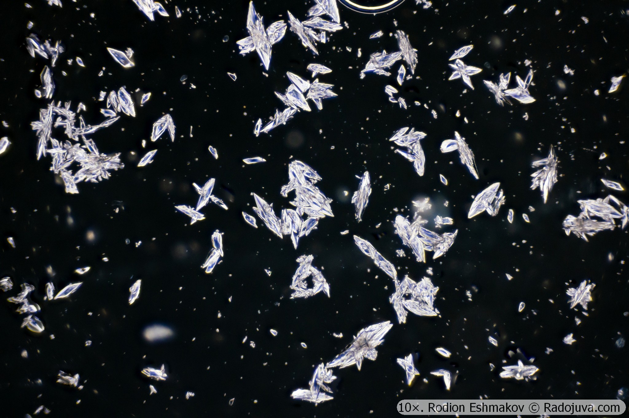
The KF-1 condenser can also be used to achieve darkfield illumination for lenses with a numerical aperture of no more than ~0.4 (typical for 20×) and a subject field no larger than ~8 mm (4× with a 30 mm image). To do this, select the largest diameter annular diaphragm (slot 90) and adjust the condenser height. A disadvantage of this darkfield illumination is the presence of color distortions associated with pronounced chromatic aberrations of the anachromatic condenser. Examples of photographs taken with 4×-20× lenses and the KF-1 condenser in darkfield mode are shown below.
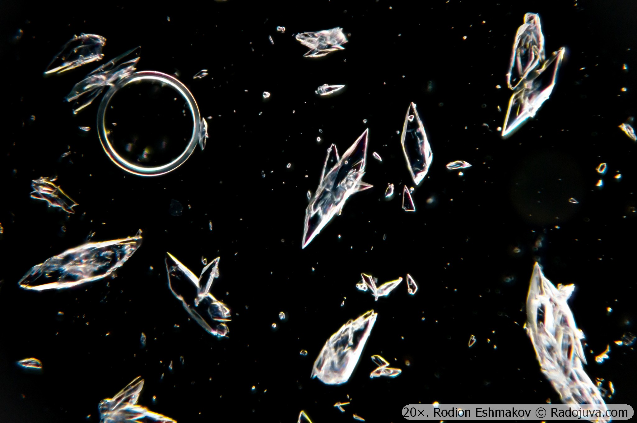
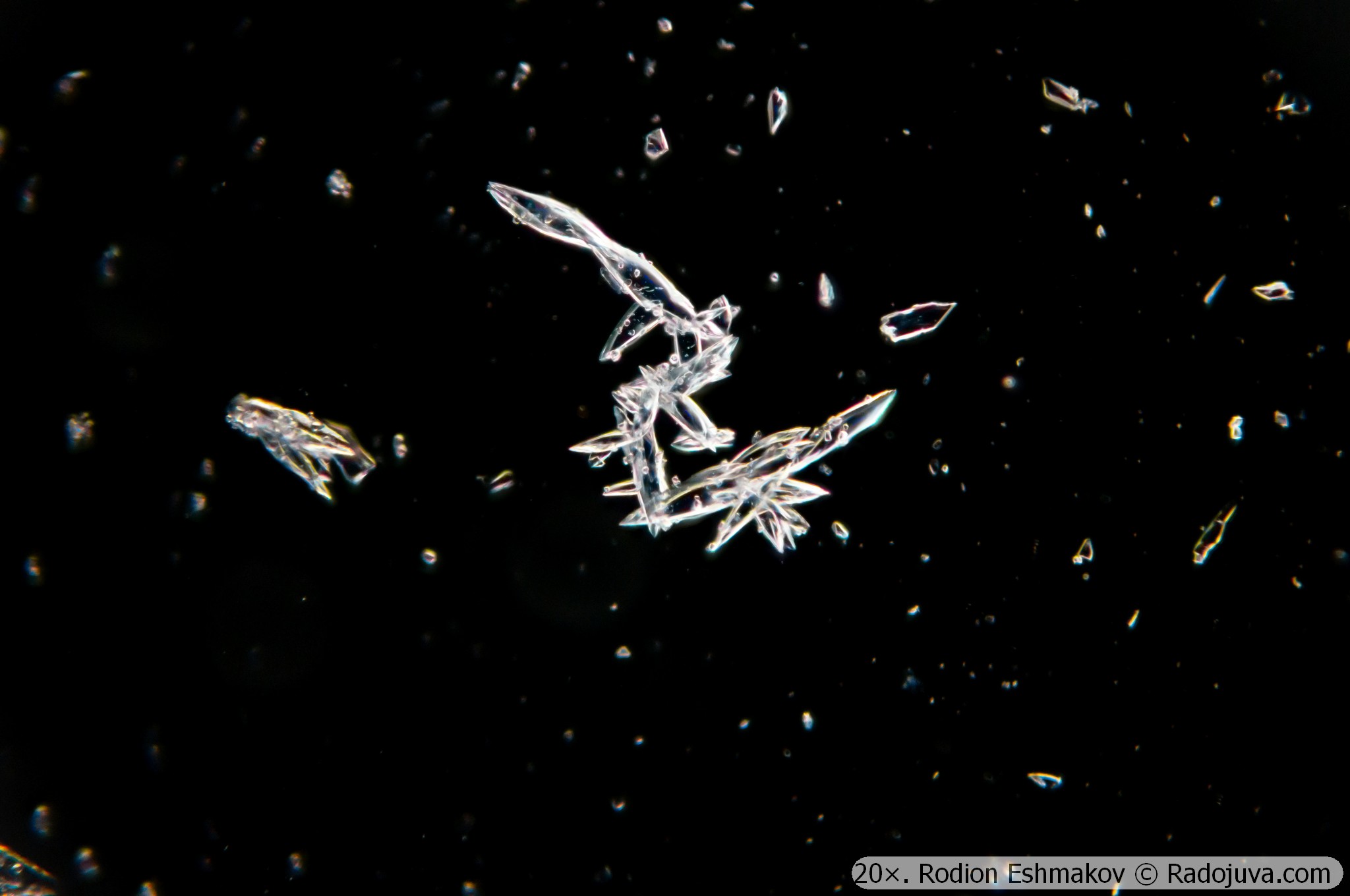
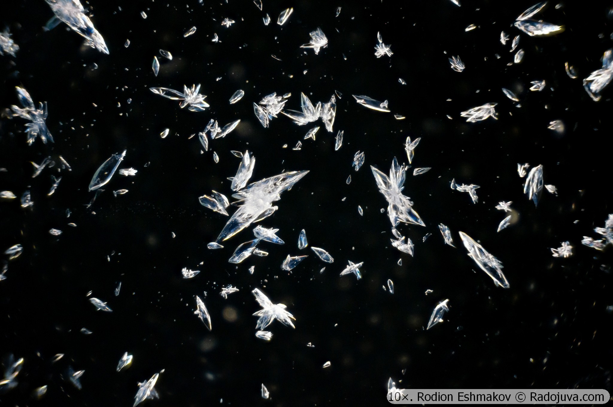
The identified advantages and disadvantages of the KF-1 condenser prompted me to calculate a condenser that would allow for seamless observations with dry objectives, including survey lenses with an 8 mm field of view and high-aperture lenses with a numerical aperture of 0.9. This condenser would also be suitable for dark-field illumination without color distortion. The calculation takes into account the actual dimensions of the illumination system. Aspherical elements were excluded from the calculation, as their cost is prohibitive for small-scale production. The result is a four-lens achromat-aplanat condenser that meets the specified requirements. Modern materials with ultra-high refraction (n>1.9) were used in the calculation. A light source with hemispherical geometry is optimal for this condenser, but not required.
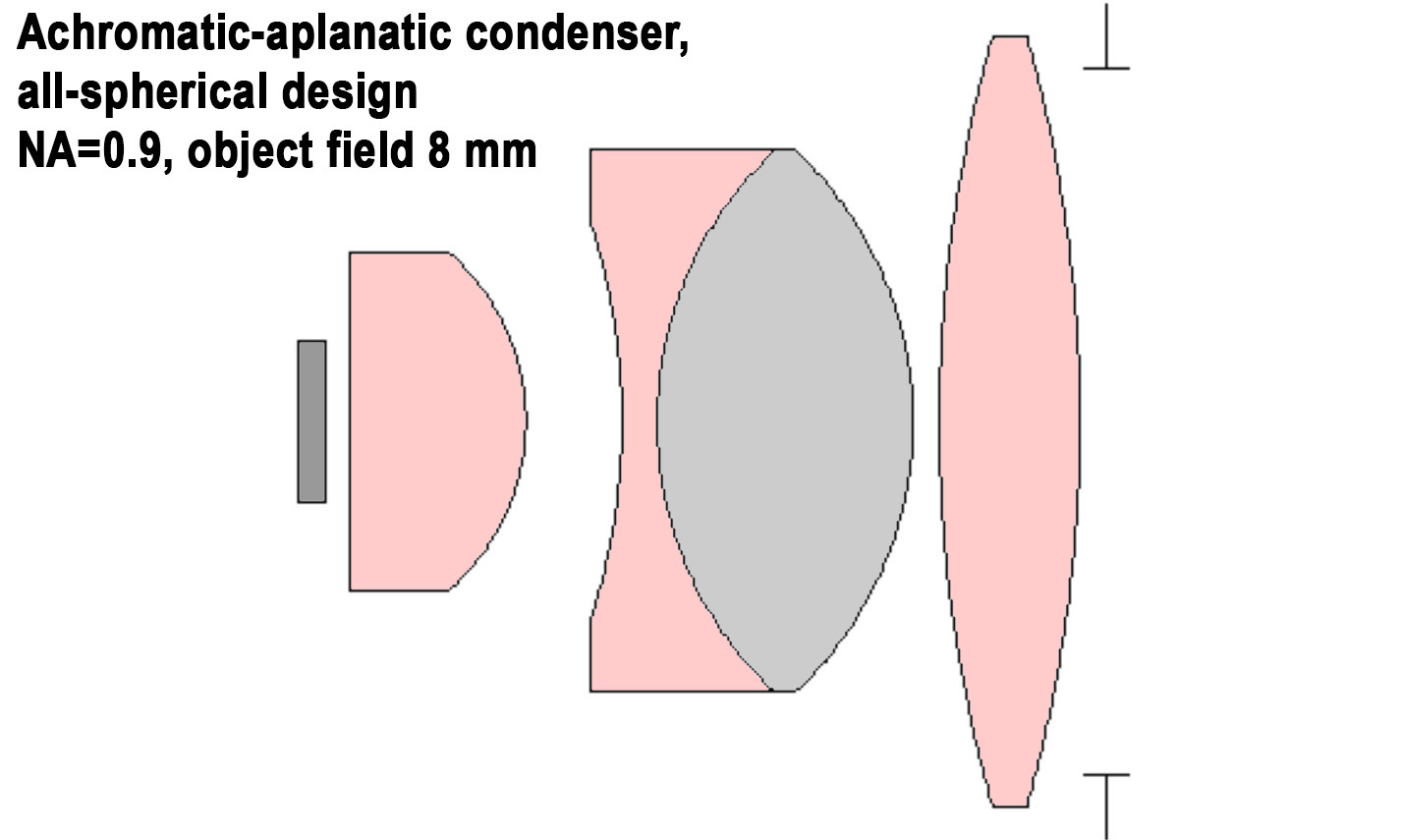
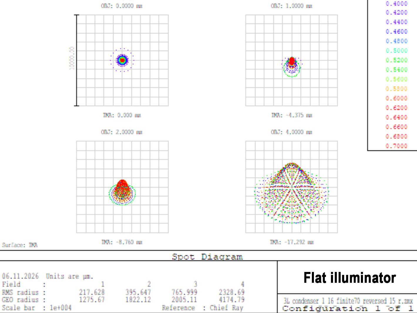
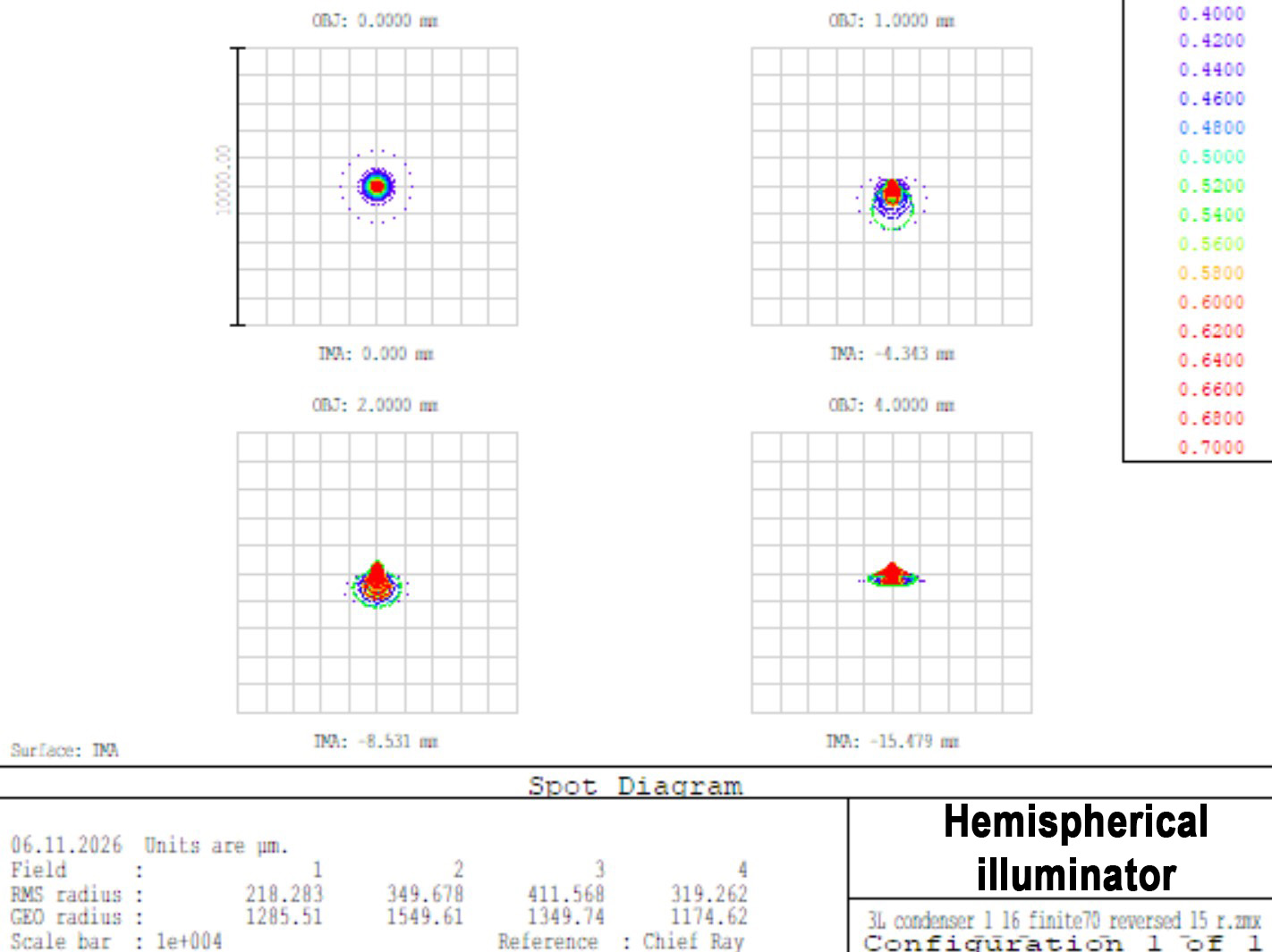
For comparison, an Abbe condenser with a numerical aperture of 0.7 under the same conditions.
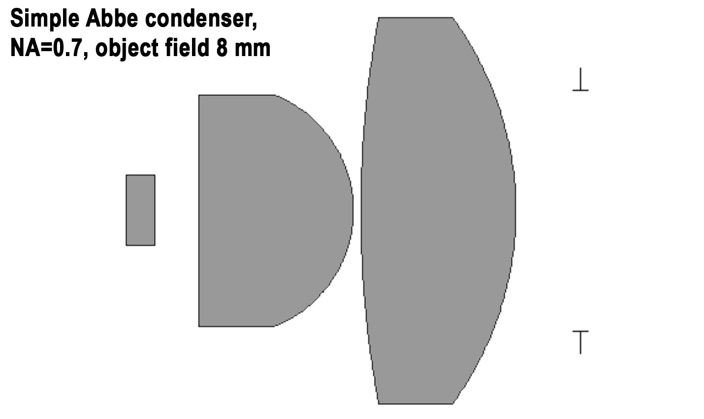
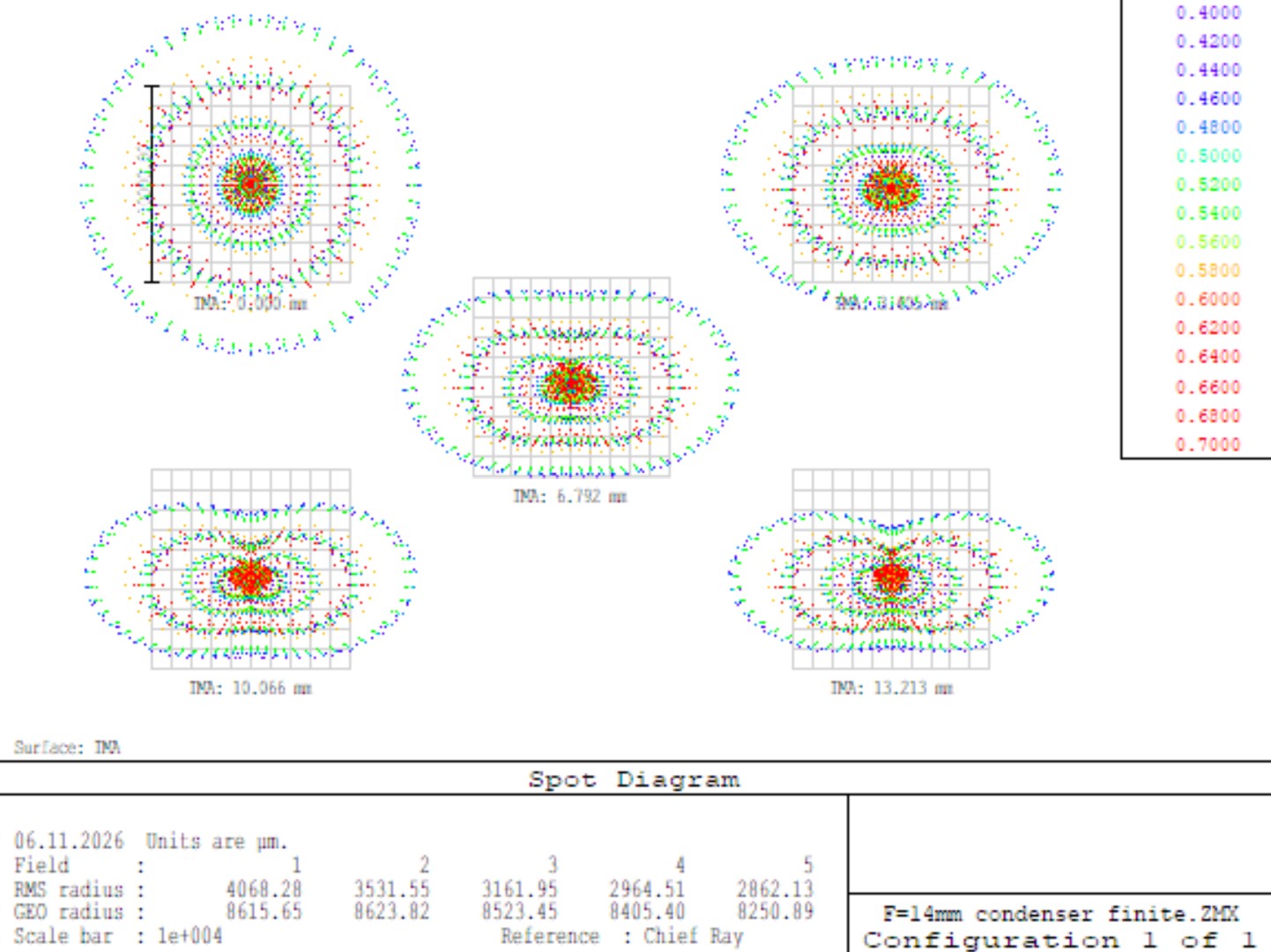
As you can see, even with a significantly smaller aperture, the Abbe condenser has a secondary spectrum literally 10 times longer, and the size of its aberration spots is 10-20 times larger than that of the achromatic-aplanatic condenser I calculated. Overall, Abbe condensers are extremely ineffective when it comes to providing high-quality illumination with large-aperture objectives—they generate a large amount of stray light, which can negatively impact image contrast. And, as noted earlier, they are also not very convenient to use as darkfield lenses, and are practically impossible to use with high-aperture objectives.
Lenses for the KF-1: General Information
The KF-1 phase contrast device comes with four lenses: the F OM-5 10×0.3, the F OM-27 20×0.4, the F MS 40×0.65, and the F MI OM-41 90×1.25. The lenses in the KF-1 kit are identified by the red letter "F" on their barrels. The lenses in the KF-4 kit had a black letter "F" on their barrels, although all of them, except for the 90×1.25, are compatible with the KF-1. The KF-1 and KF-4 kit lenses comply with the RMS standard—a 20.2 mm thread and a parfocal distance of 33 mm. The lenses for the KF-4M are made according to the DIN standard with a parfocal distance of 45 mm.
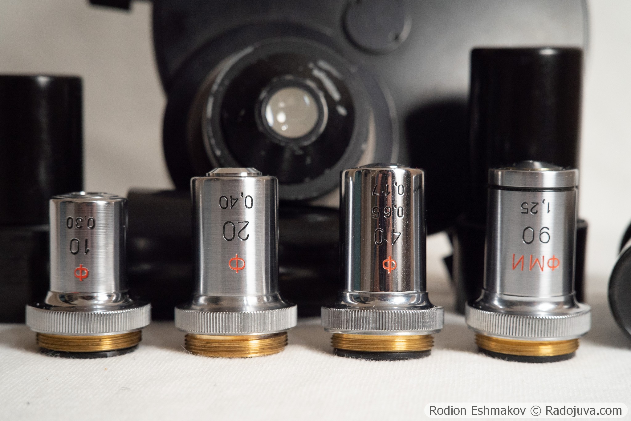
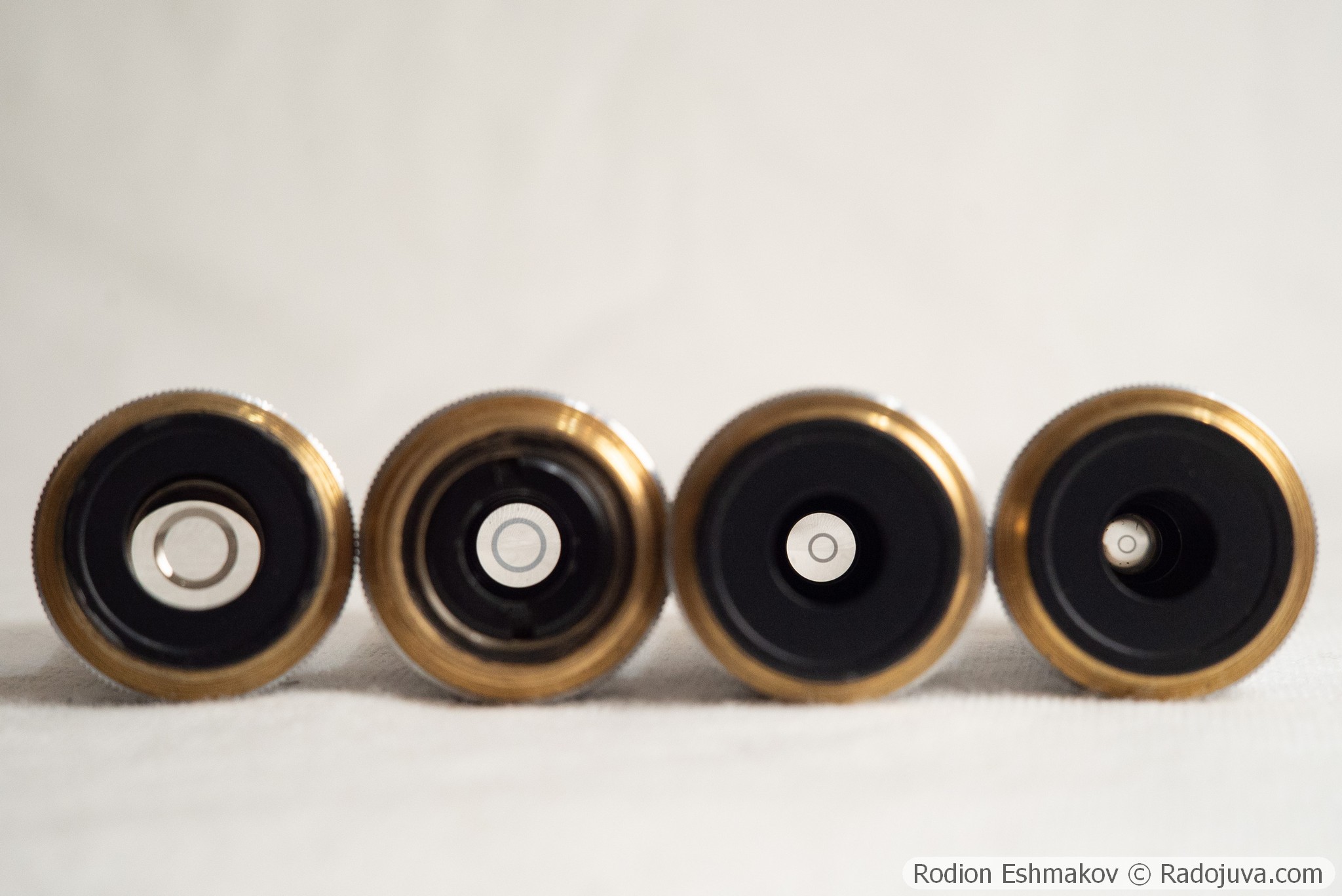
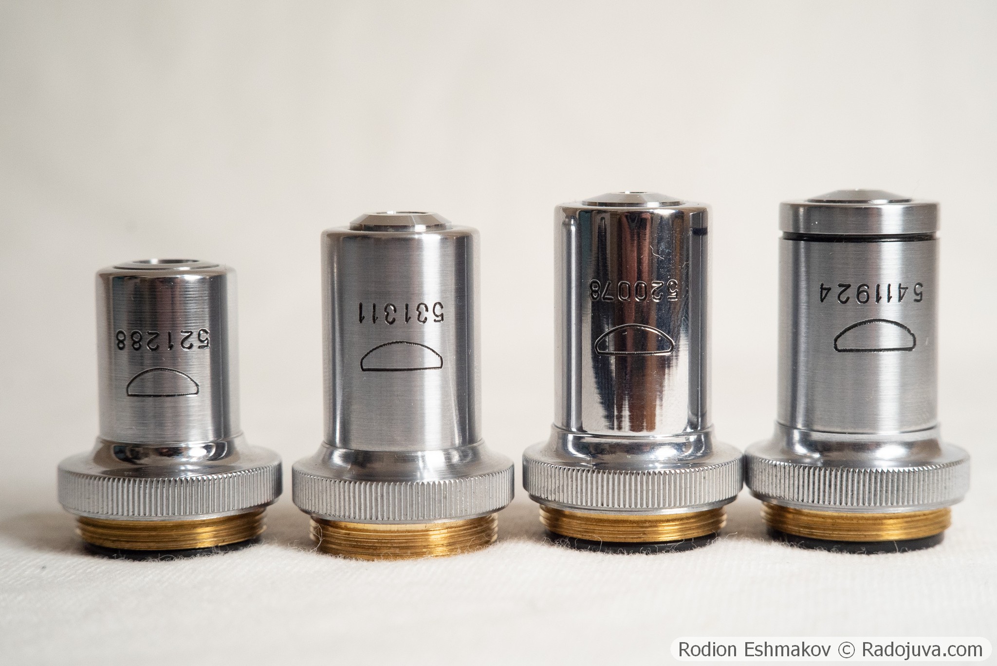
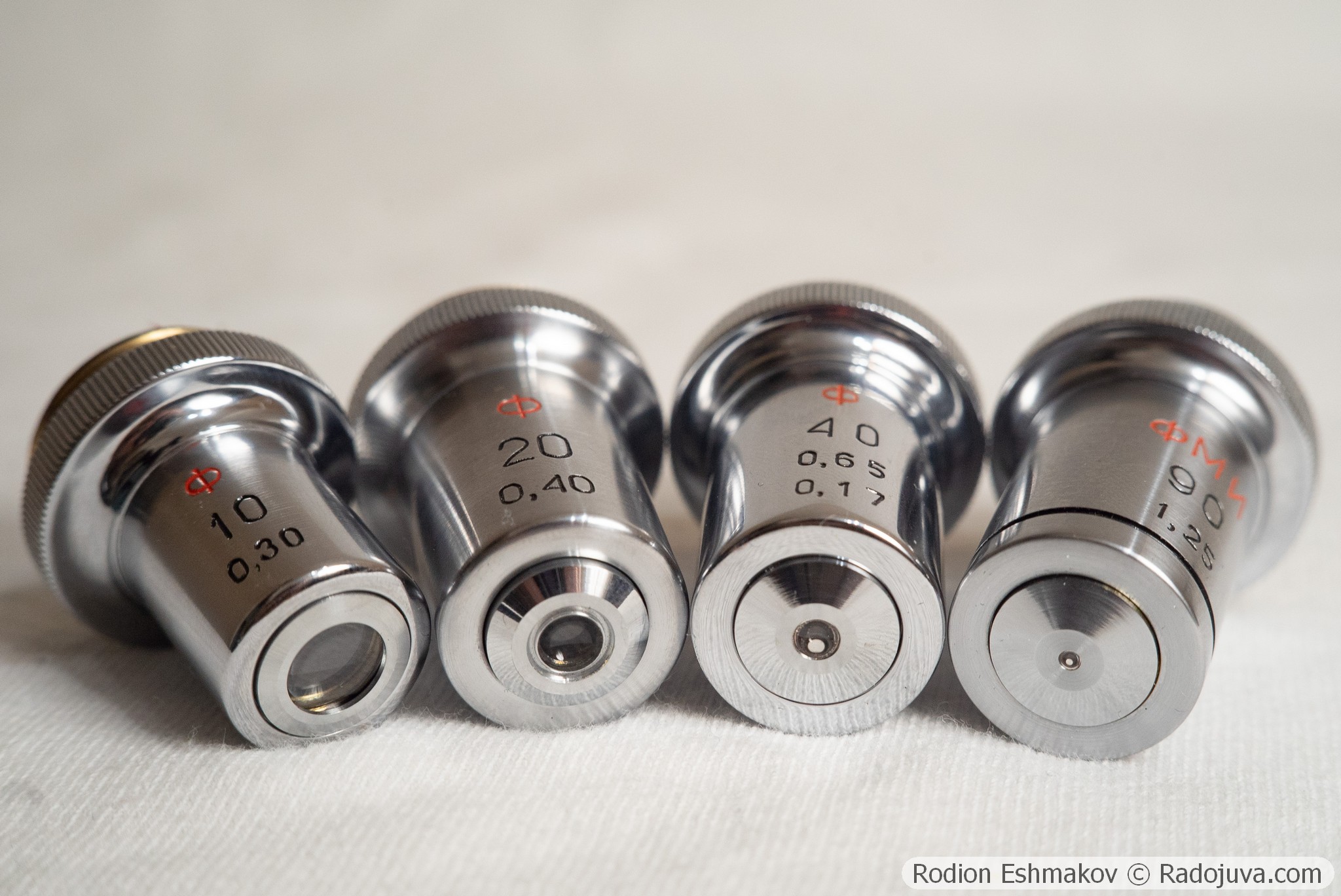
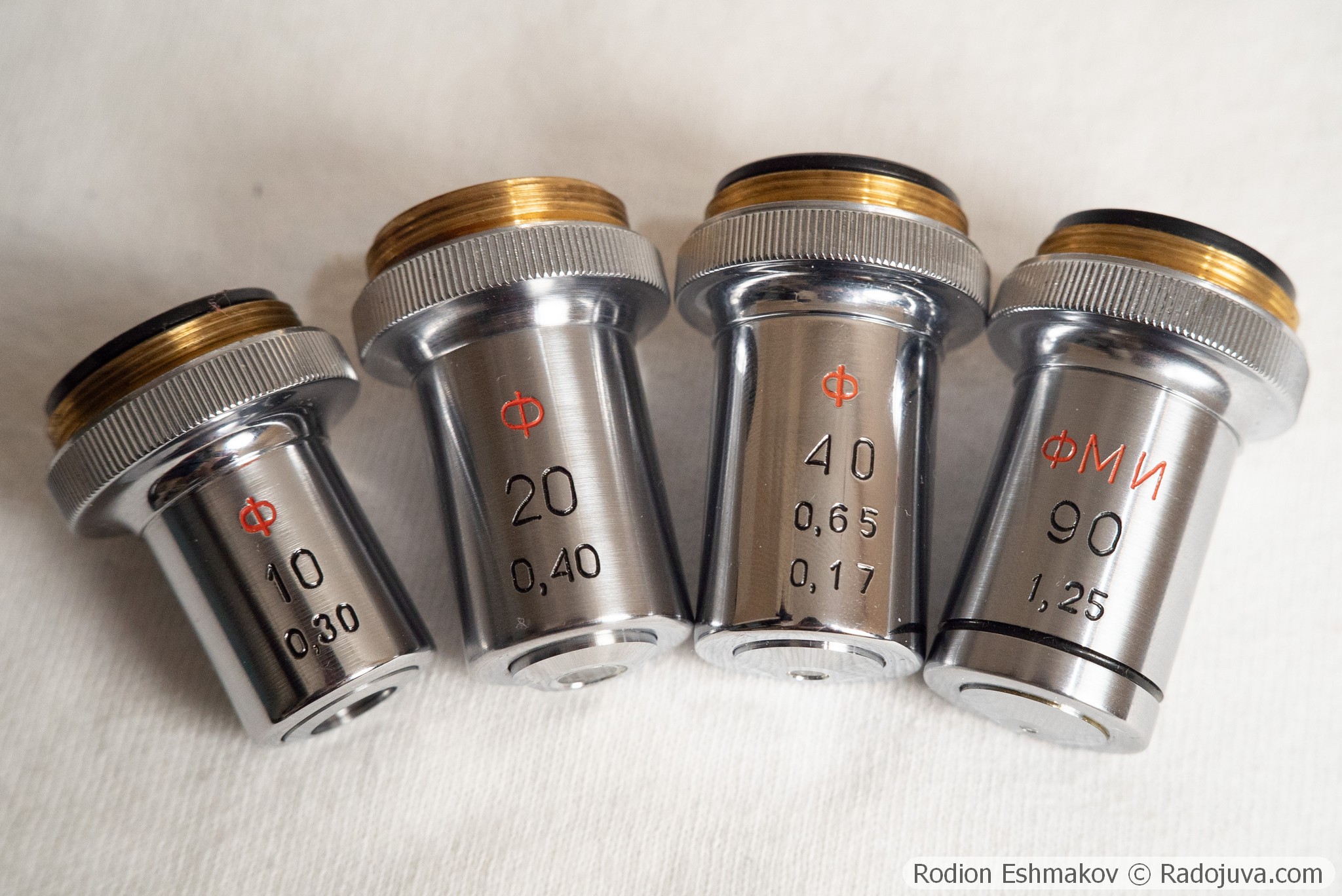
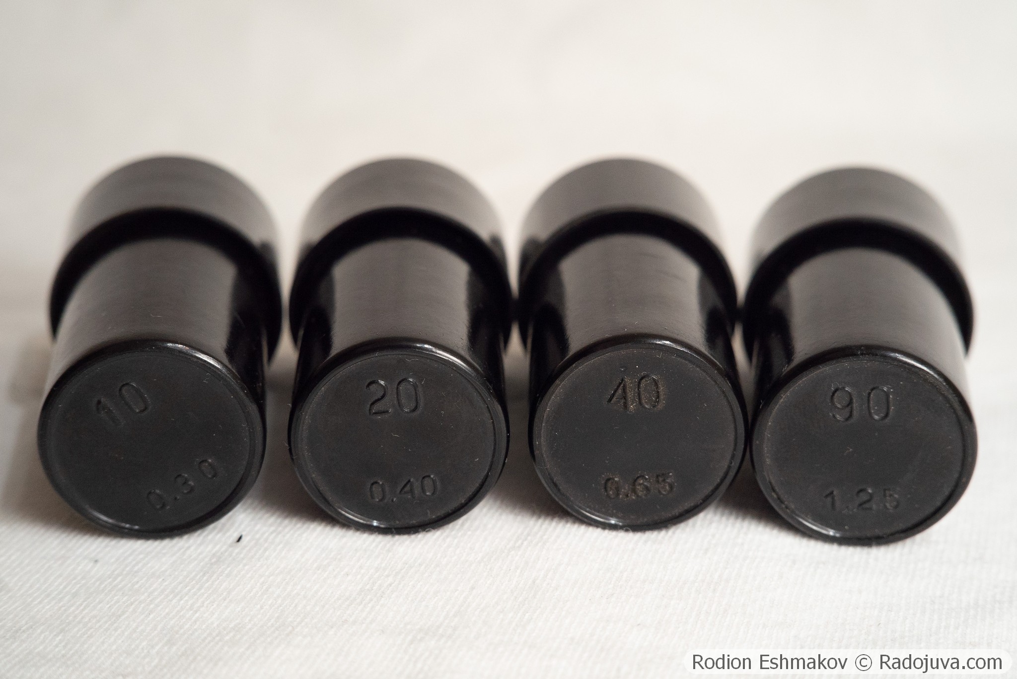
The KF-1 and KF-4 objectives are based on the standard Progress/LOMO OM-5 10×0.3 , OM-27 20×0.4 , MS 40×0.65 , and OM-41 90×1.25 MI lenses. Like the standard versions, the phase-contrast MS and OM-41 lenses do not have a spring to protect the lens from damage to the front element upon impact with the specimen. The objectives included with the KF-1 lens have different types of phase rings—they are likely manufactured using different methods. A very common problem with phase-contrast lenses is fading (oxidation) of the phase rings, which makes it impossible to implement the phase contrast method. This defect can sometimes be difficult to visually identify, but fading and high transparency of the rings should raise concerns when purchasing.
The water immersion objectives stand out: the achromatic F VI OM-23 40×0.75 and the apochromatic Apochromatic VI F 70×1.25 (also known as the F APO VI 70×1.23). They are not included with the KF-1, but they also have a red letter F on the barrel and are compatible with the KF-1 condenser. To use the 40×0.75 objective, you need a ring in slot 40, while for the 70×1.25 objective, you need a ring in slot 90 of the KF-1 condenser. This may be a coincidence, as these objectives were intended for use with a zoom condenser with a ring insert. Both objectives are parfocal with the KF-1's kit lenses.
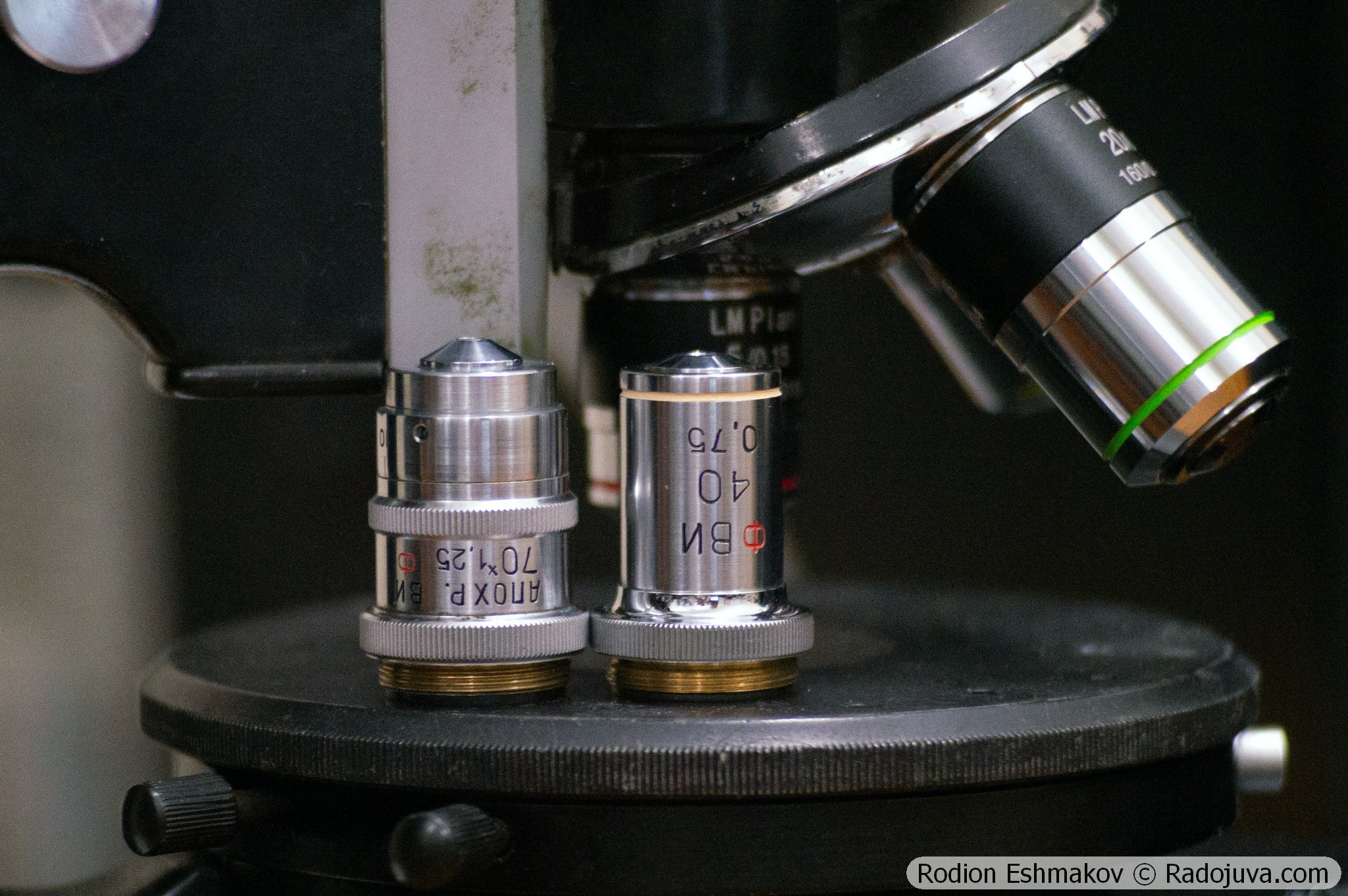
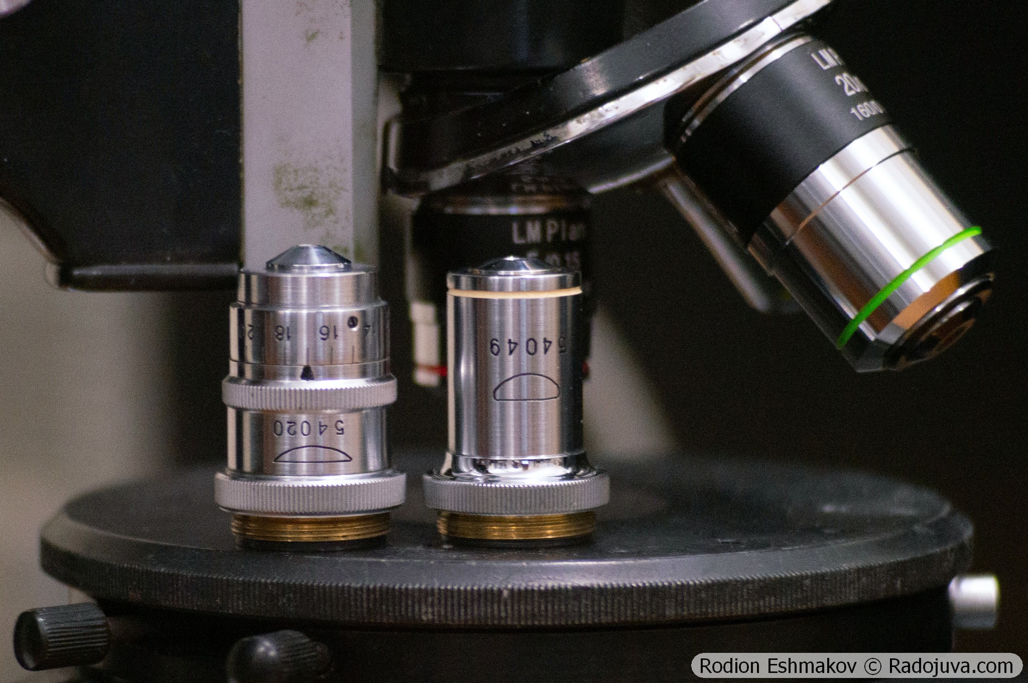
All objectives for the KF-1 and KF-4, as well as the F VI OM-23 40×0.75 and F APO VI 70×1.25 water immersion objectives, are uncoated, and their internal blackening and light protection are extremely poor. The phase ring itself also contributes to reduced image contrast. As a result, all these objectives perform extremely poorly in standard brightfield conditions. Due to this, and the fact that lateral chromatic aberration is not corrected, I do not recommend using these objectives for brightfield observations.
Lenses for KF-1: 10×0.3 F OM-5
Optical design – 5 lenses in 3 groups, high-aperture aplanat;
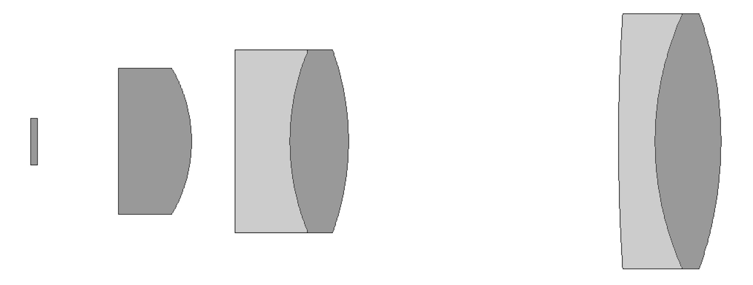
Drawing of the optical scheme of F OM-5 10×0.3.
Type of correction – achromat;
Tube distance – 160 mm;
Parfocal distance – 33 mm;
Magnification: 10×;
Numerical aperture – 0.3 (phase ring 0.2);
Working distance – 7.2 mm;
Cover glass thickness: 0-0.17 mm;
Mounting type – RMS standard (4/5” × 1/36” thread);
Lateral chromatic aberration – corrected with a compensating eyepiece (such as LOMO 4×, K10×/18) or ignored;
Features - microscopic lens, does not have an iris diaphragm and a focusing mechanism.
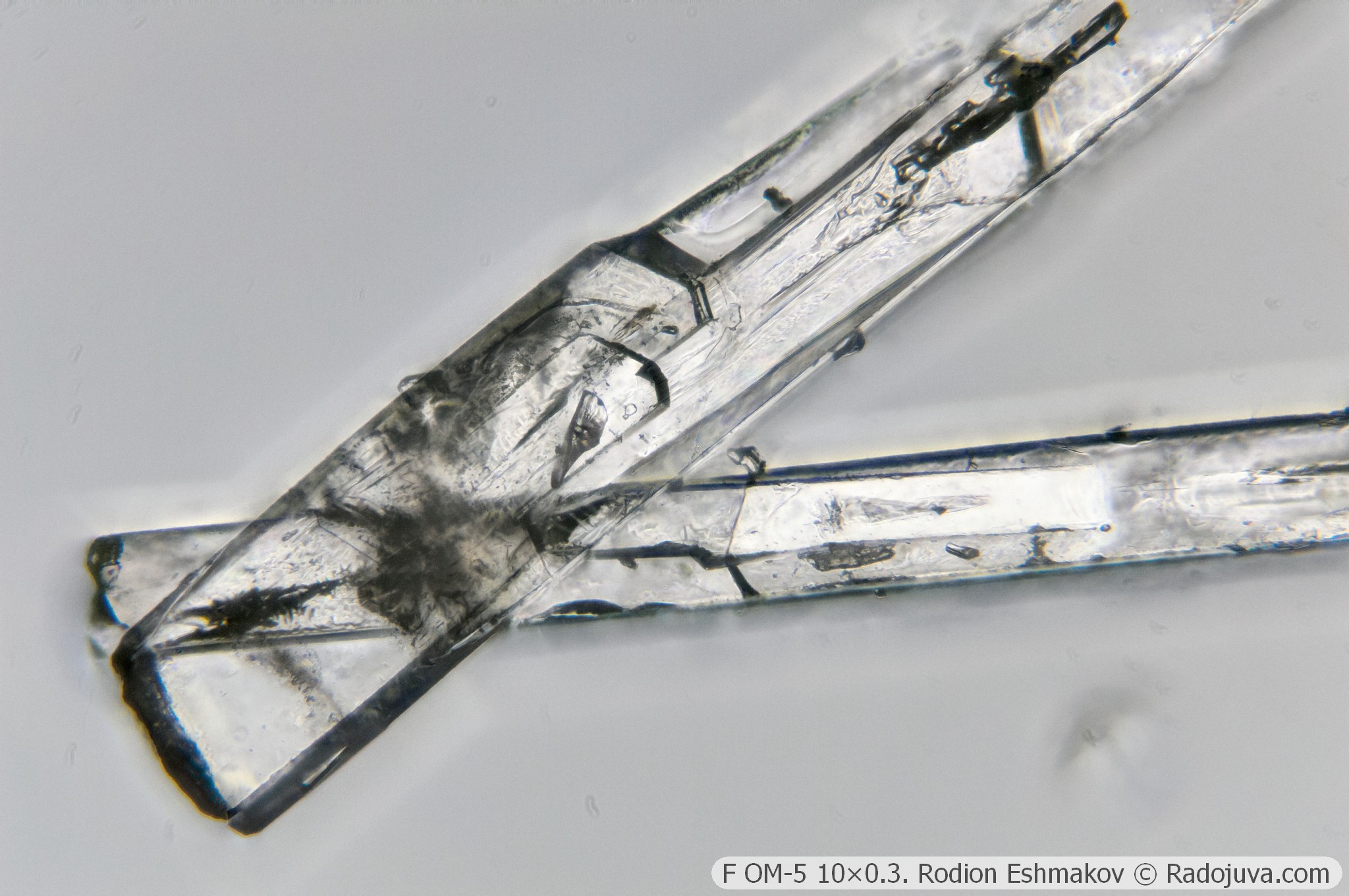
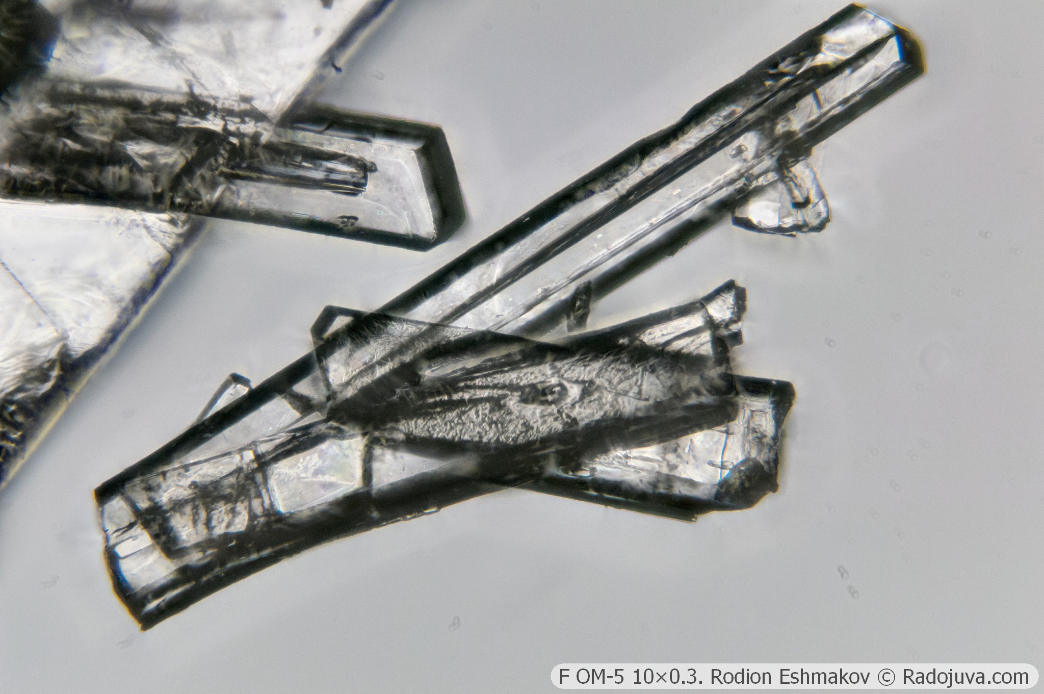
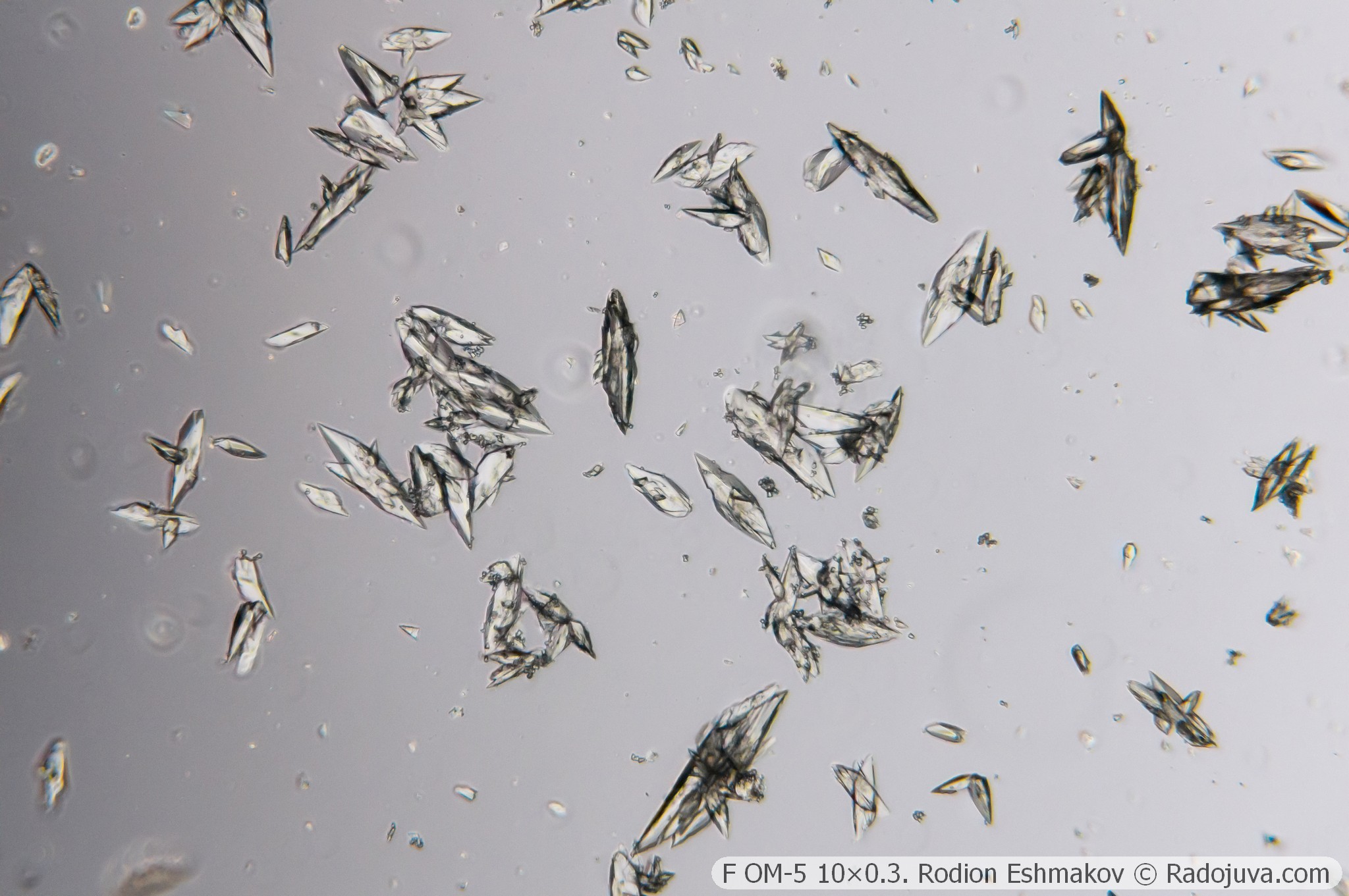
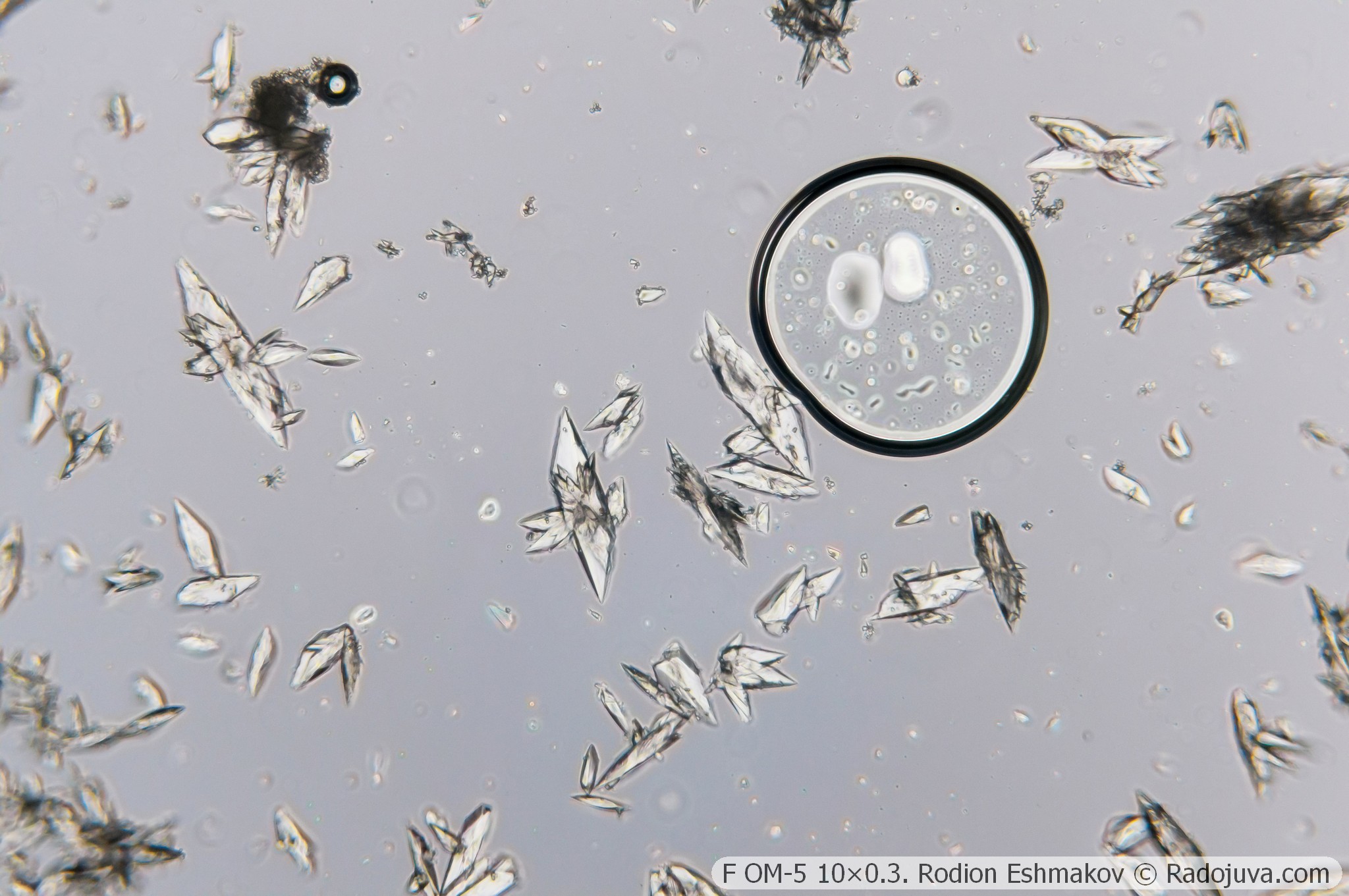
The standard 10×0.3 lens included with the KF-1 is perhaps the sharpest for photography, thanks to its acceptable combination of numerical aperture (including the phase ring aperture) and magnification. While the lens is truly excellent in the center of the field, its performance quickly deteriorates at the edges due to very high levels of astigmatism and lateral chromatic aberration.
There's essentially no way to compensate for the lateral chromatic aberration of the F OM-5 10×0.3: the K-series compensating eyepieces overcompensate (like the K5× ML-2 eyepiece I use as a relay component). There are some unverified reports that Huygens eyepieces can act as compensating eyepieces for this lens, but they themselves exhibit an unacceptable range of aberrations.
Apparently, the field curvature makes it impossible to achieve a uniform phase contrast effect beyond the ~20mm image field. The lens's phase ring colors the background brown.
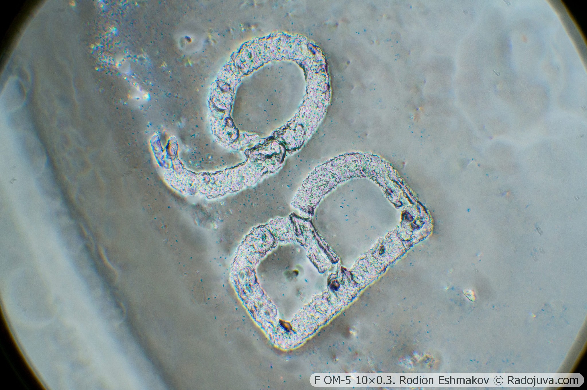
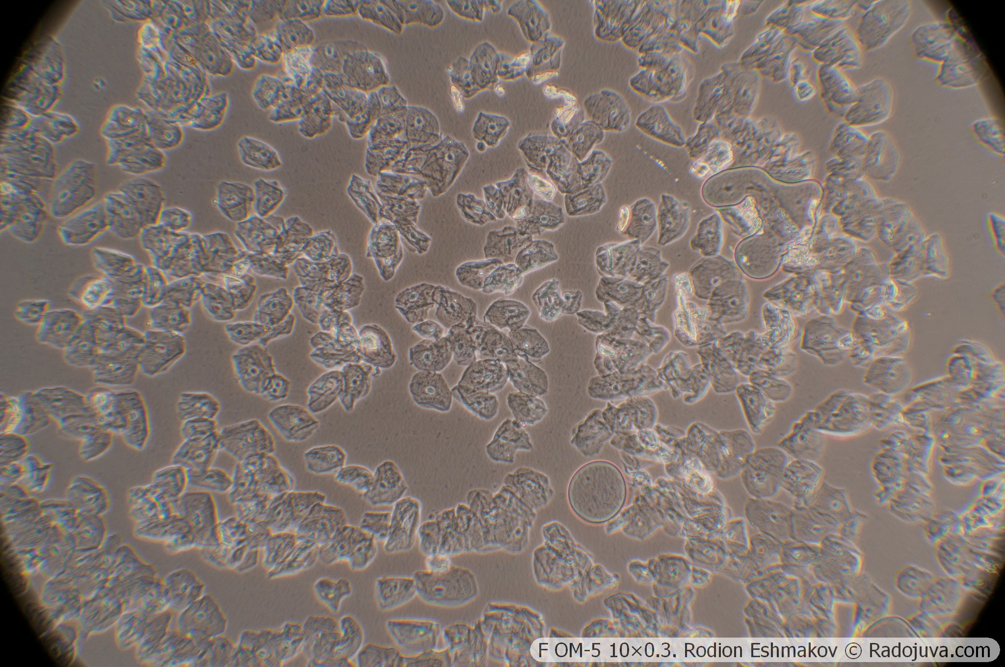
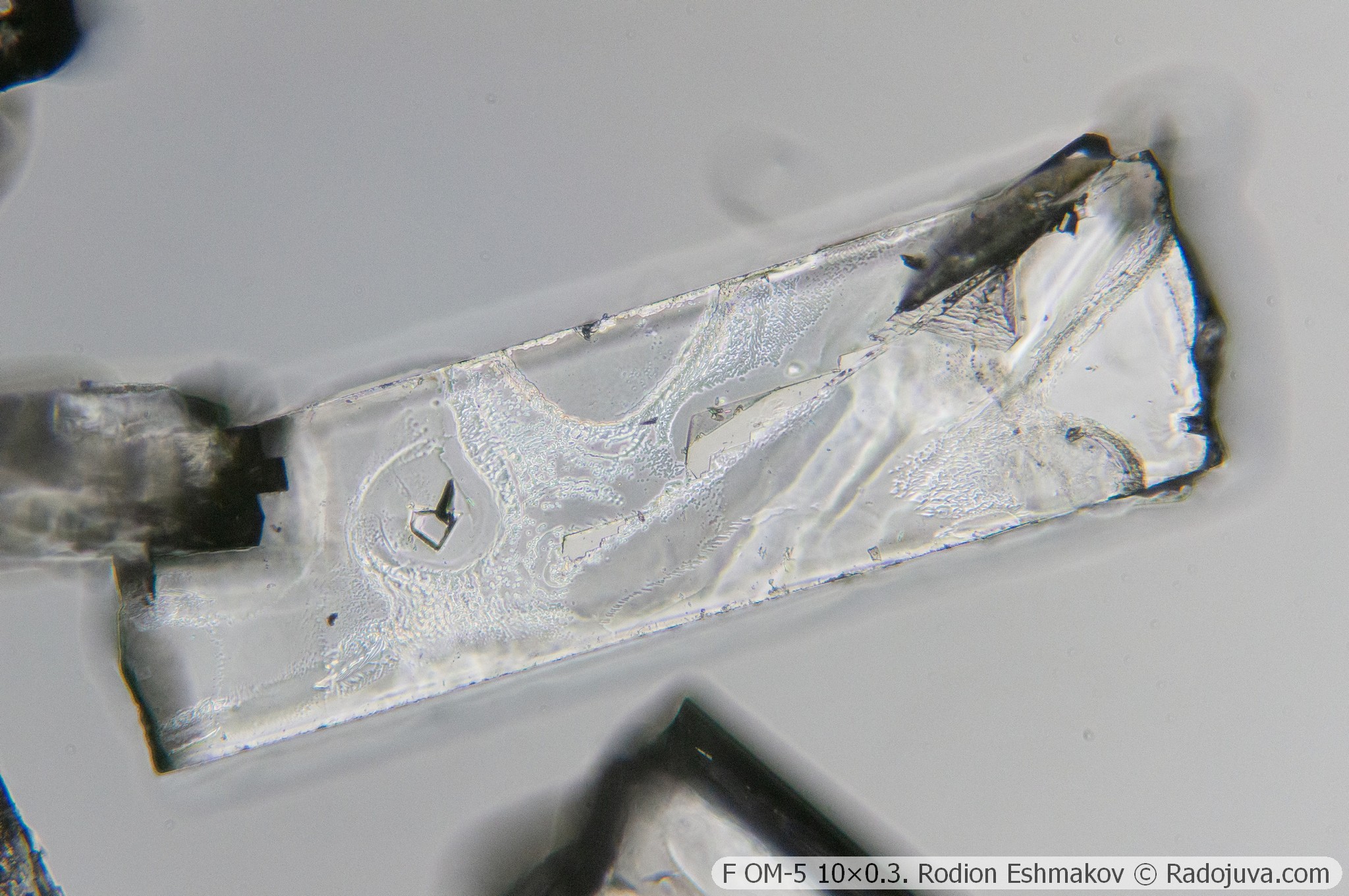
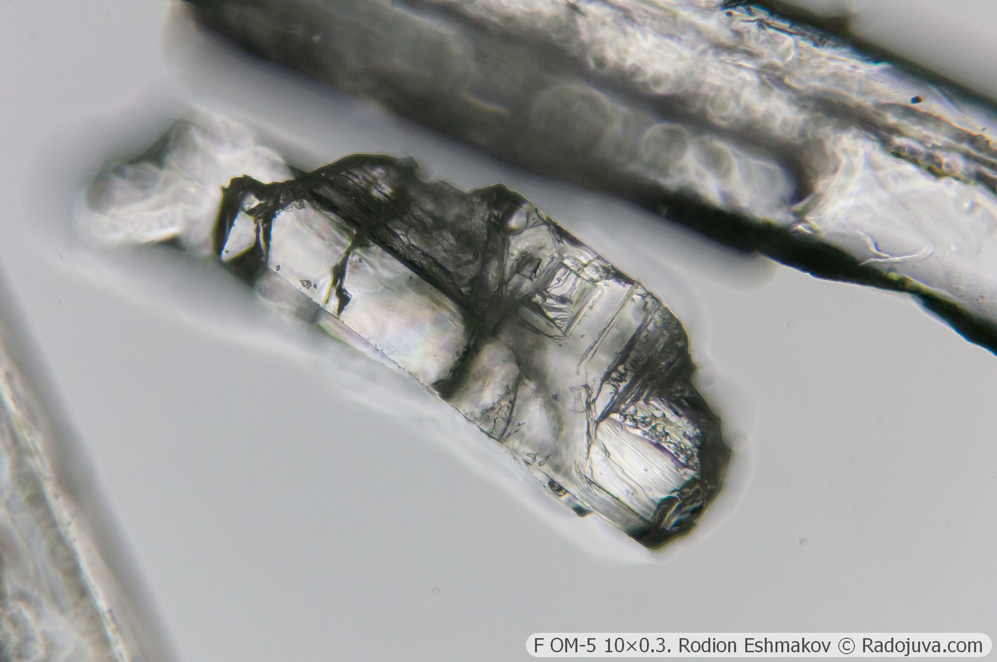
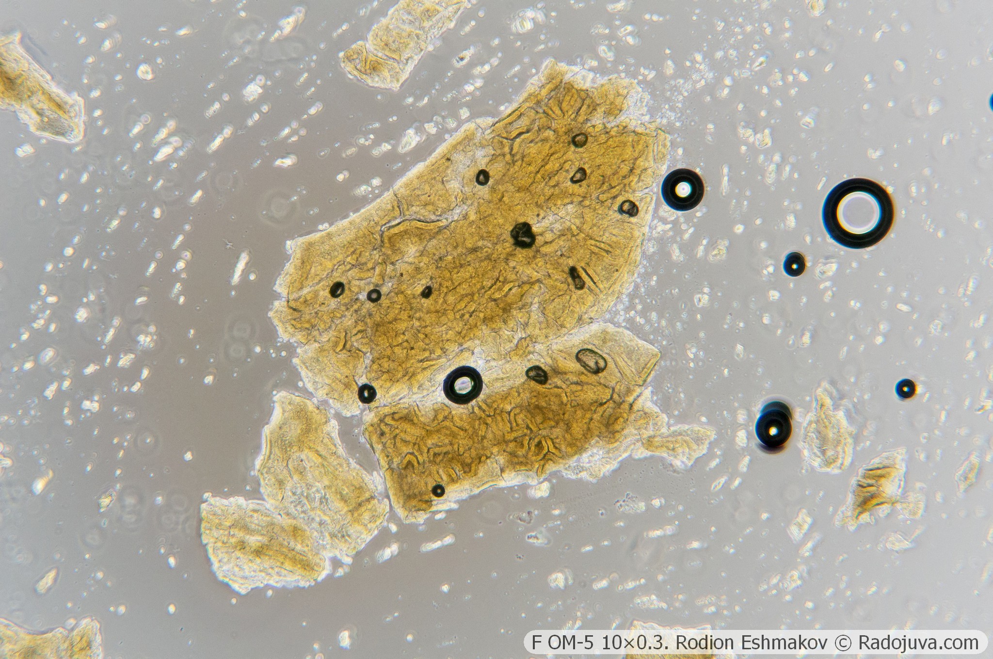
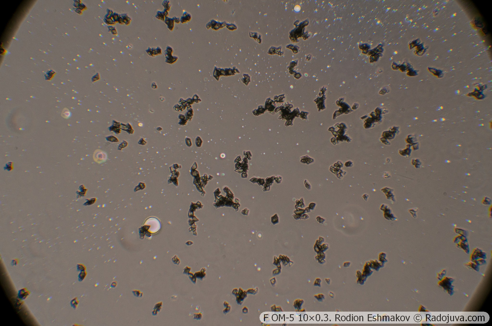
Below are examples of photos taken with the Sony NEX-6 and the F OM-5 10×0.3 lens in prime focus and using the relay from the K5× ML-2 eyepiece and the WitrueHD 75/2.8 10 Mpix lens.
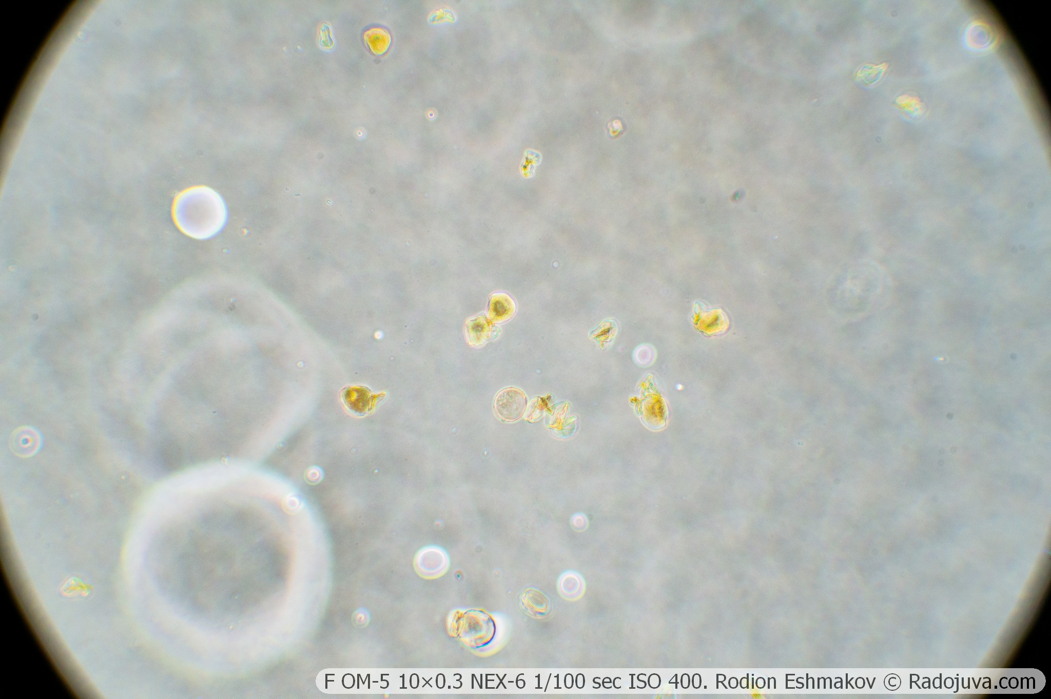
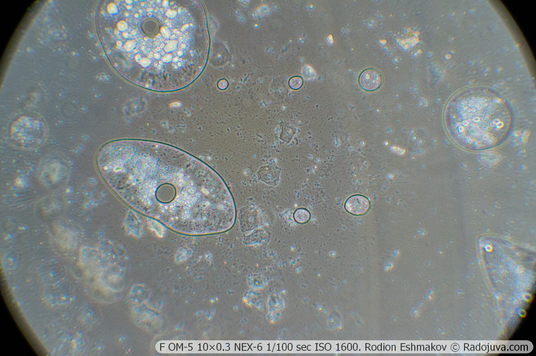
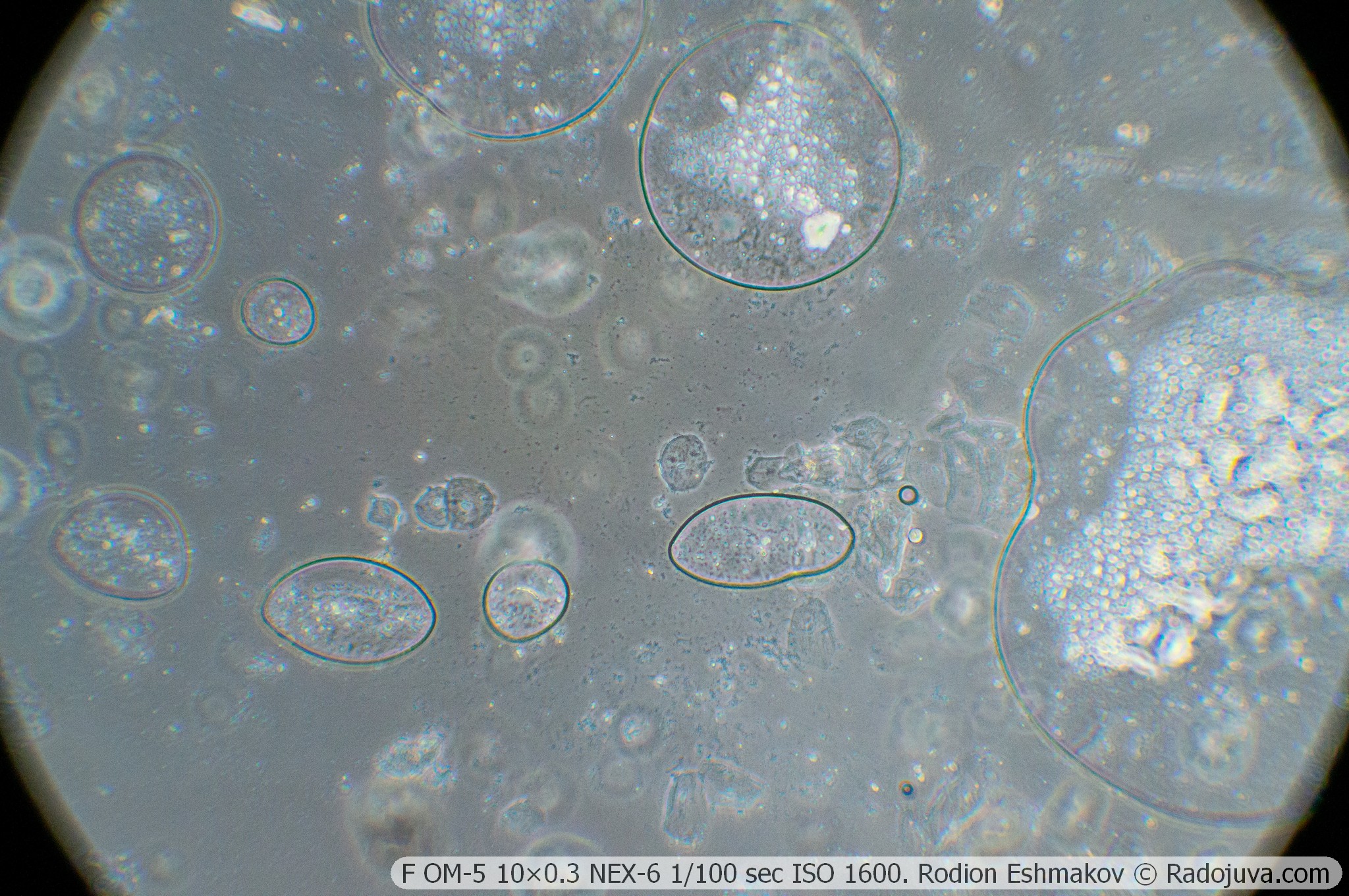
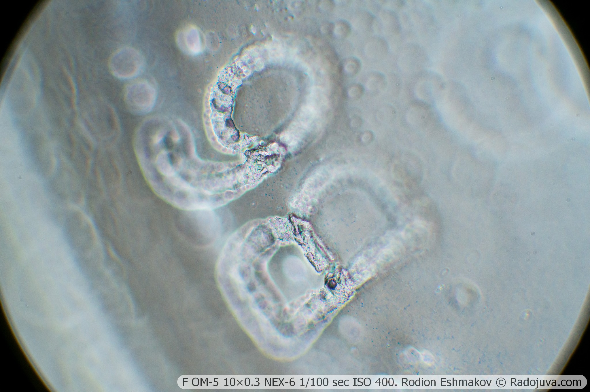
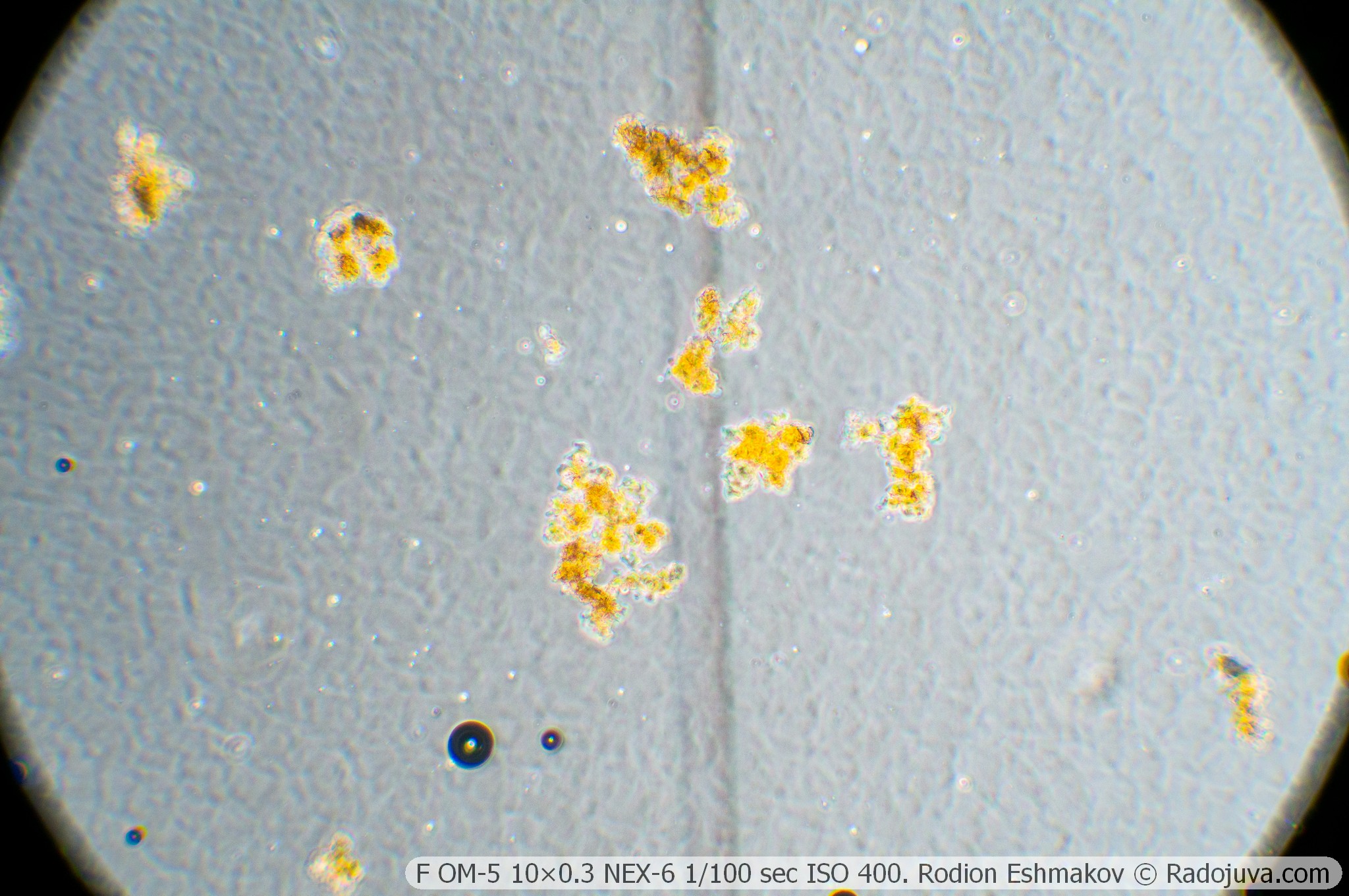
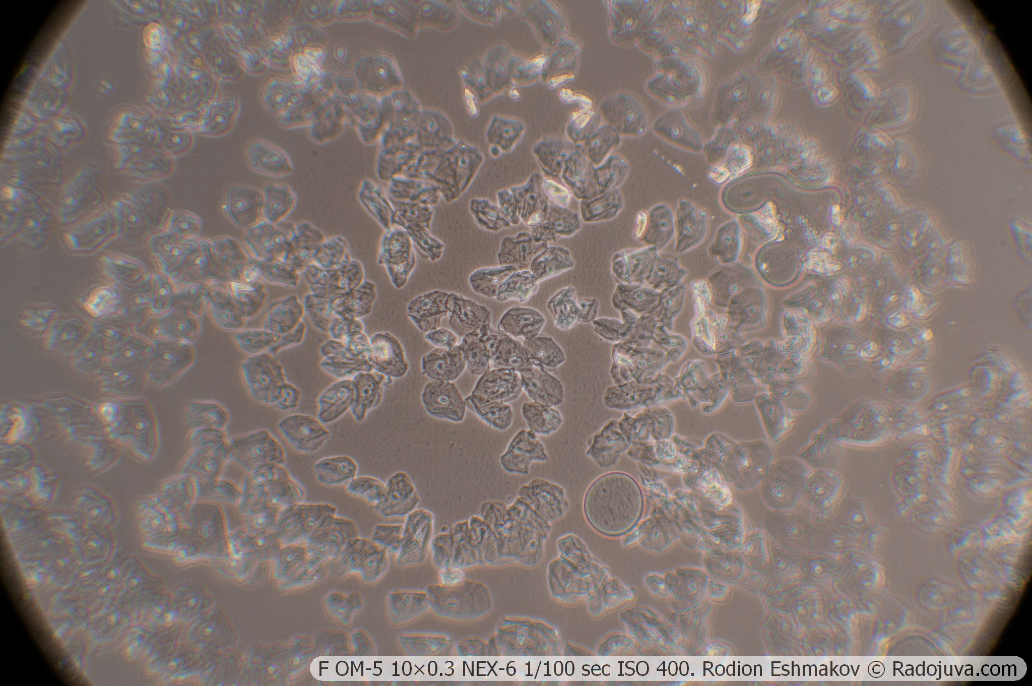
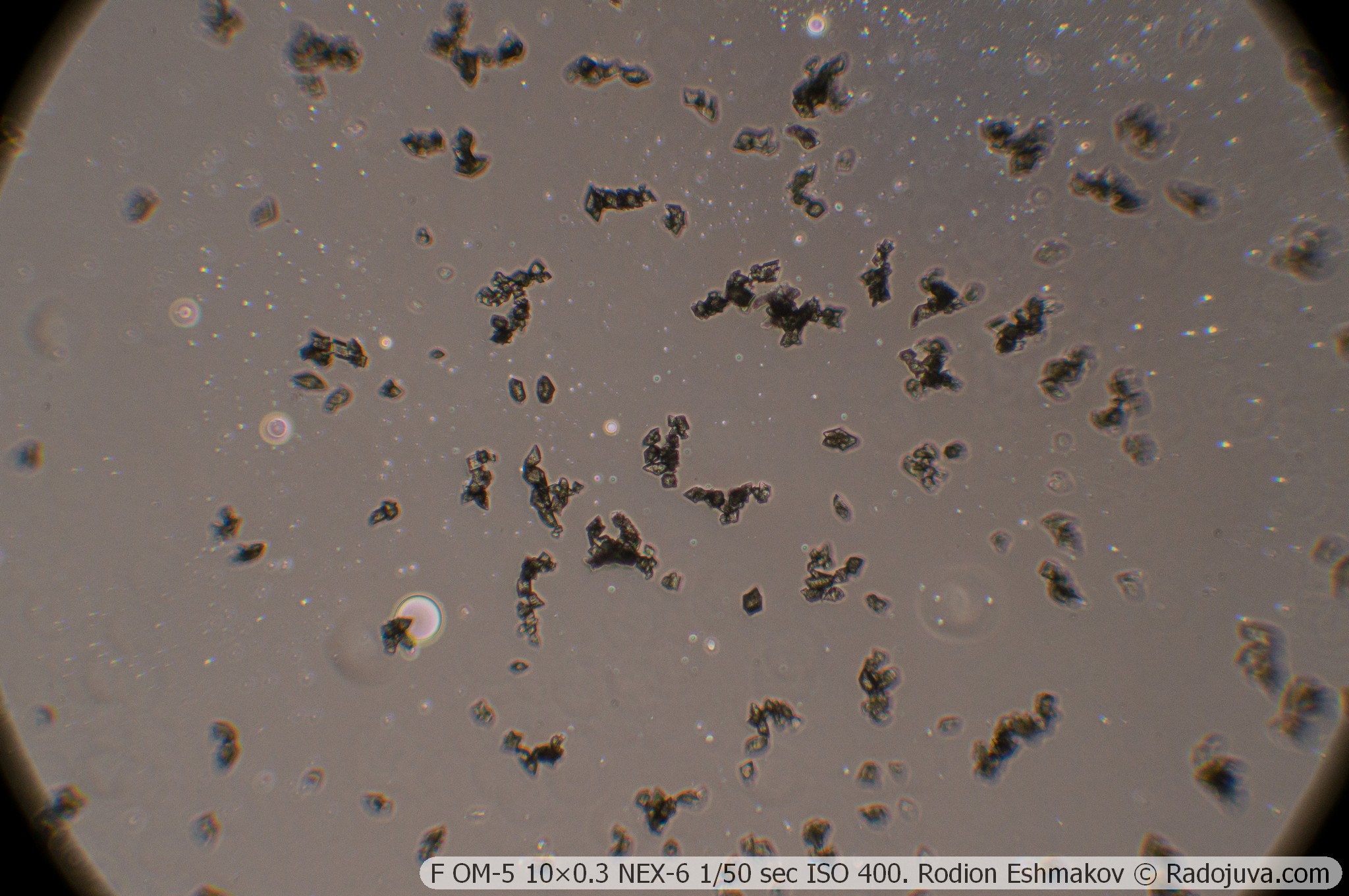
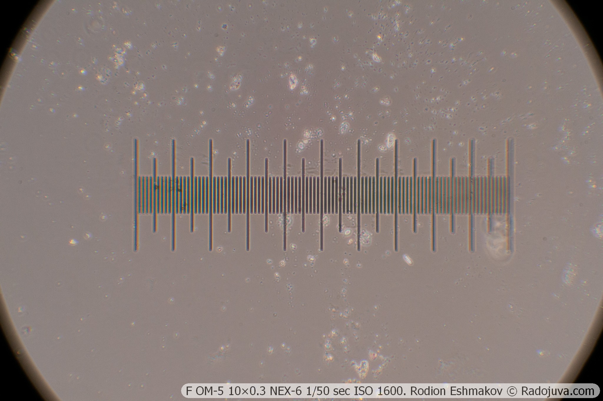
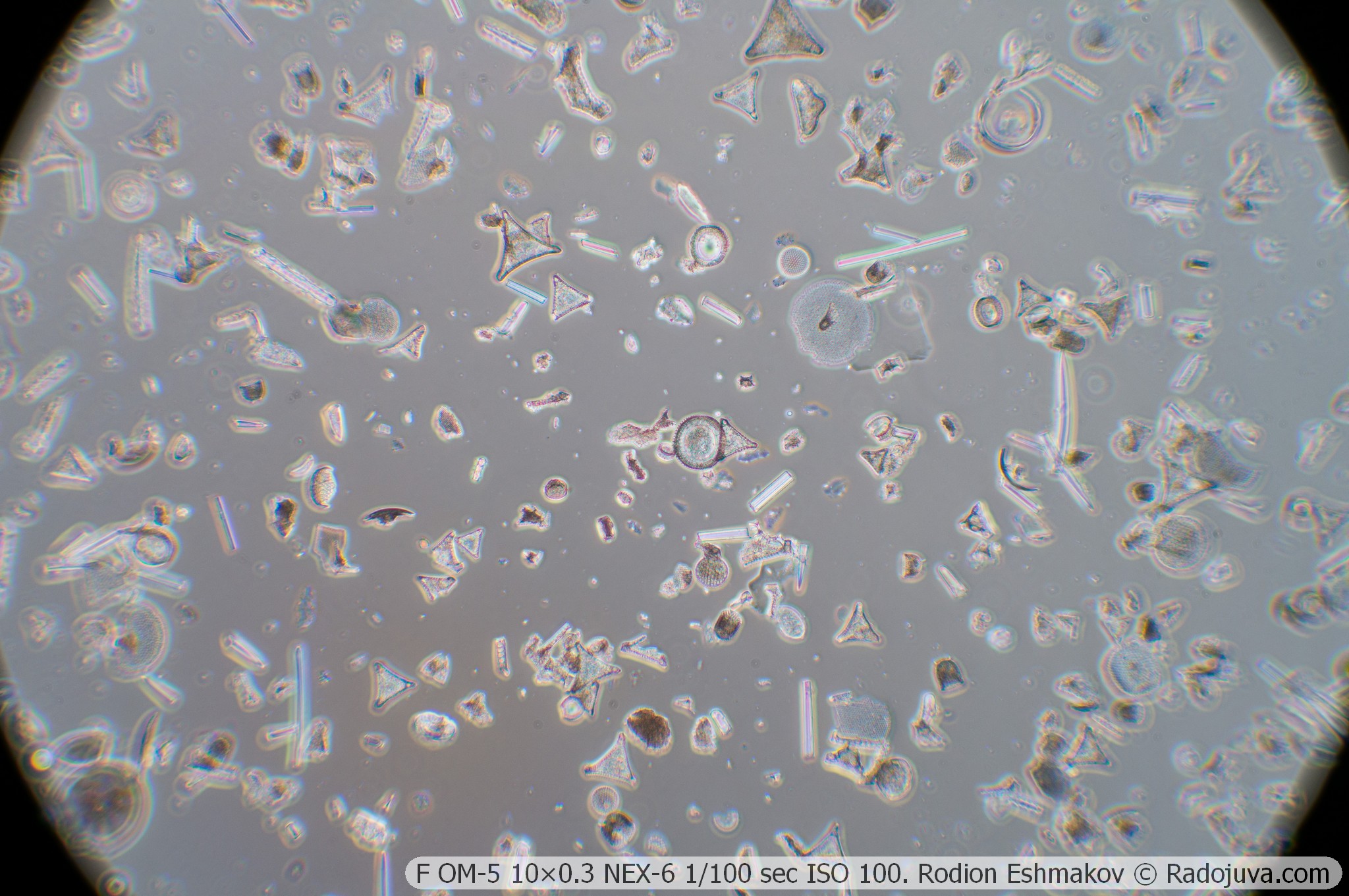
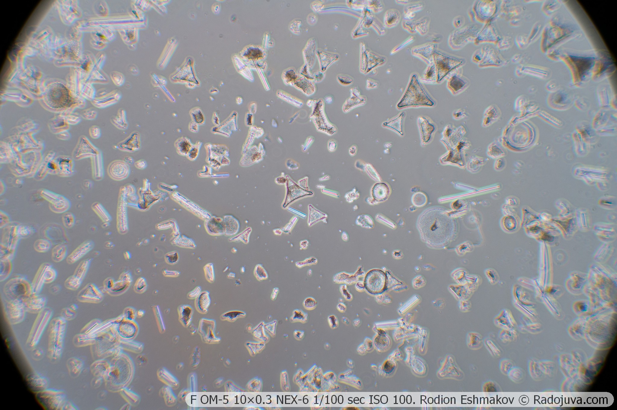
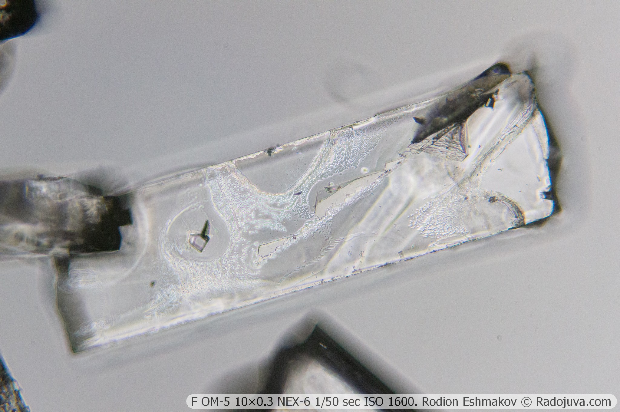
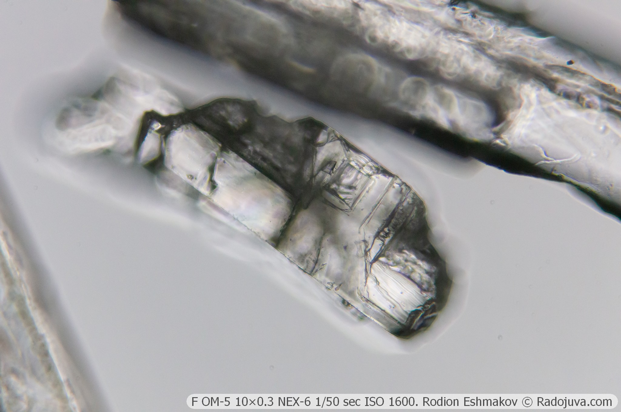
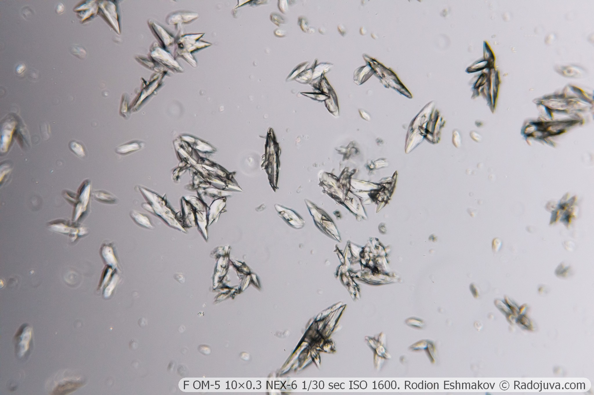
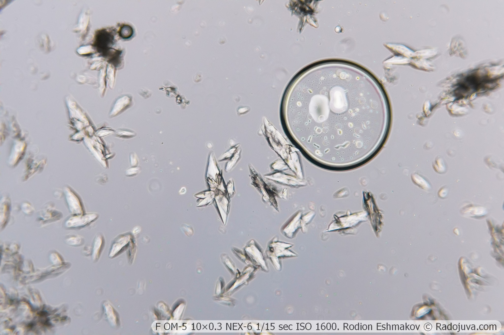
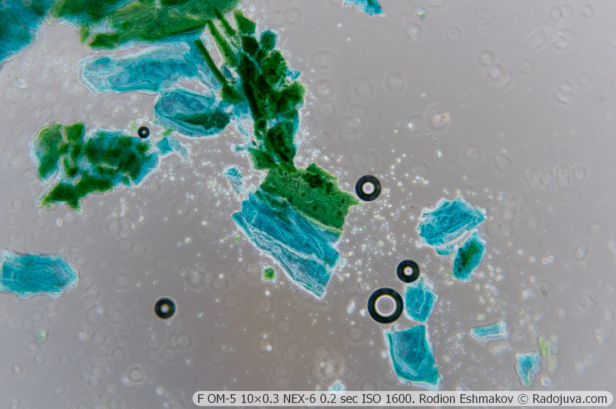
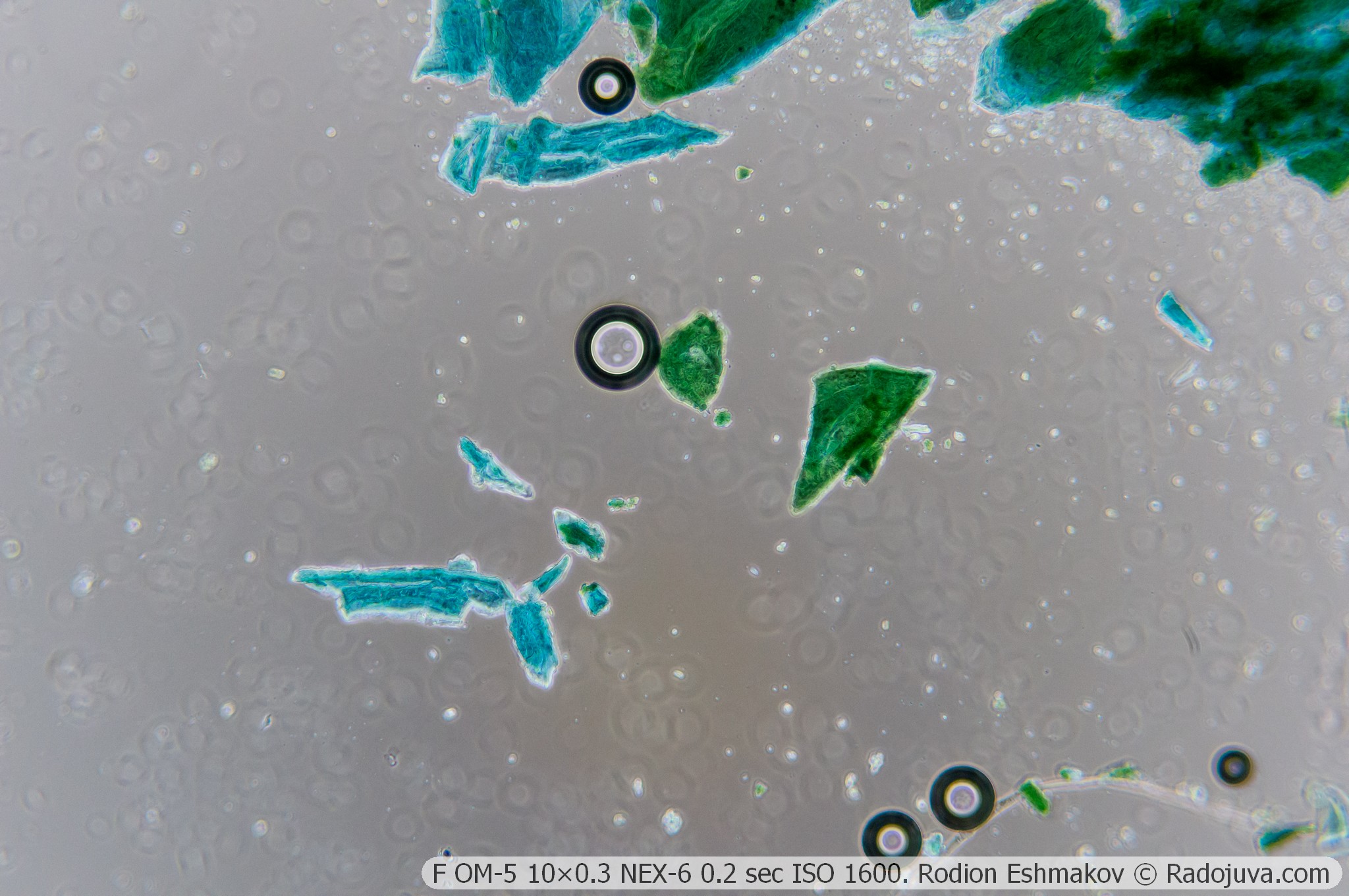
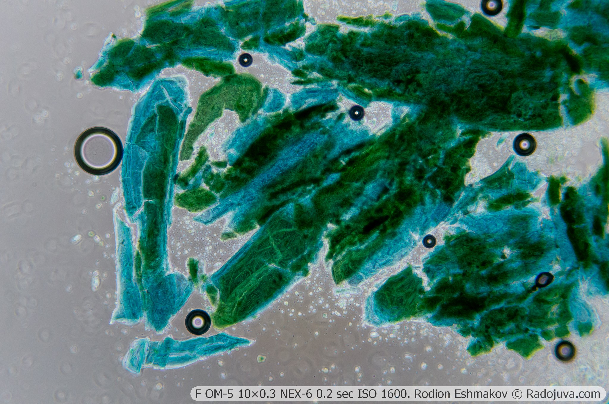
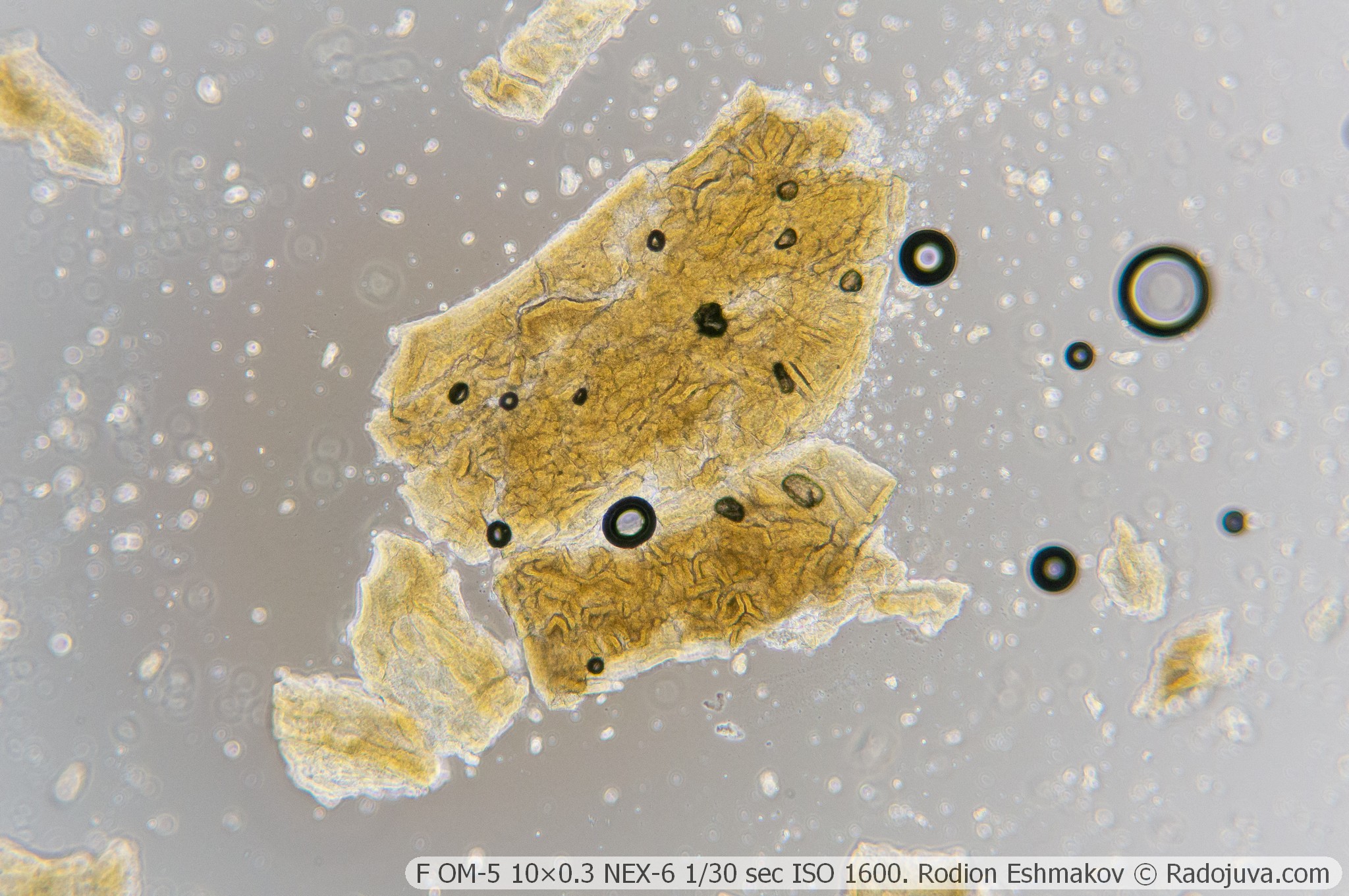
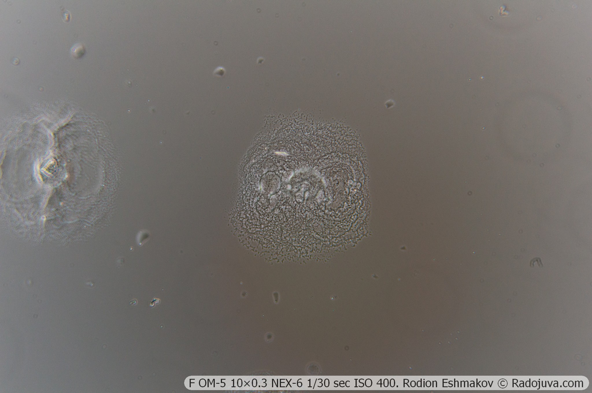
Below are examples of photos with stacking.
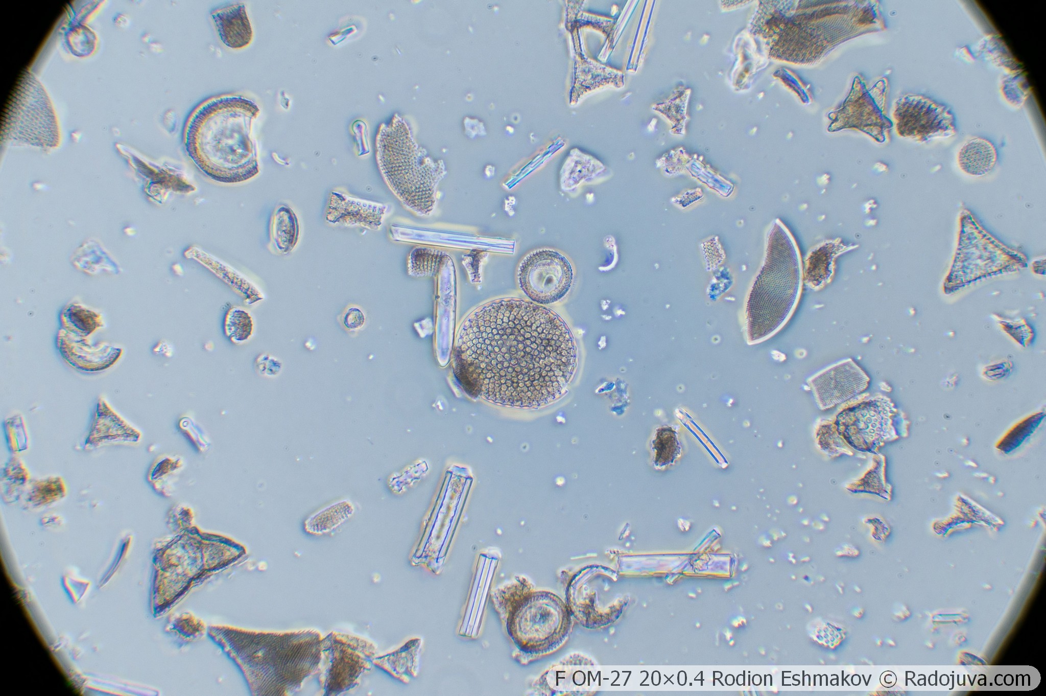
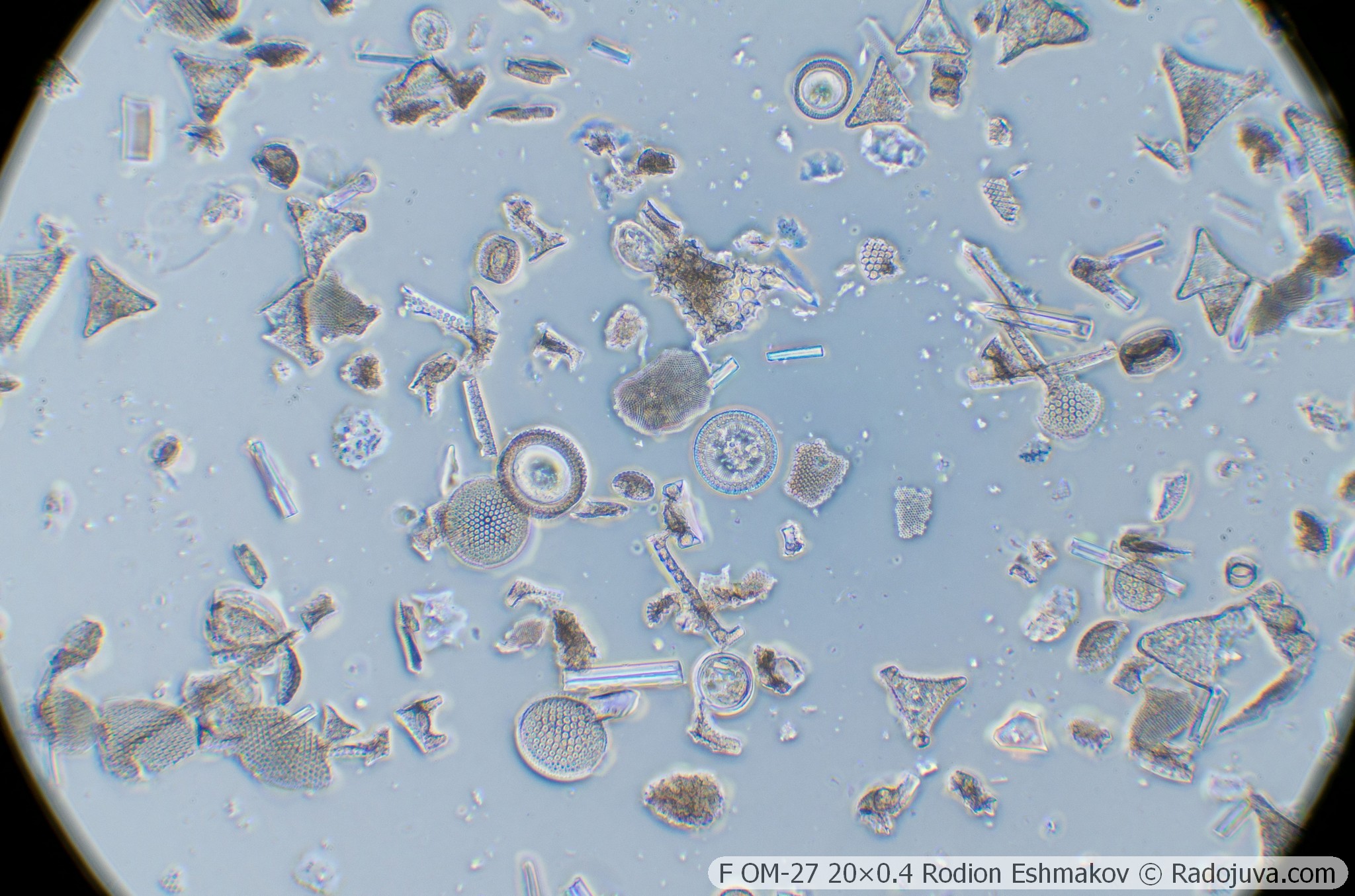
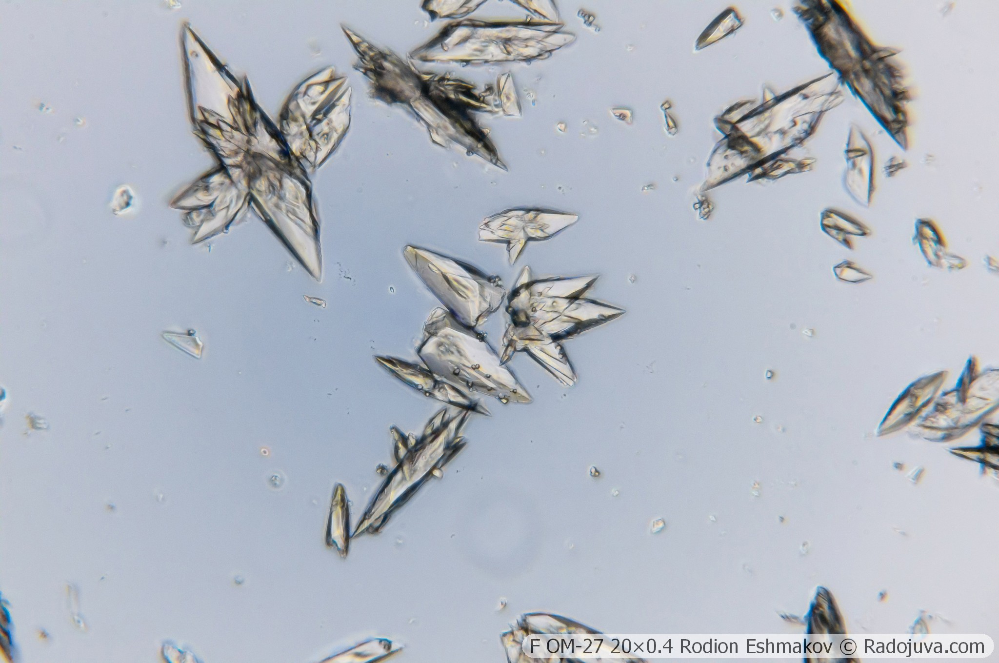
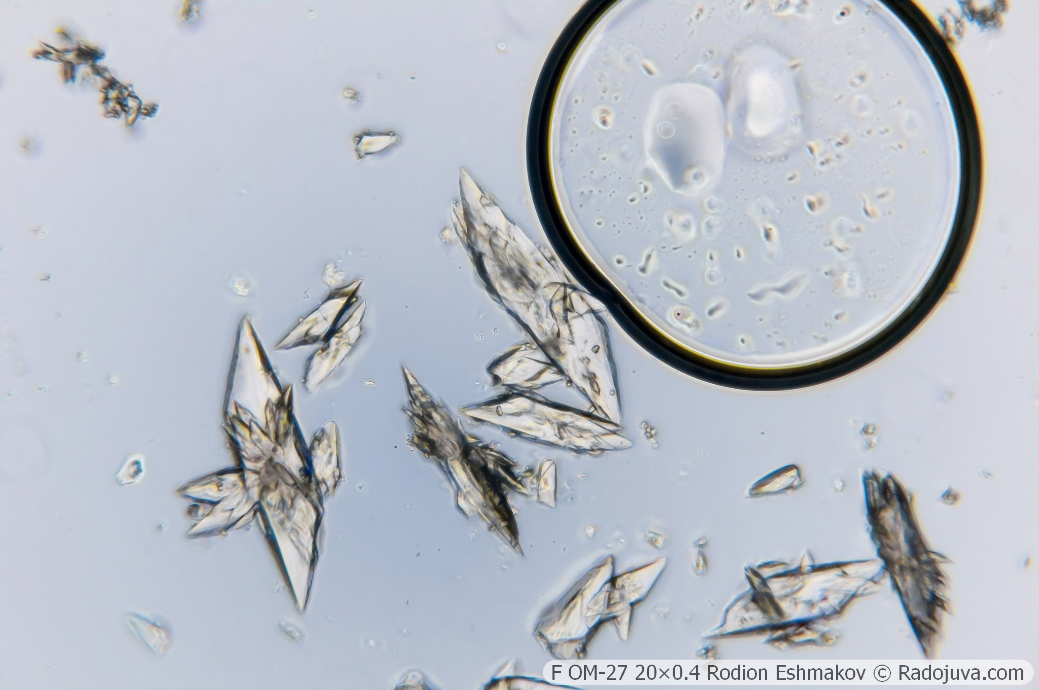
Lenses for KF-1: 20×0.4 F OM-27
Optical design – 5 lenses in 3 groups, high-aperture aplanat;
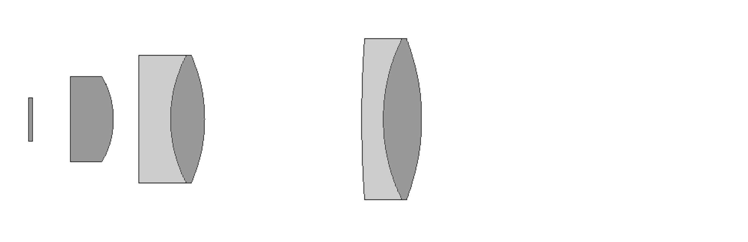
Drawing of the optical scheme of F OM-27 20×0.4.
Type of correction – achromat;
Tube distance – 160 mm;
Parfocal distance – 33 mm;
Magnification: 20×;
Numerical aperture – 0.4 (phase ring 0.25);
Working distance – 1.7 mm;
Cover glass thickness – 0.17 mm;
Mounting type – RMS standard (4/5” × 1/36” thread);
Lateral chromatic aberration – corrected with a compensating eyepiece (such as LOMO 4×, K10×/18) or ignored;
Features - microscopic lens, does not have an iris diaphragm and a focusing mechanism.
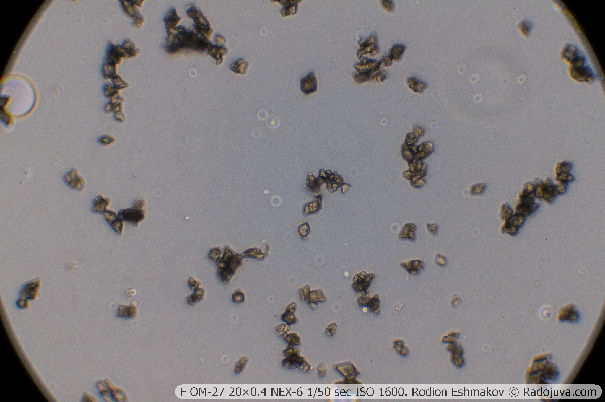
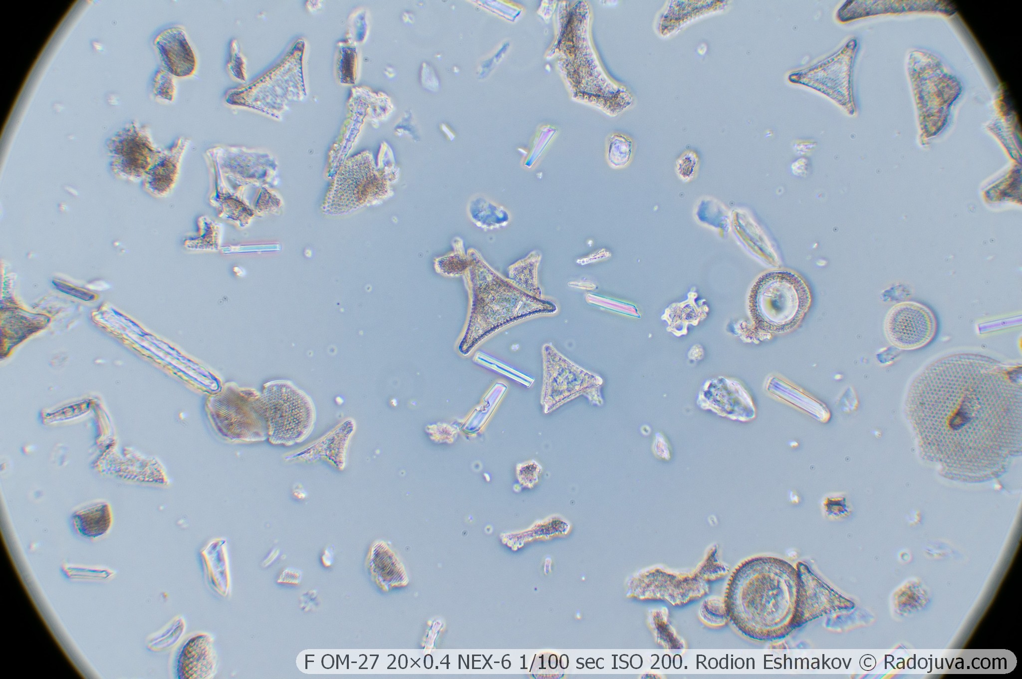
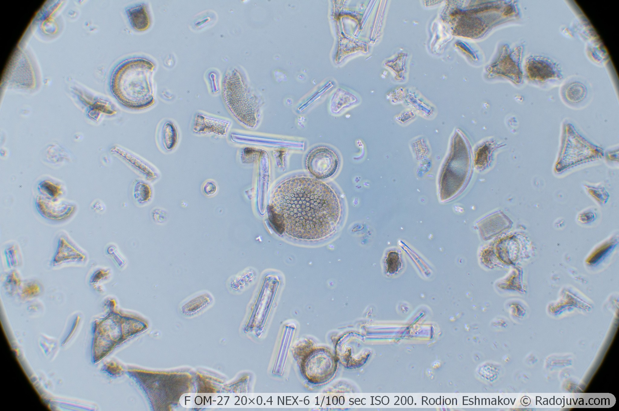
This lens is based on the OM-27 20×0.4 achromat, which isn't exactly a stellar lens, but offers acceptable image quality in the central part of the field. As with the OM-5 10×0.3 achromat, sharpness deteriorates significantly as you move away from the center due to astigmatism, field curvature, and lateral chromatic aberration. There's no compensation for lateral chromatic aberration either.
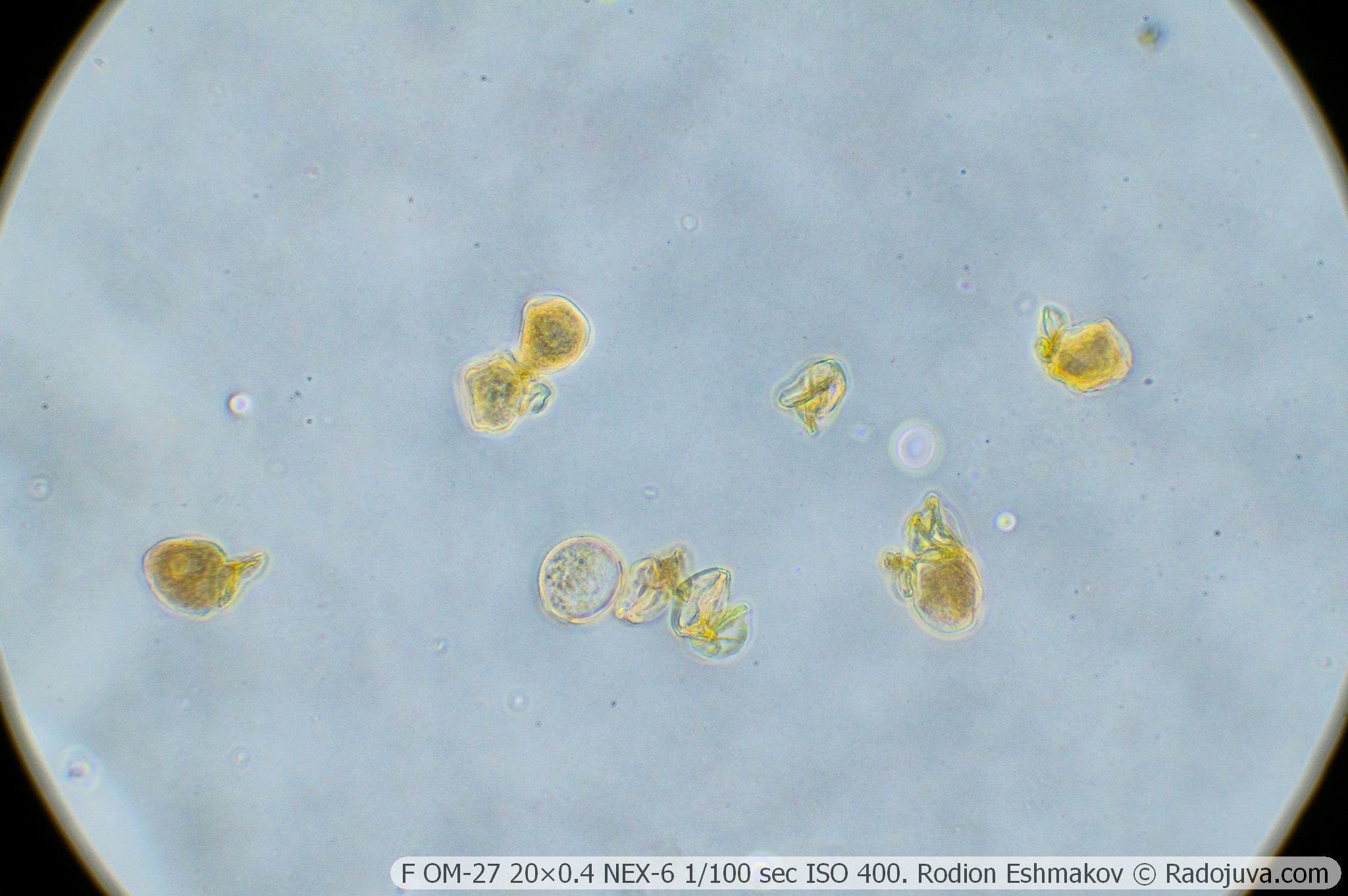
When using prime focus with an APS-C sensor, an uneven gray-blue background is observed, which is due to field aberrations of the lens. When shooting with the K5× ML-2 relay lens and the WitrueHD 75/2.8 10 MP lens with a usable field size of approximately 20 mm, the issue is virtually unnoticeable. In the KF-1 kit, I would probably consider the OM-27 to be the most convenient lens – it has a decent magnification, tolerable image quality, and a more uniform background compared to the 10×0.3.
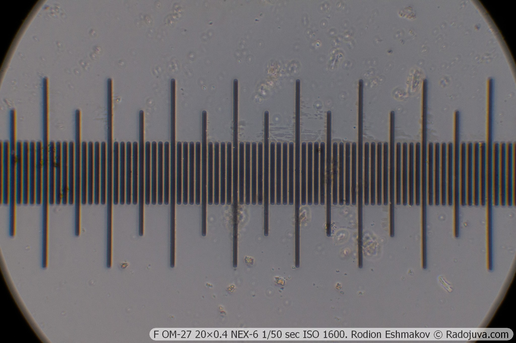
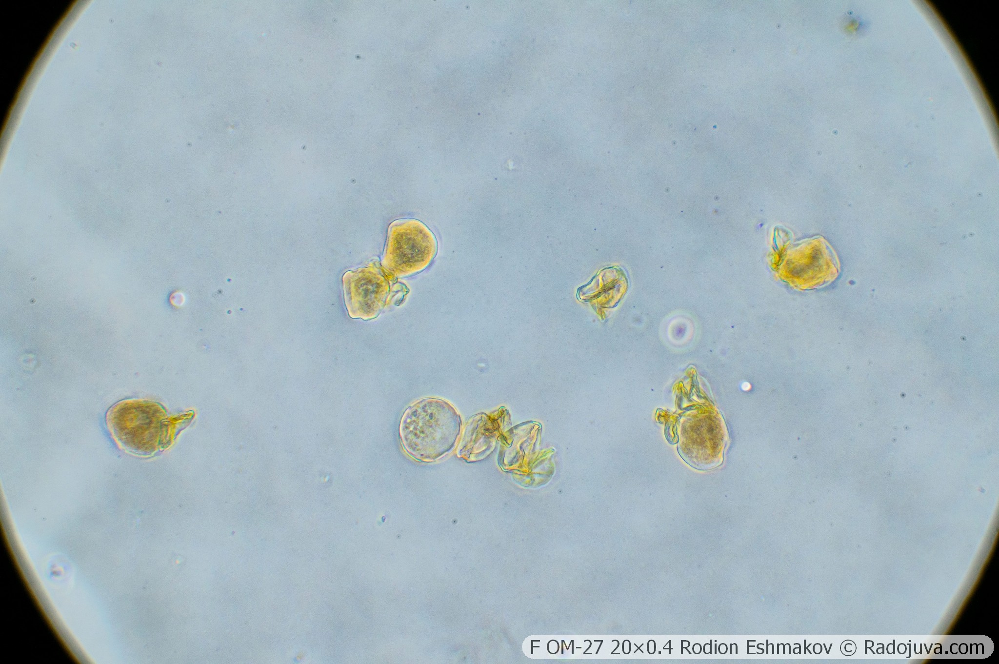
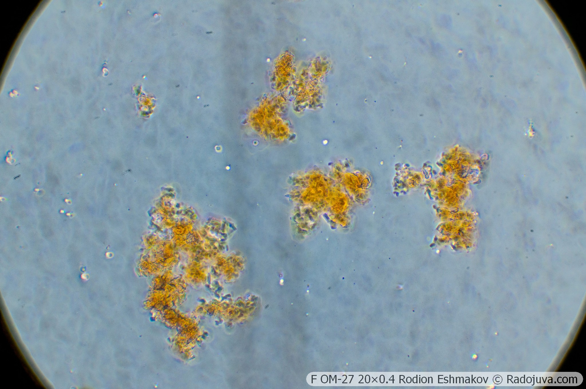
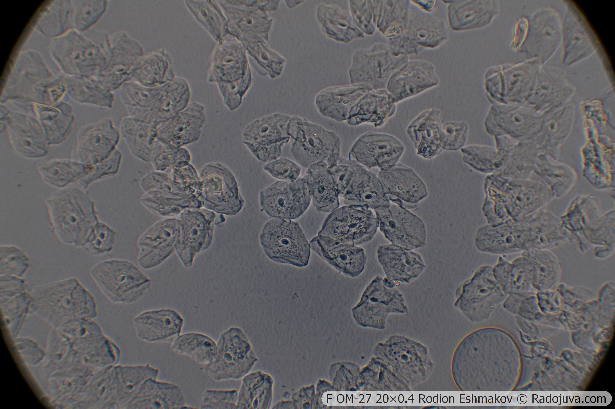
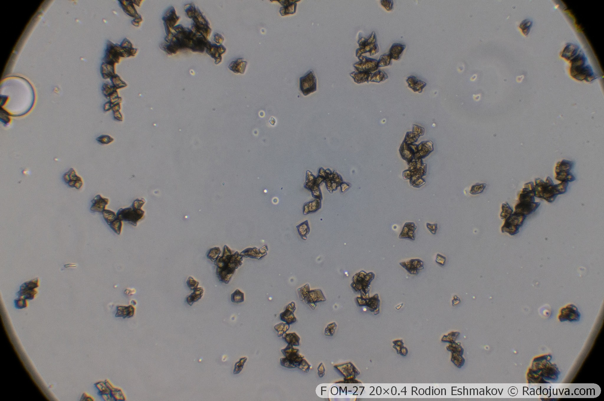
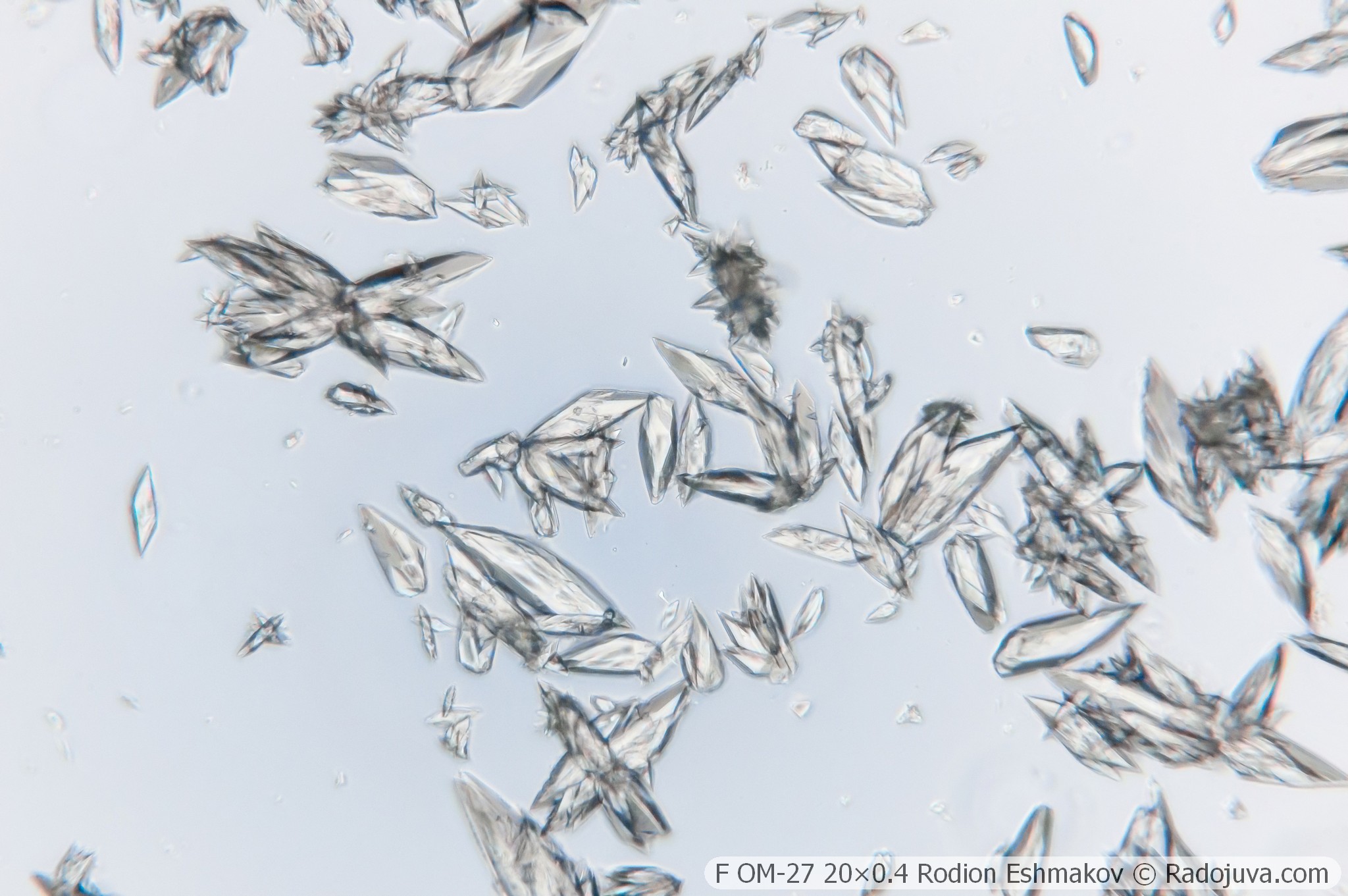
Below are examples of photos taken with the Sony NEX-6 and the F OM-27 20×0.4 lens in prime focus and using the relay from the K5× ML-2 eyepiece and the WitrueHD 75/2.8 10 Mpix lens.
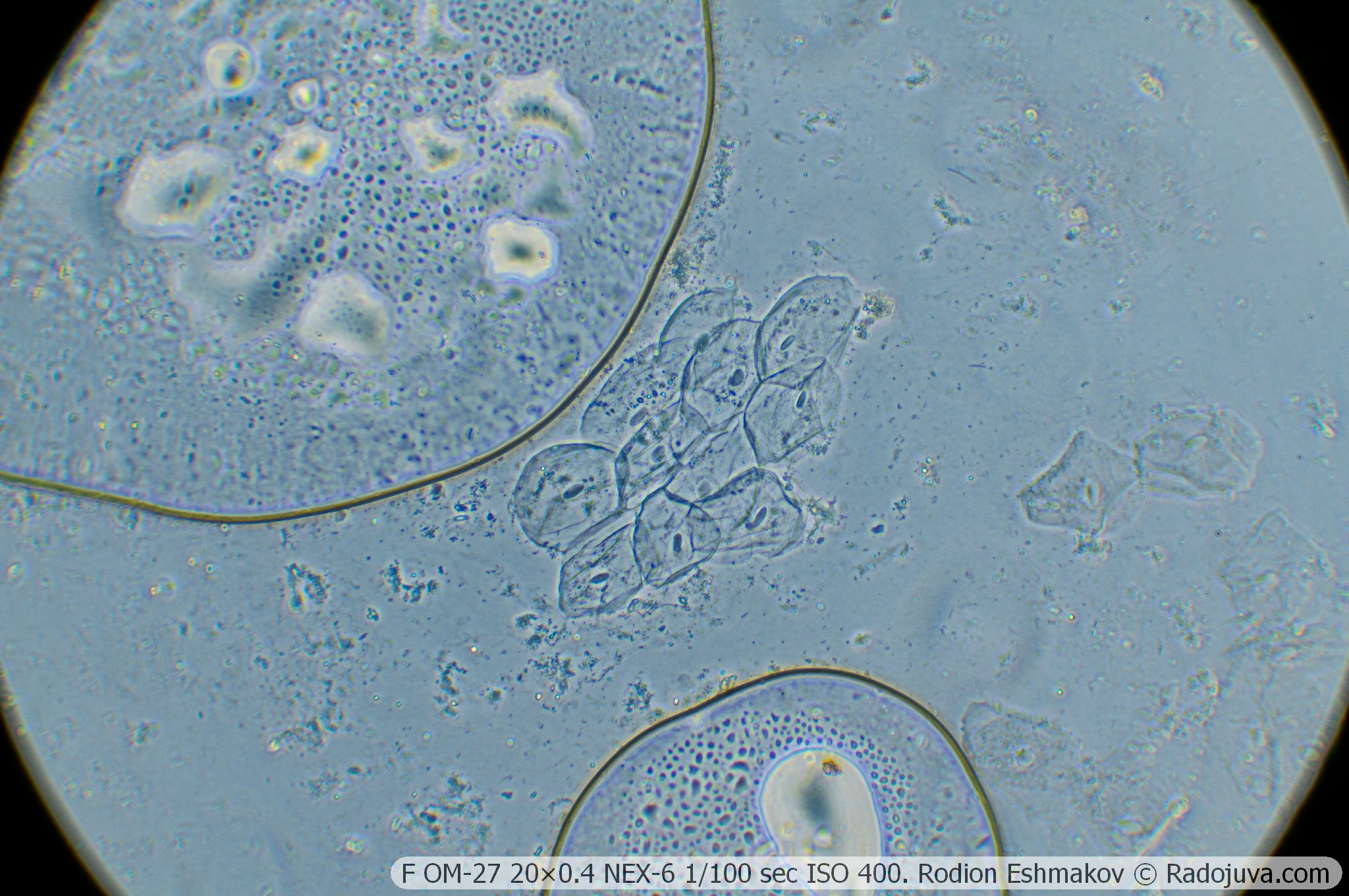
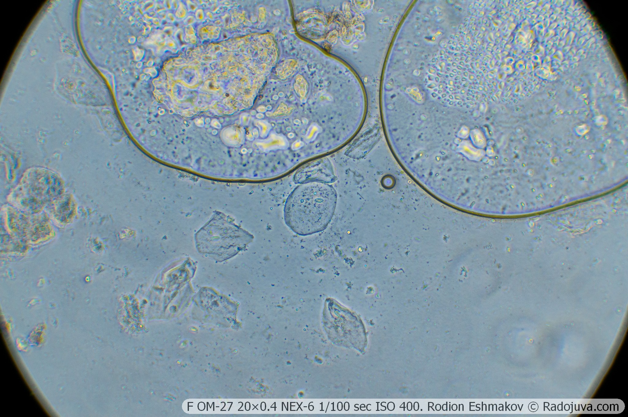
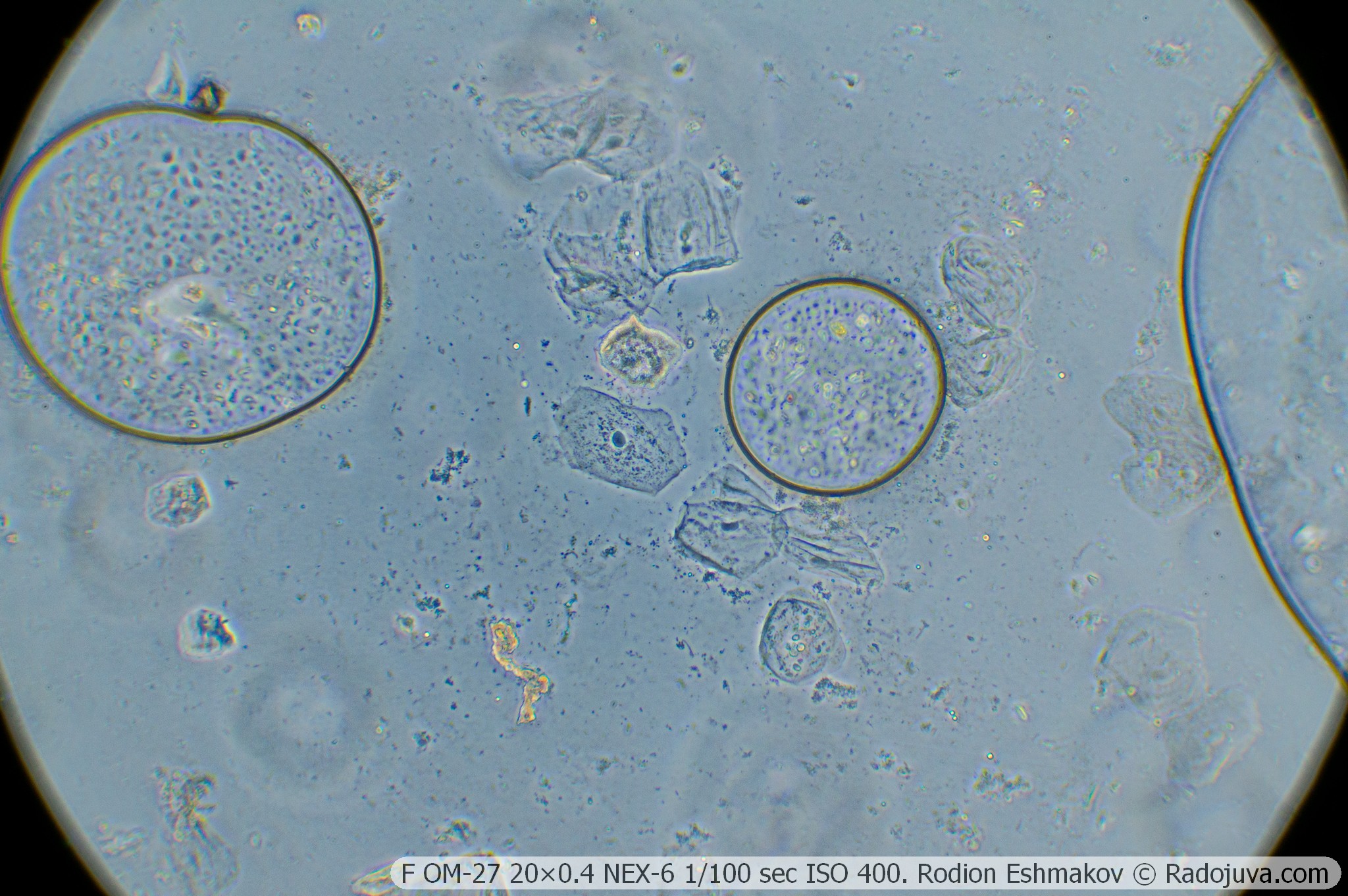
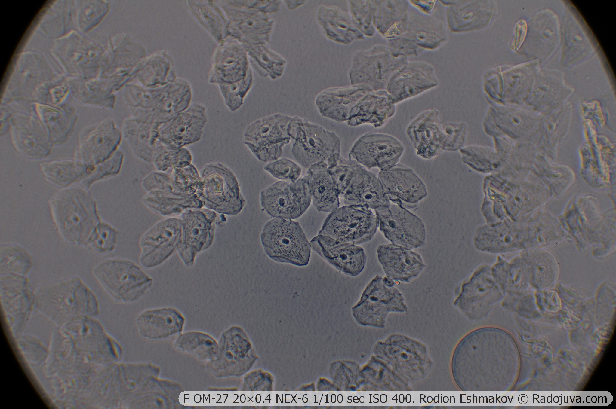
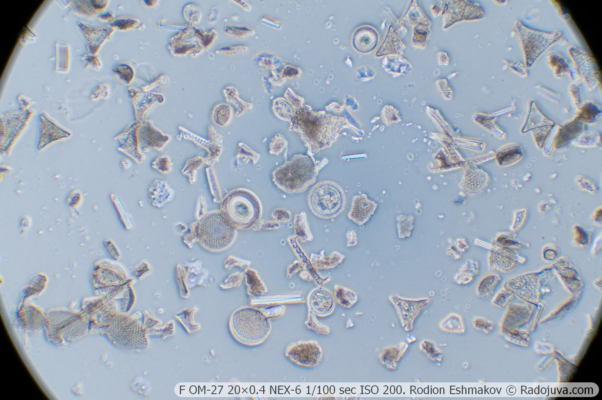
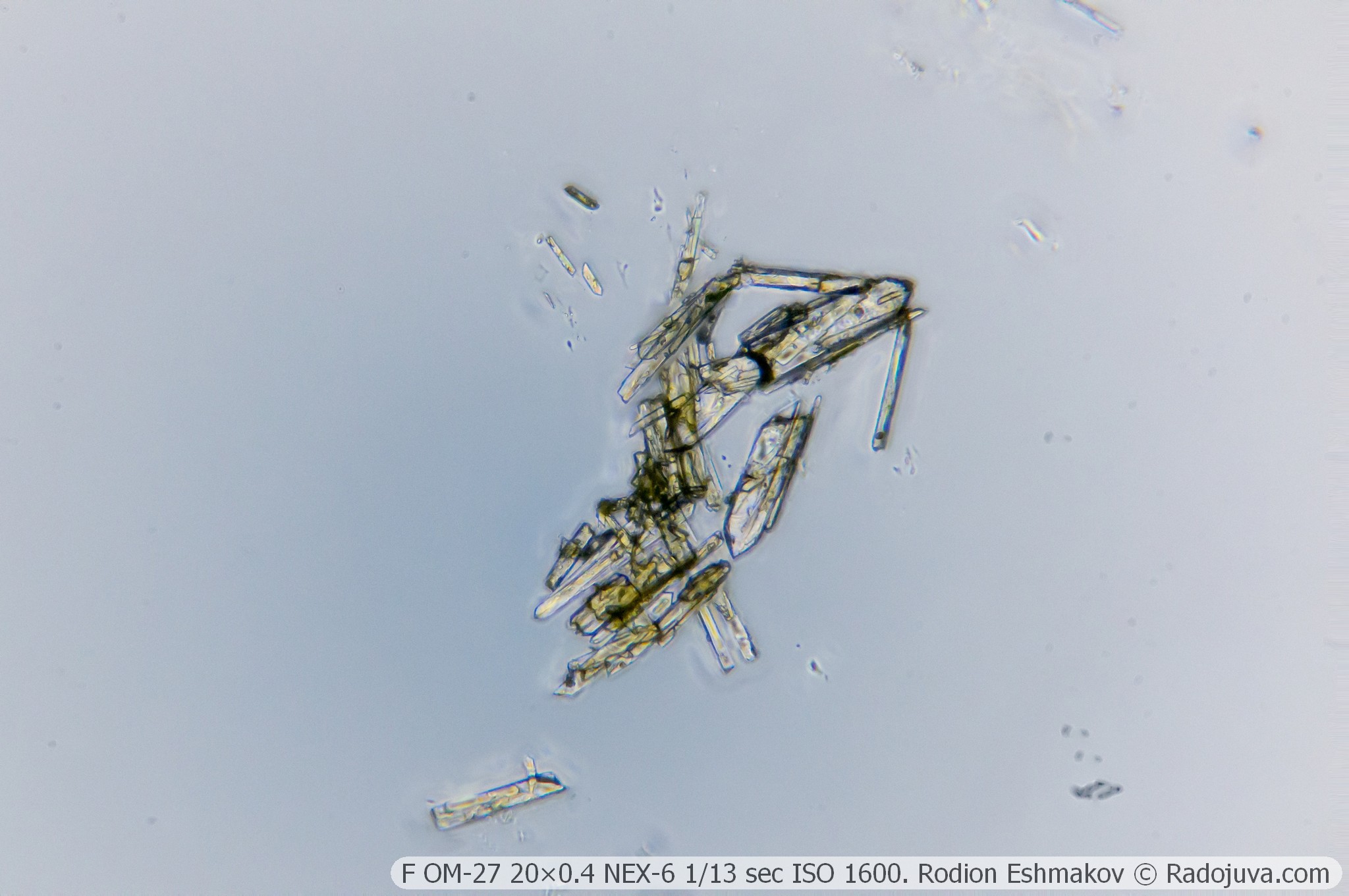
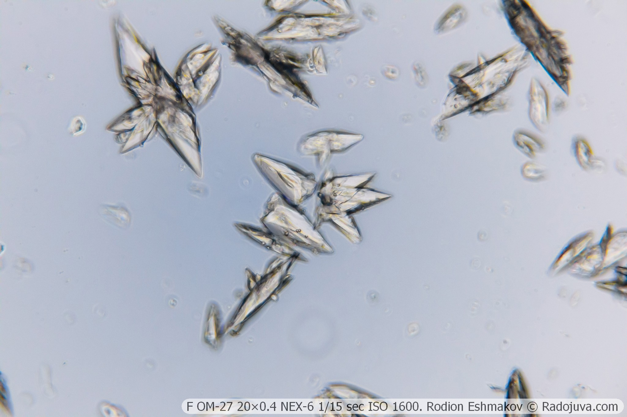
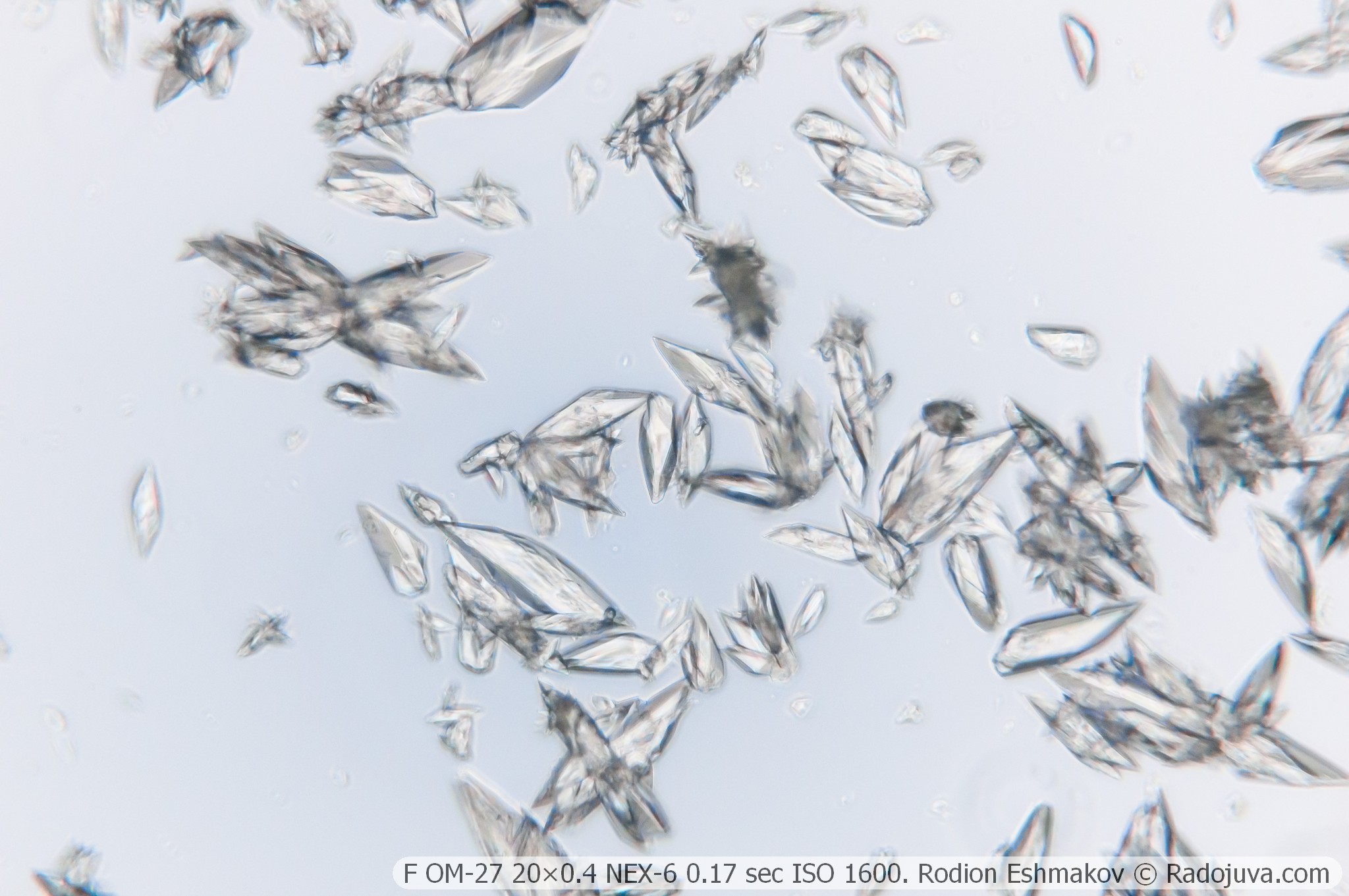
Below are examples of photos with stacking.
Lenses for KF-1: 40×0.65 F MS
Optical design – 5 lenses in 3 groups, high-aperture aplanat;
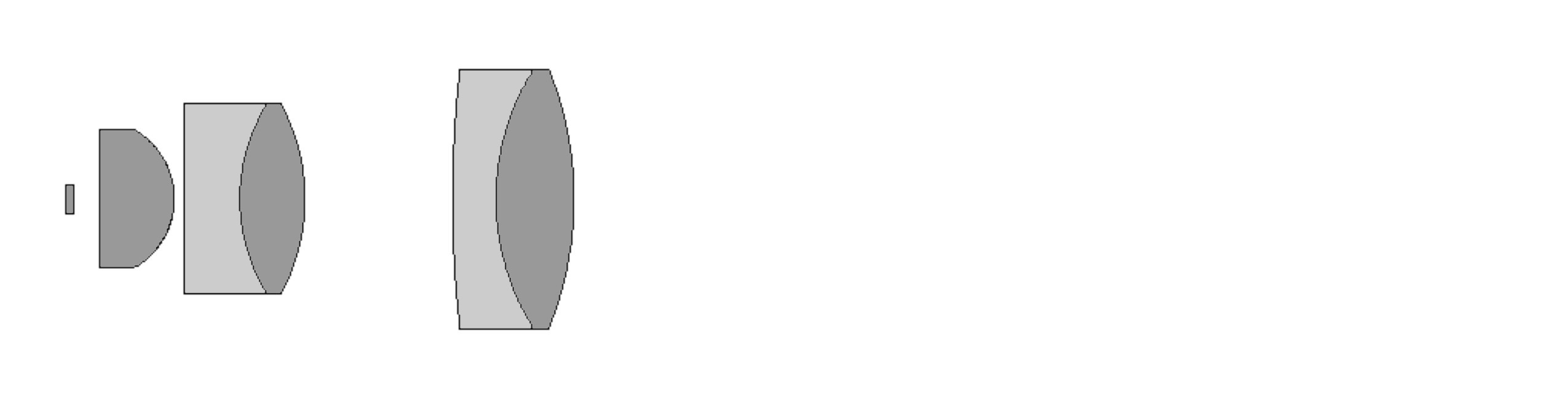
Drawing of the optical scheme of the F MS 40×0.65.
Type of correction – achromat;
Tube distance – 160 mm;
Parfocal distance – 33 mm;
Magnification: 40×;
Numerical aperture – 0.65 (phase ring 0.26);
Working distance – 0.55 mm;
Cover glass thickness – 0.17 mm;
Mounting type – RMS standard (4/5” × 1/36” thread);
Lateral chromatic aberration – corrected with a compensating eyepiece (such as LOMO K5×);
Features - microscopic lens, does not have an iris diaphragm and a focusing mechanism.
The MS objective is one of the worst 40x microscope objectives ever. High levels of spherochromatism, astigmatism, field curvature, and lateral chromatic aberration were fatal for the standard version, but are somewhat mitigated in the phase-shifted version, which operates at a very low numerical aperture. Because of this, the "smoothness" of the image is no longer as bothersome, but detail and resolution are very low. In prime focus, the objective performs abysmally, but significantly better when using the relay from the K5x ML-2 eyepiece. The background is tinted gray-blue.
The phase-detection MS is more useful for visual observations; its resolution is insufficient for photography. Using this lens offers little advantage over the 2x image upscaling of the F OM-27 20×0.4.
As you can see, the image quality of a number of KF-1 10×0.3, 20×0.4, and 40×0.65 lenses steadily declines. This is because they use the same basic optical design with increasing numerical aperture without any significant changes to the optical materials. Just as the Industar-50 50/3.5 is a decent standard lens suitable for mounting on a 50 MP camera, the Industar-37 300/4.5 is a terrible telephoto lens, so scaling the same design over such a wide range is not a good idea.
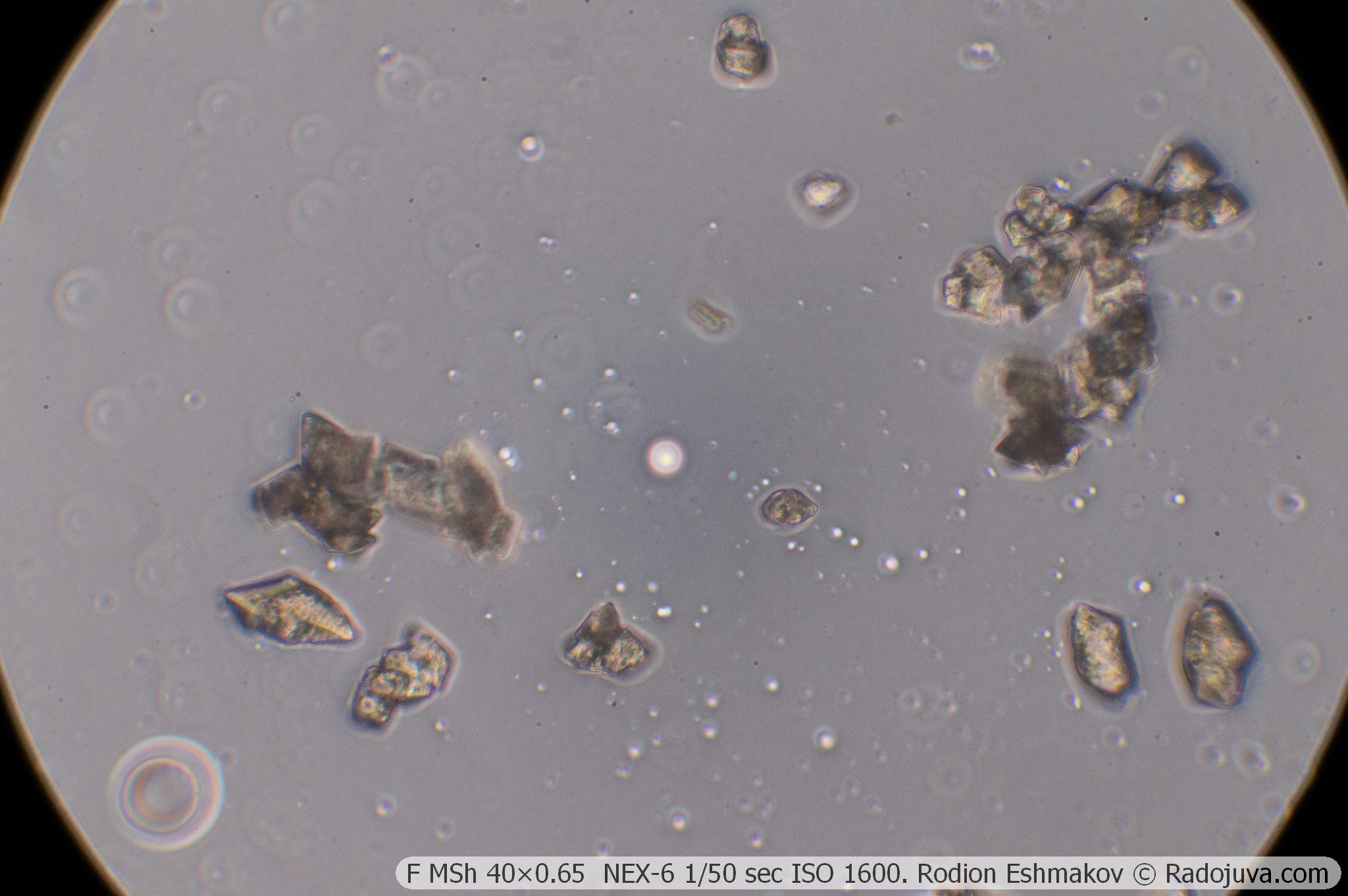
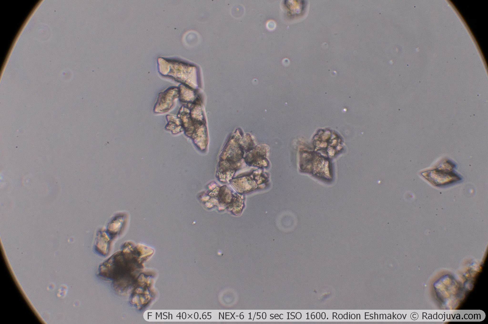
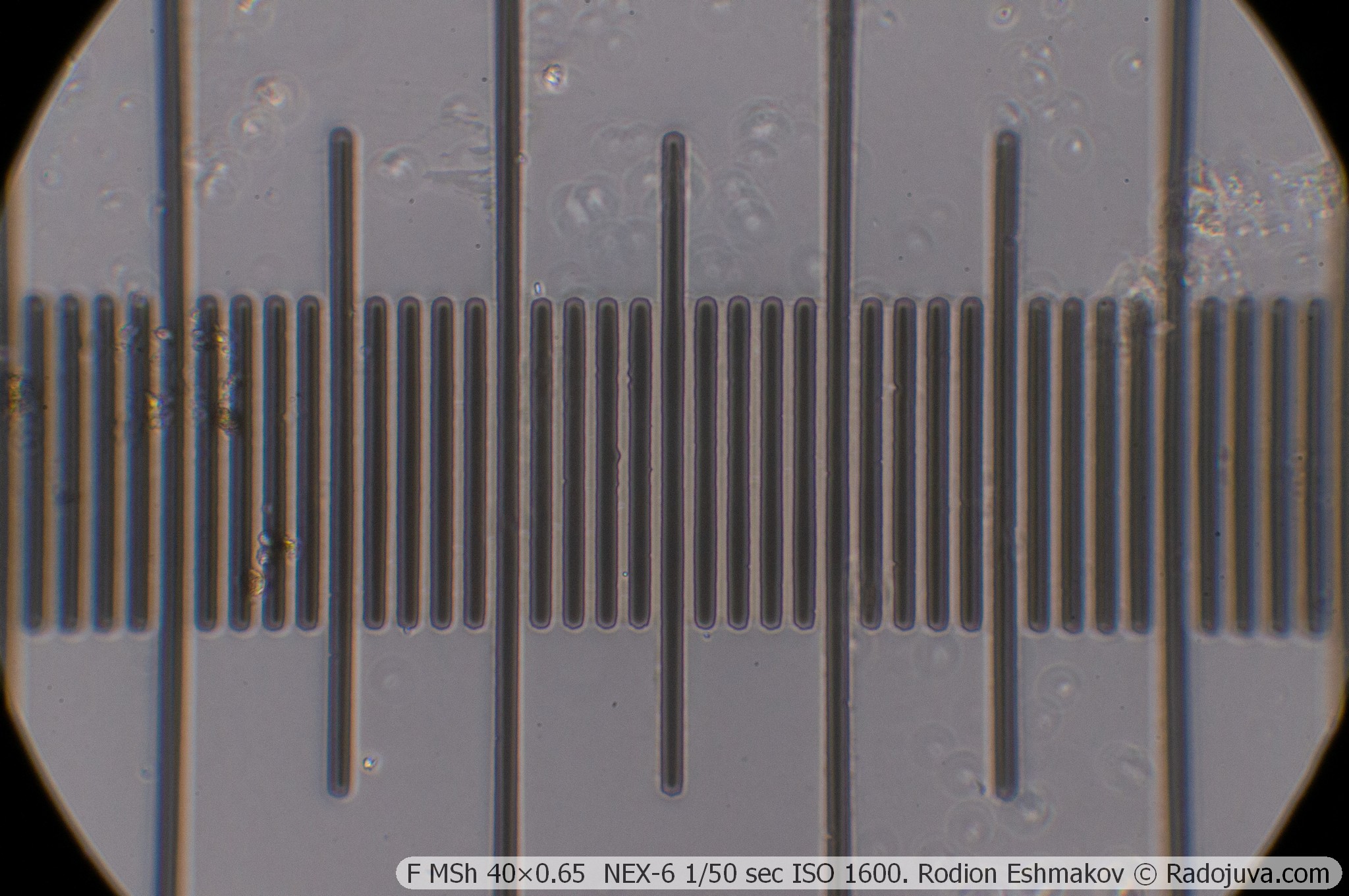
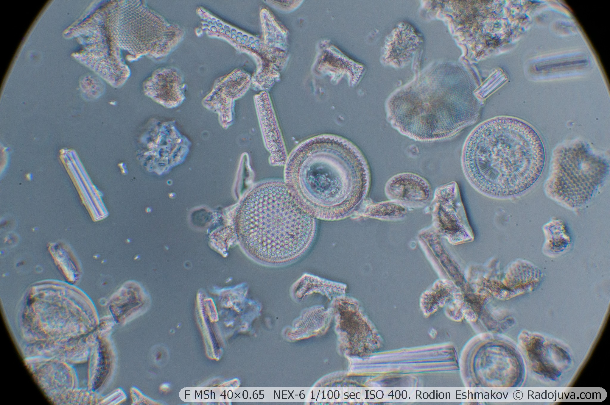
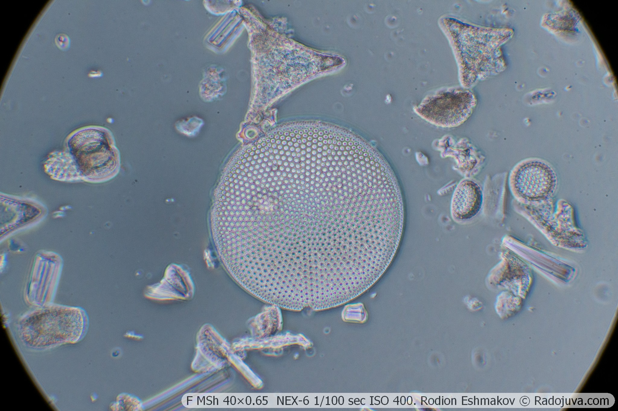
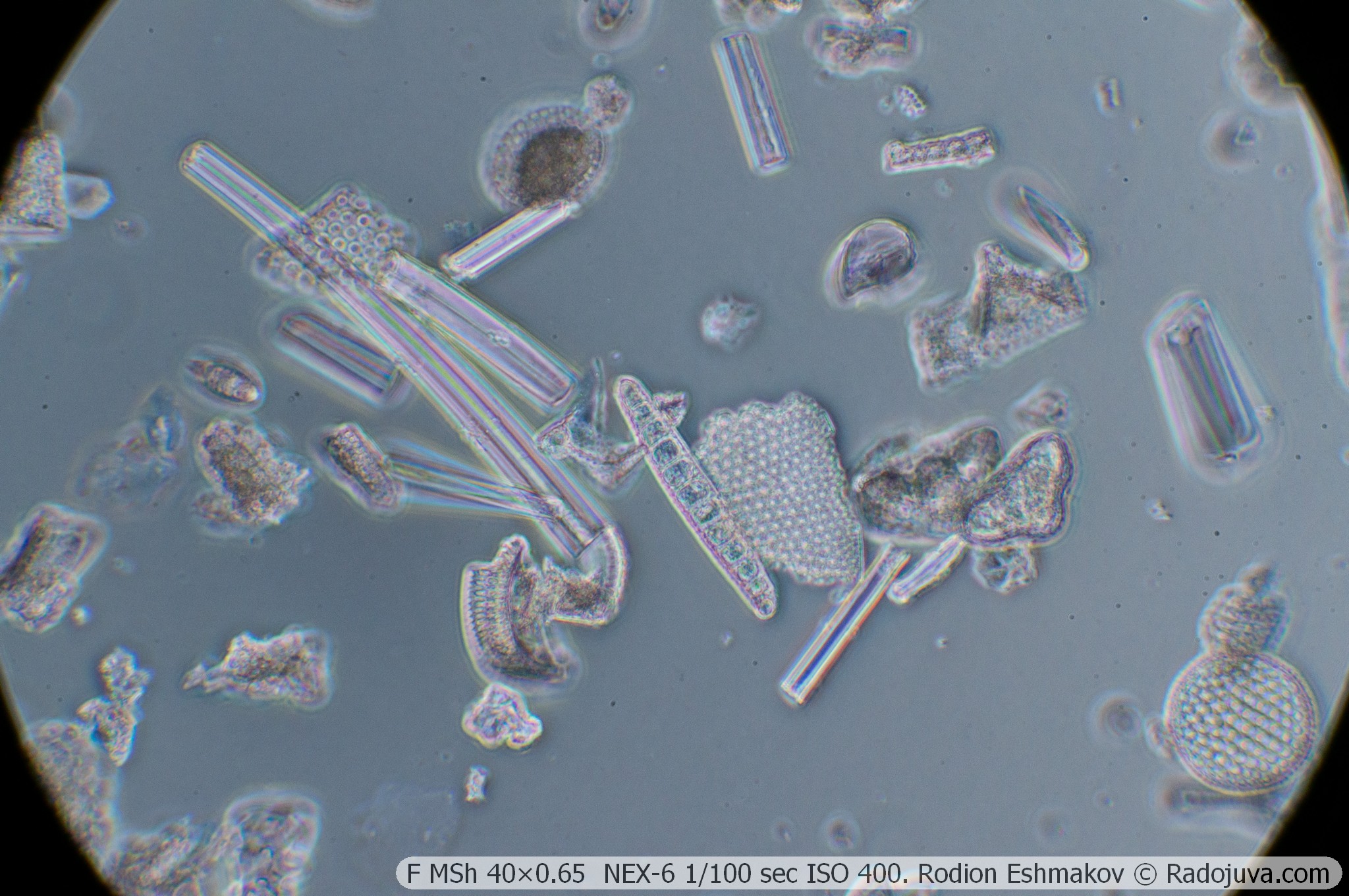
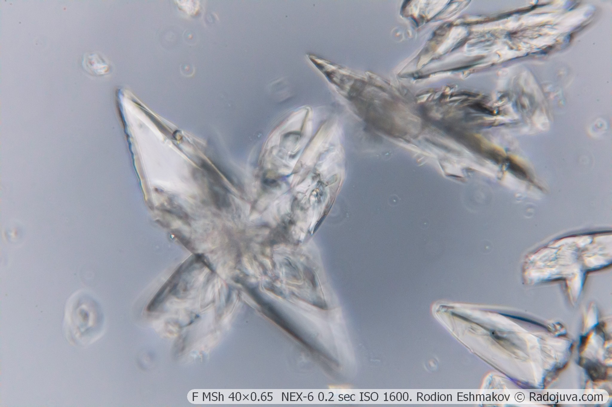
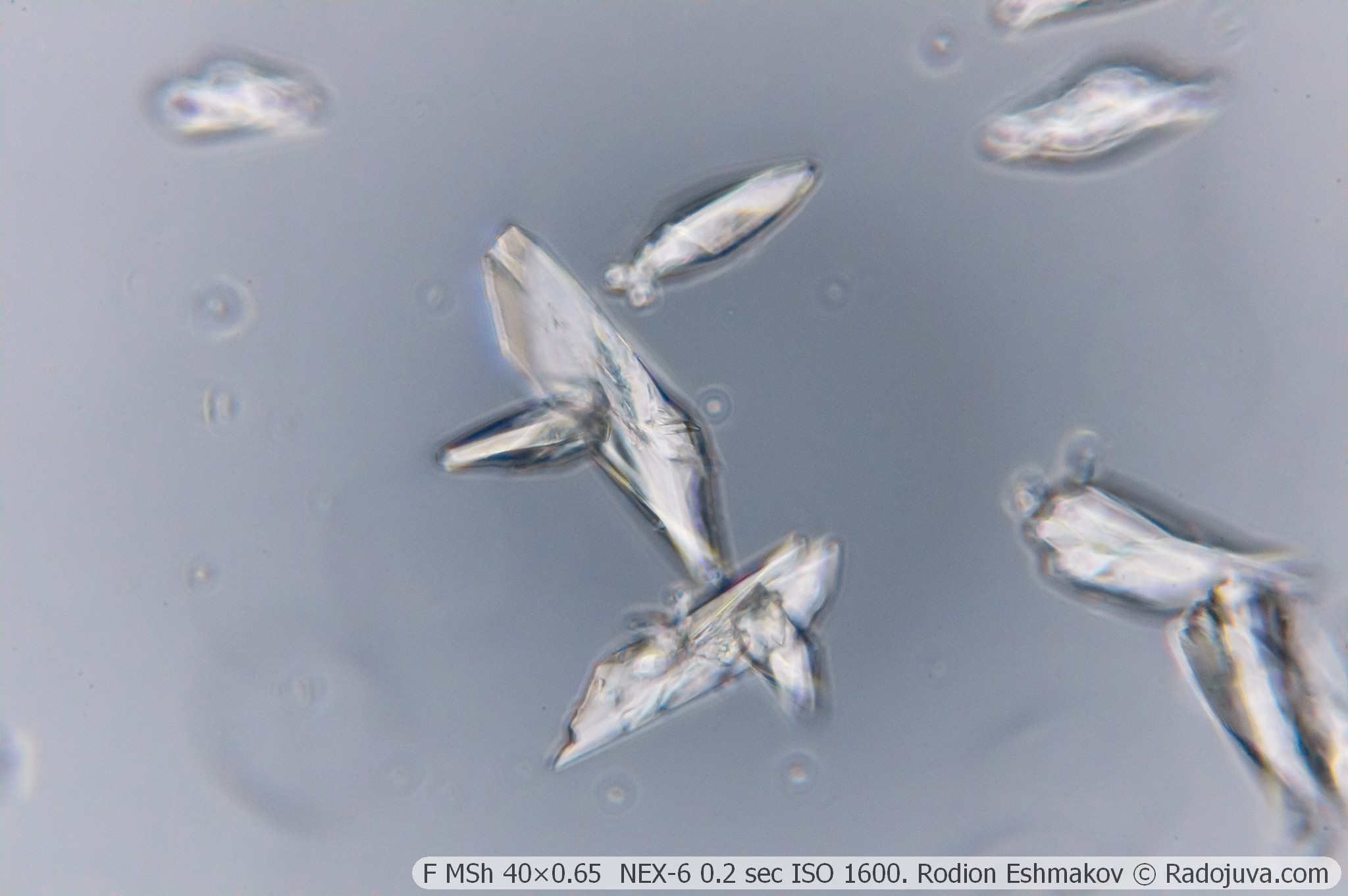
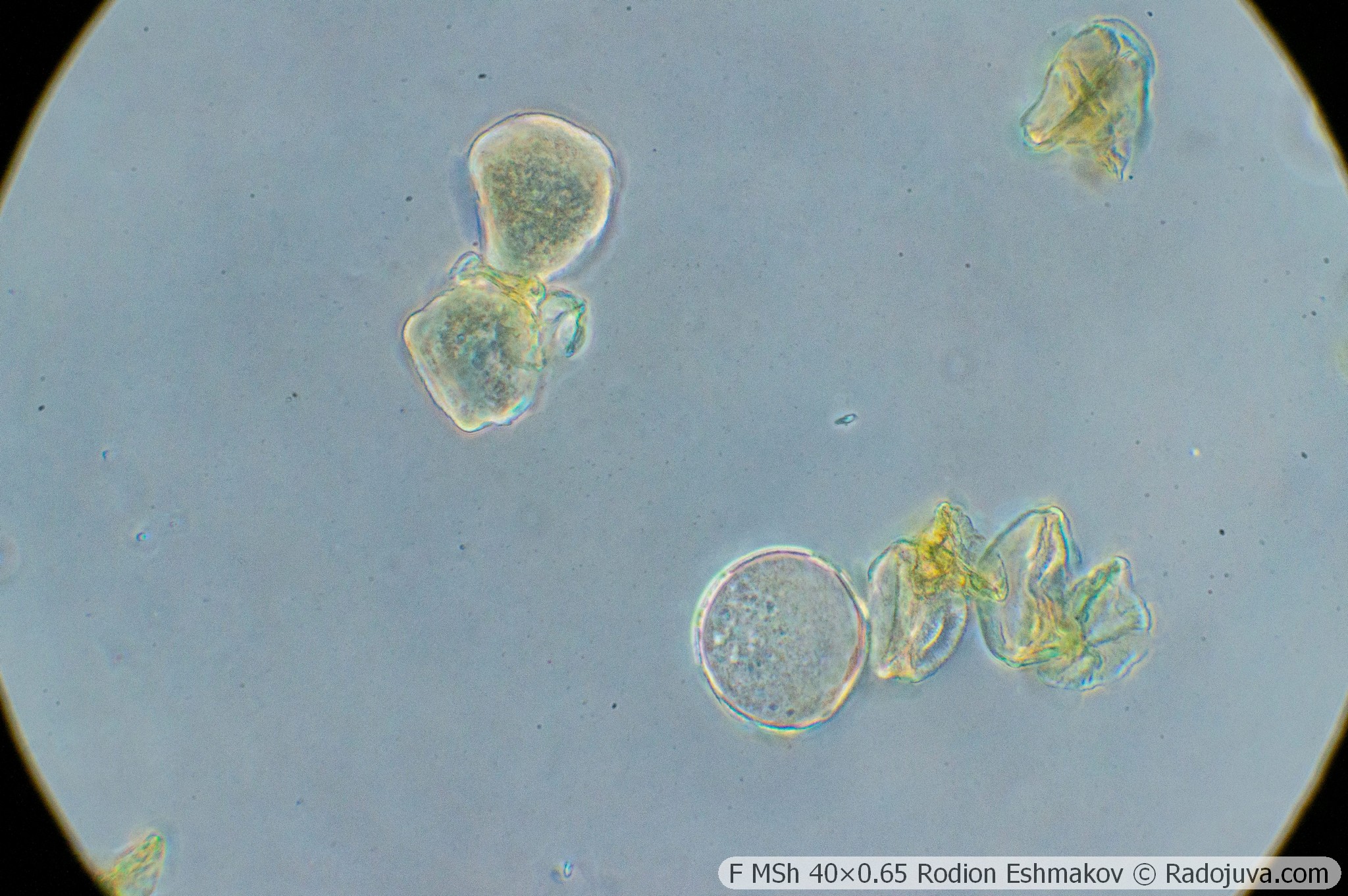
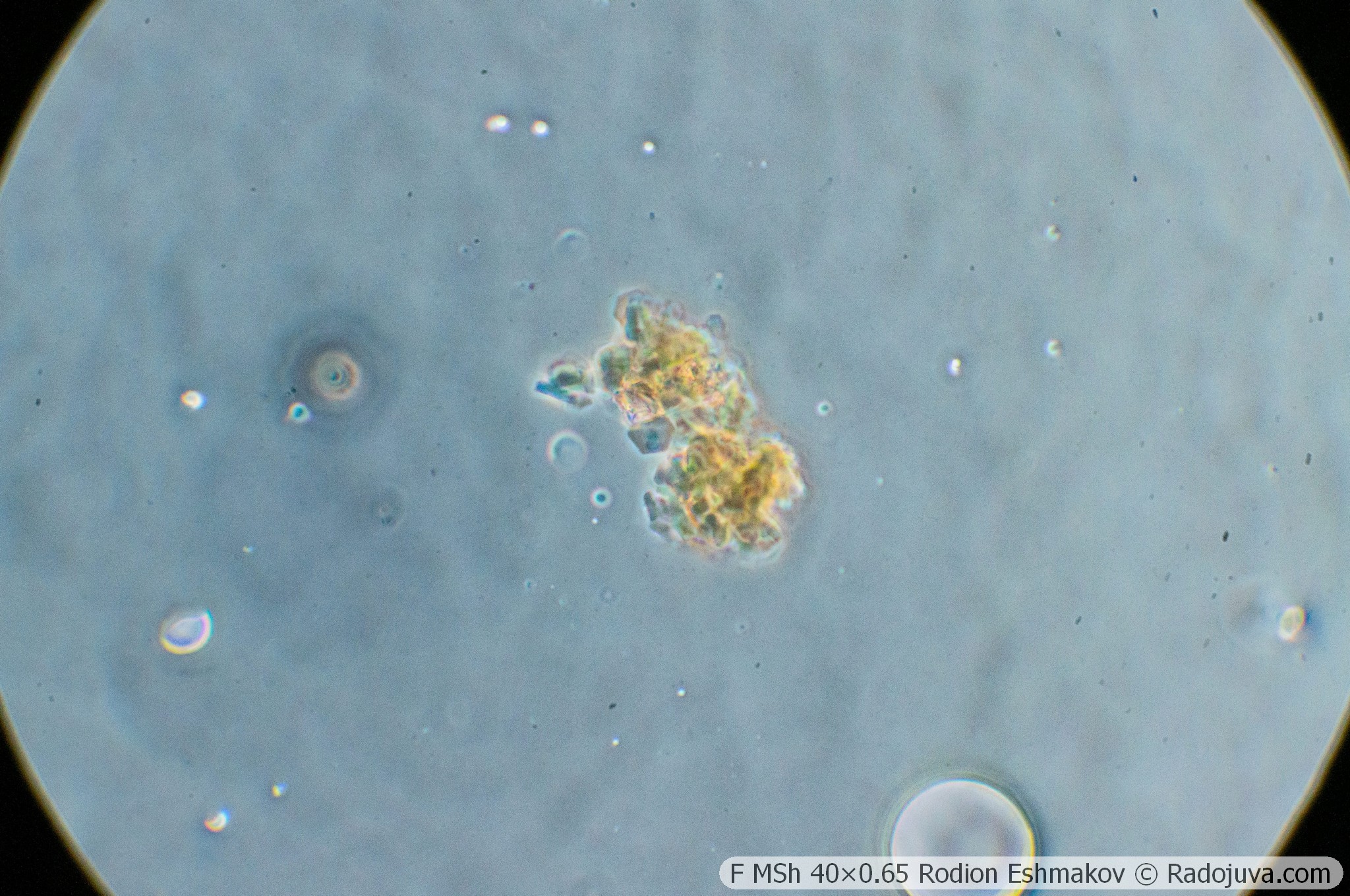
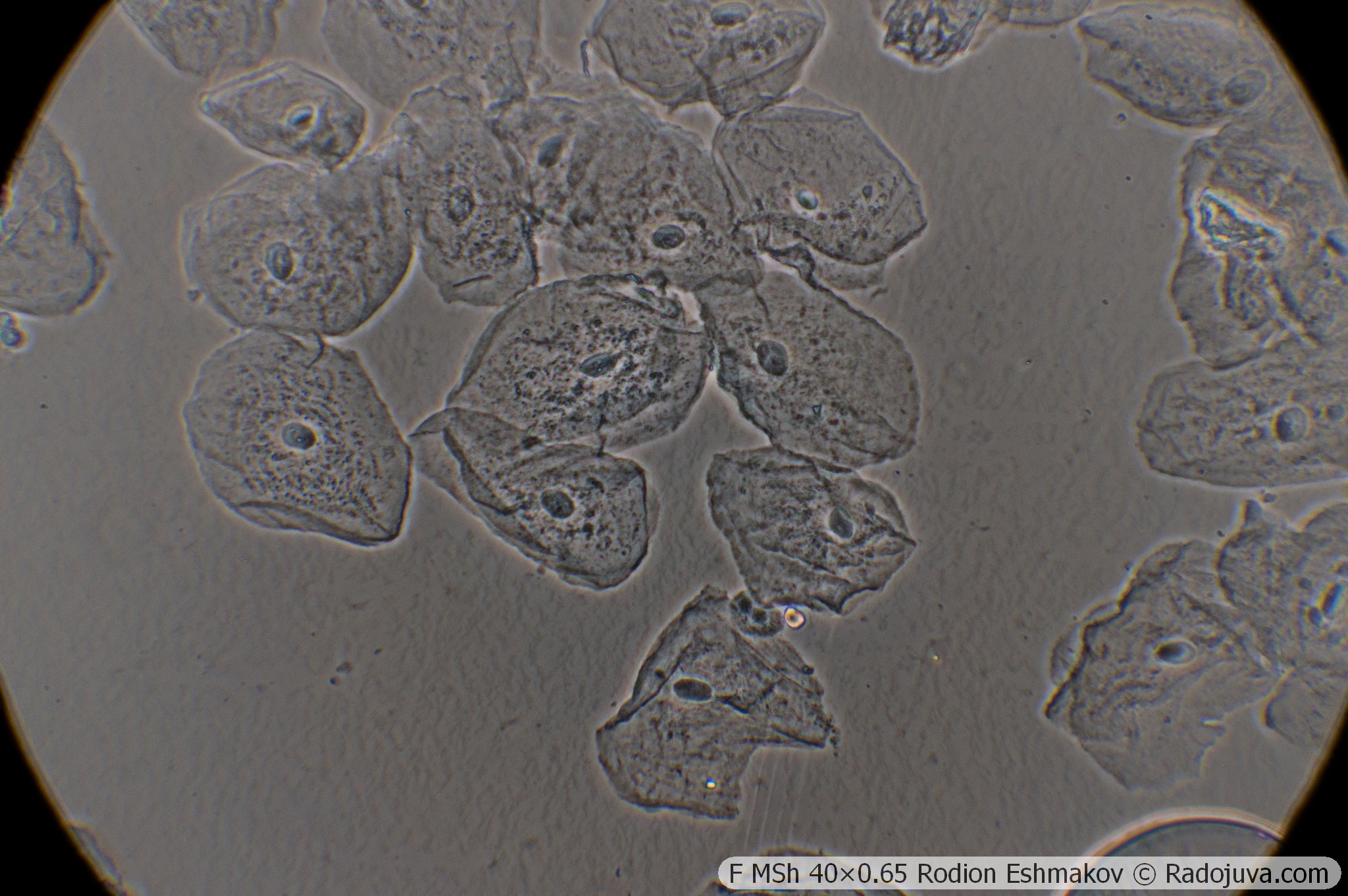
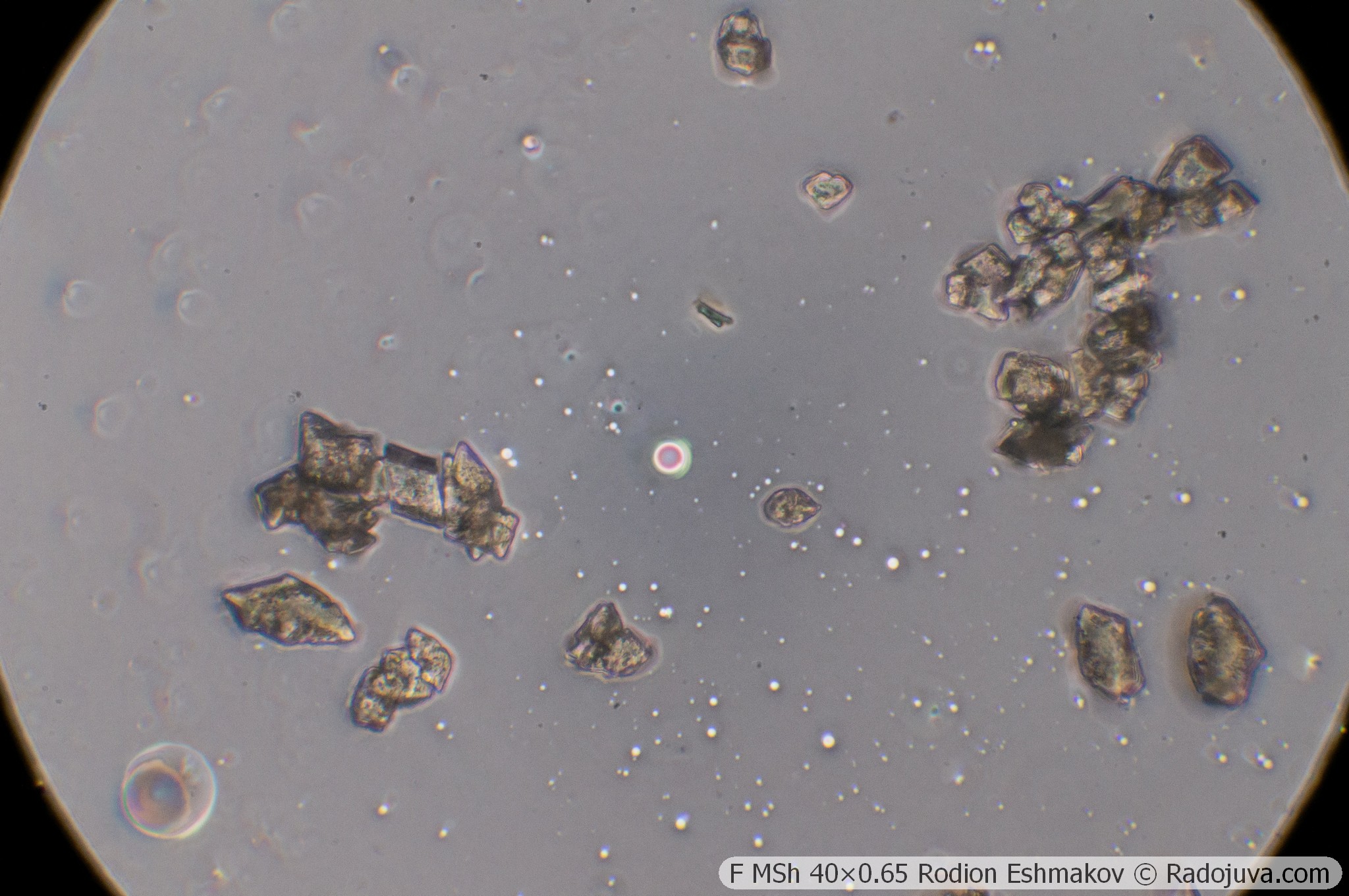
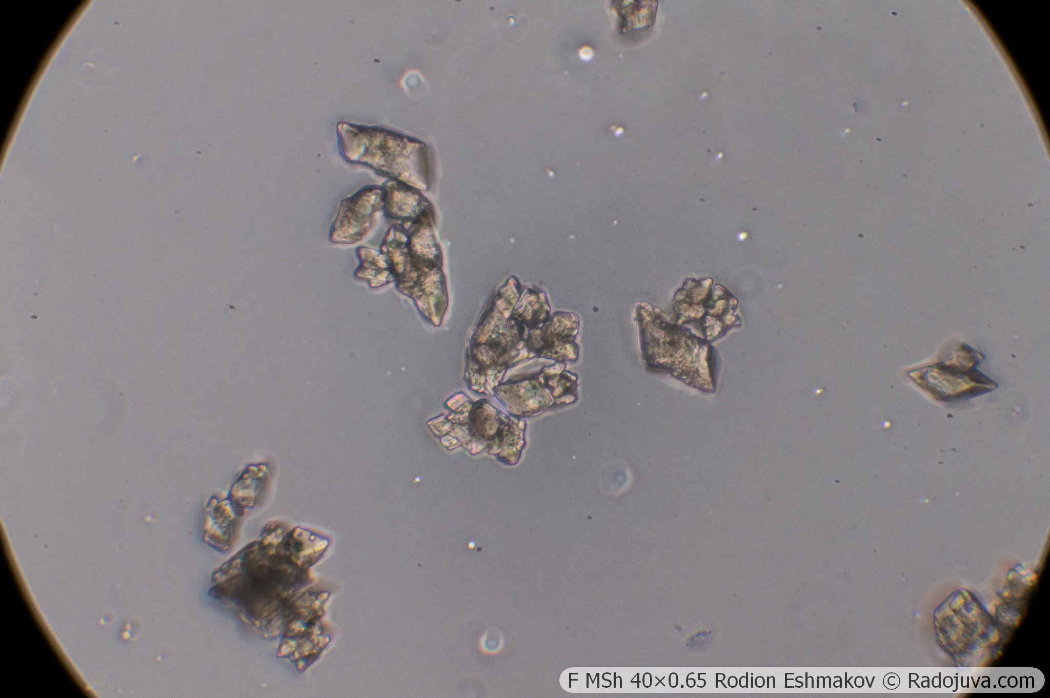
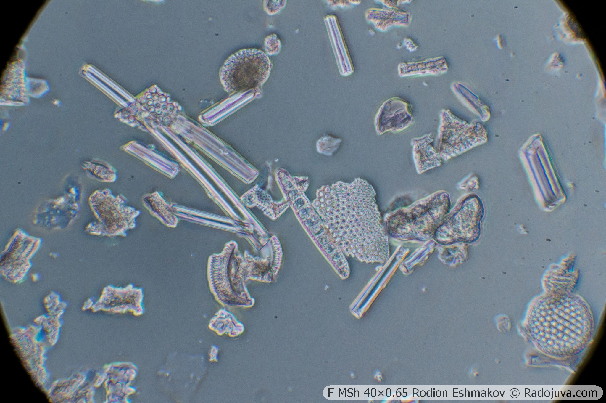
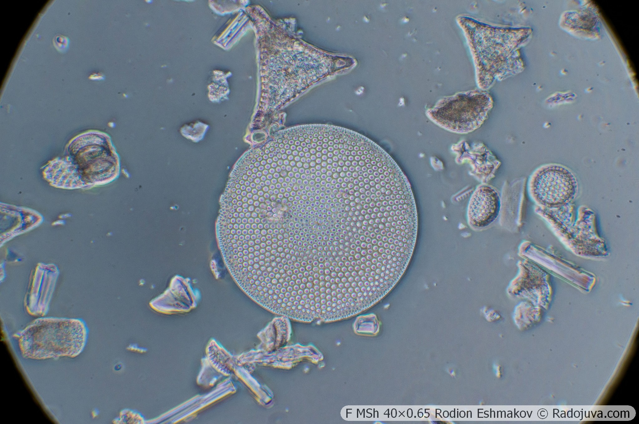
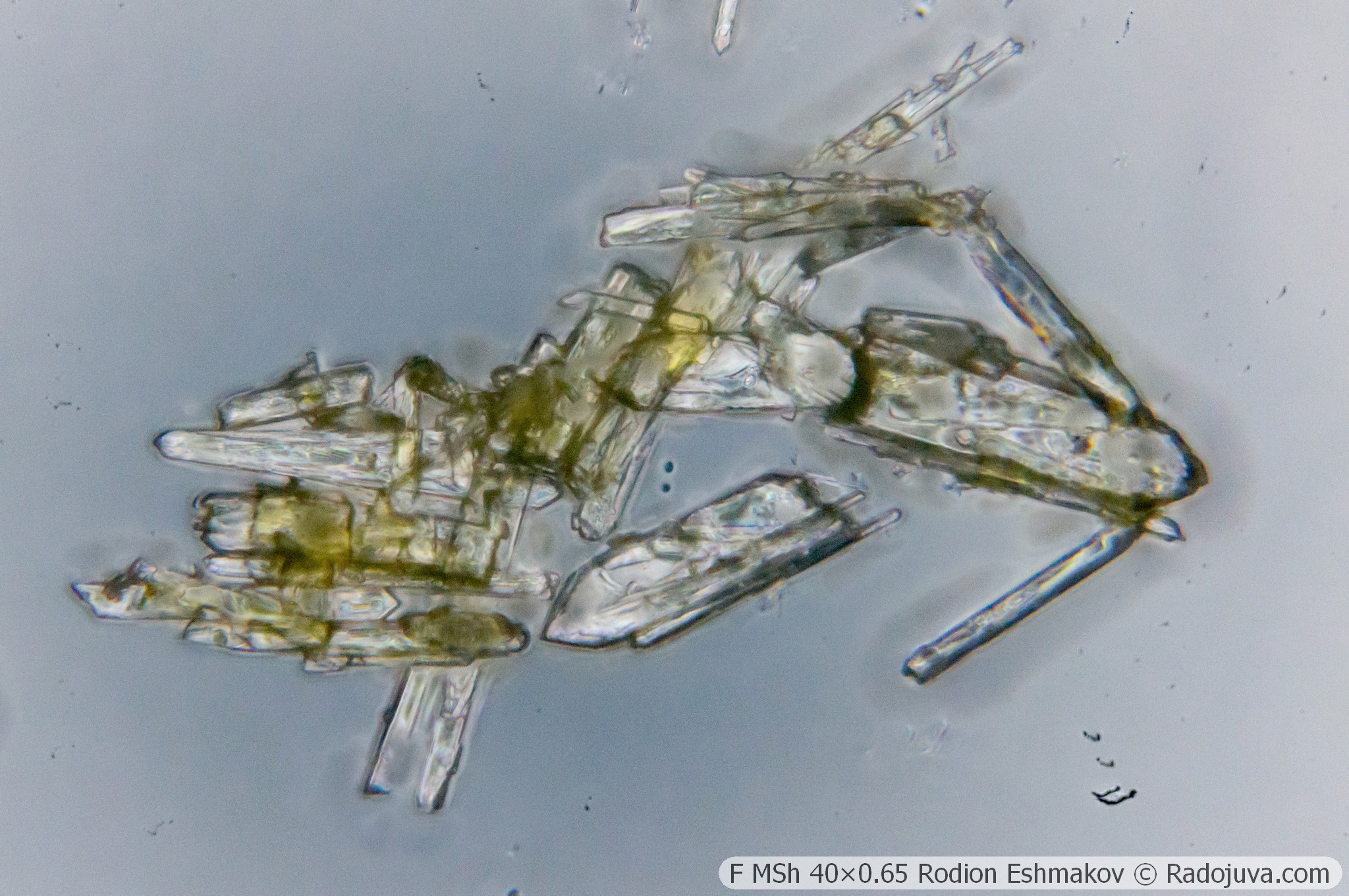
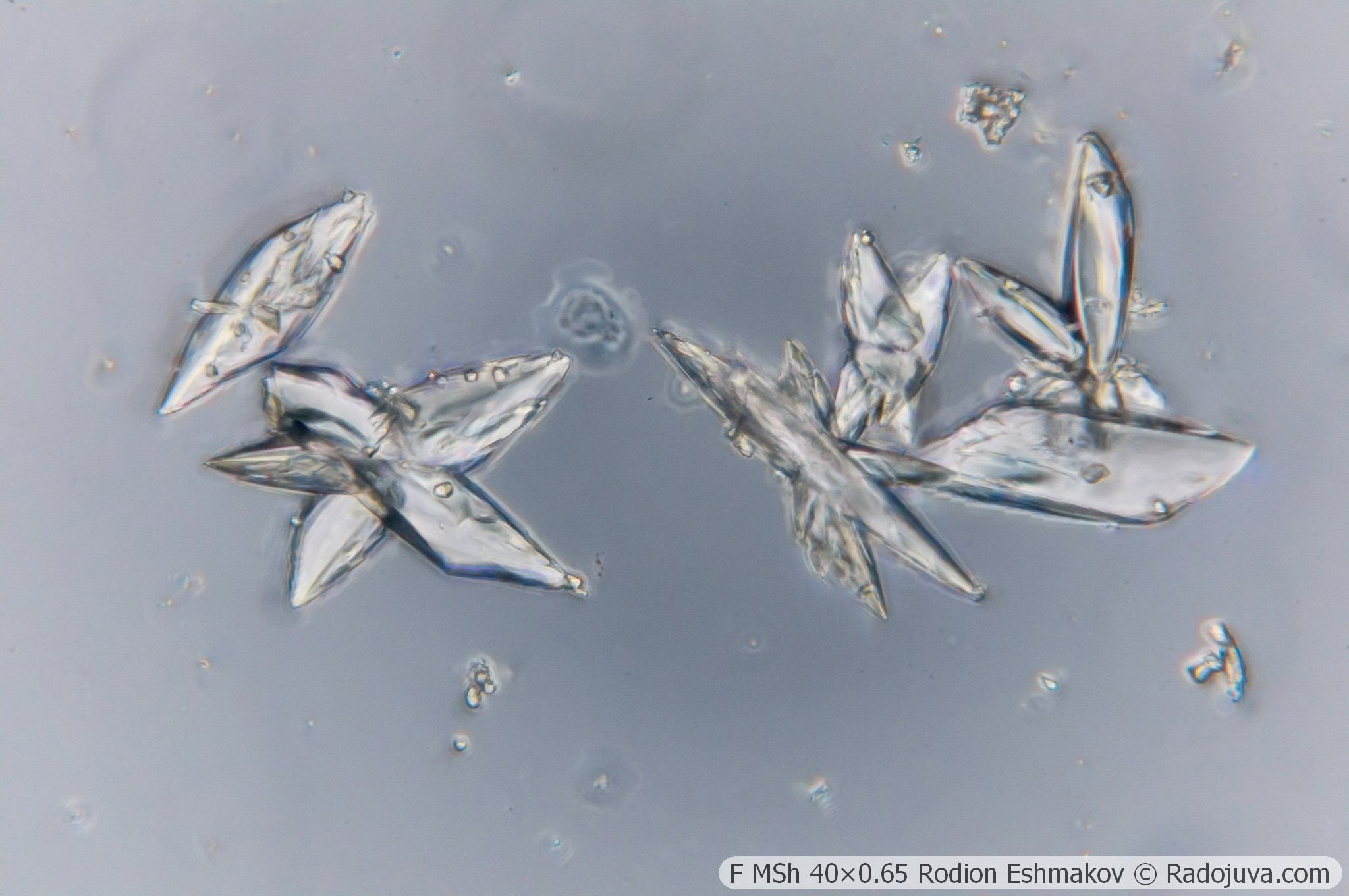
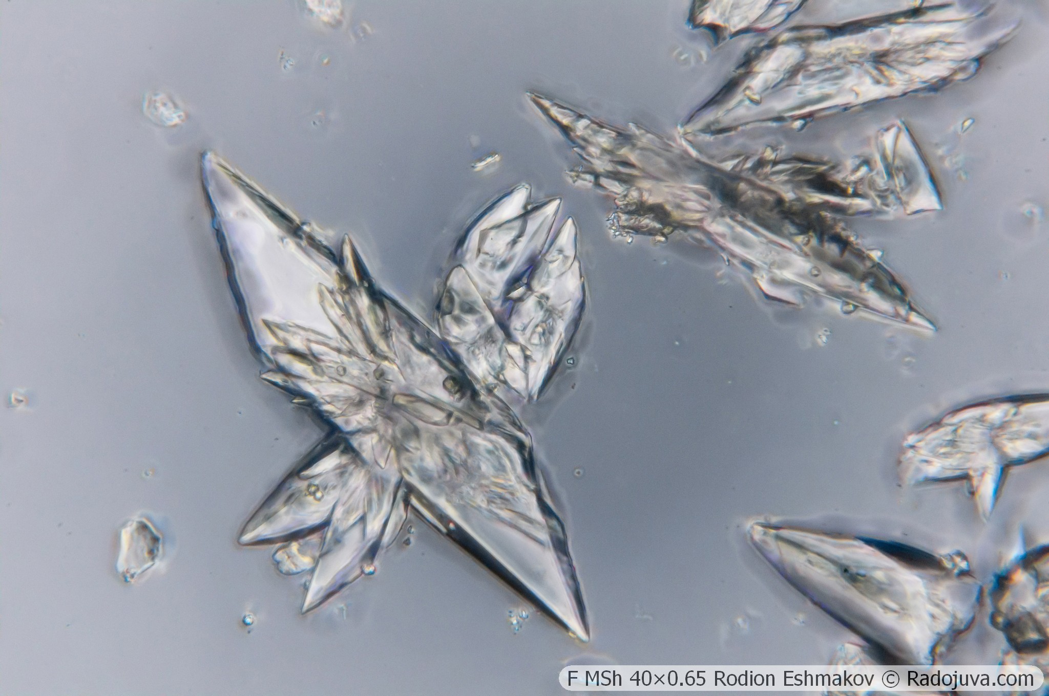
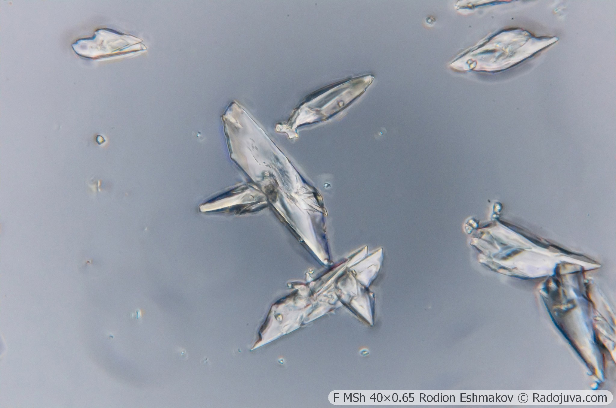
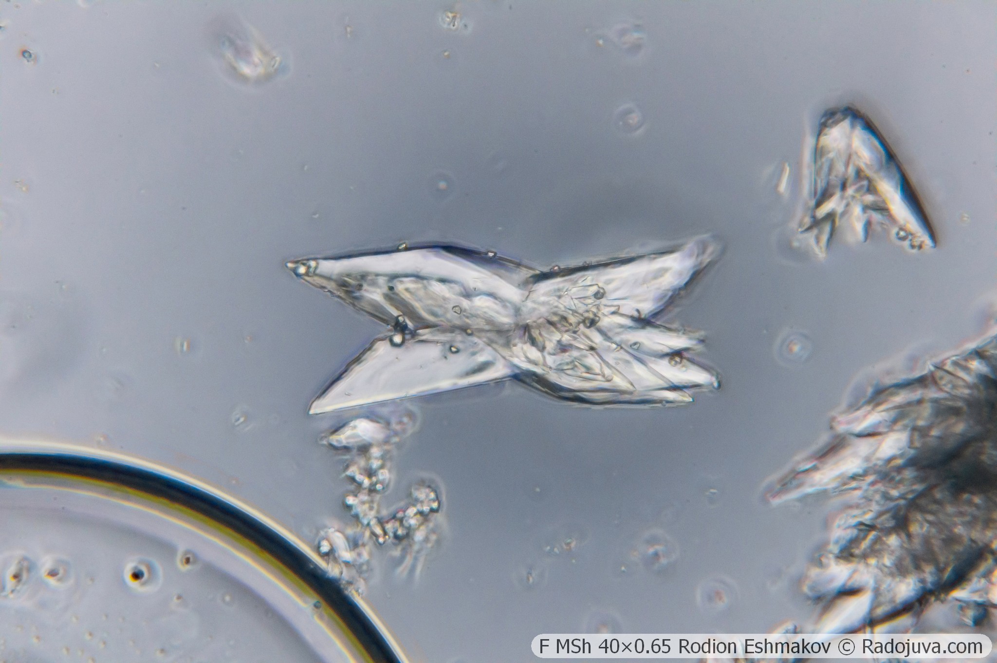
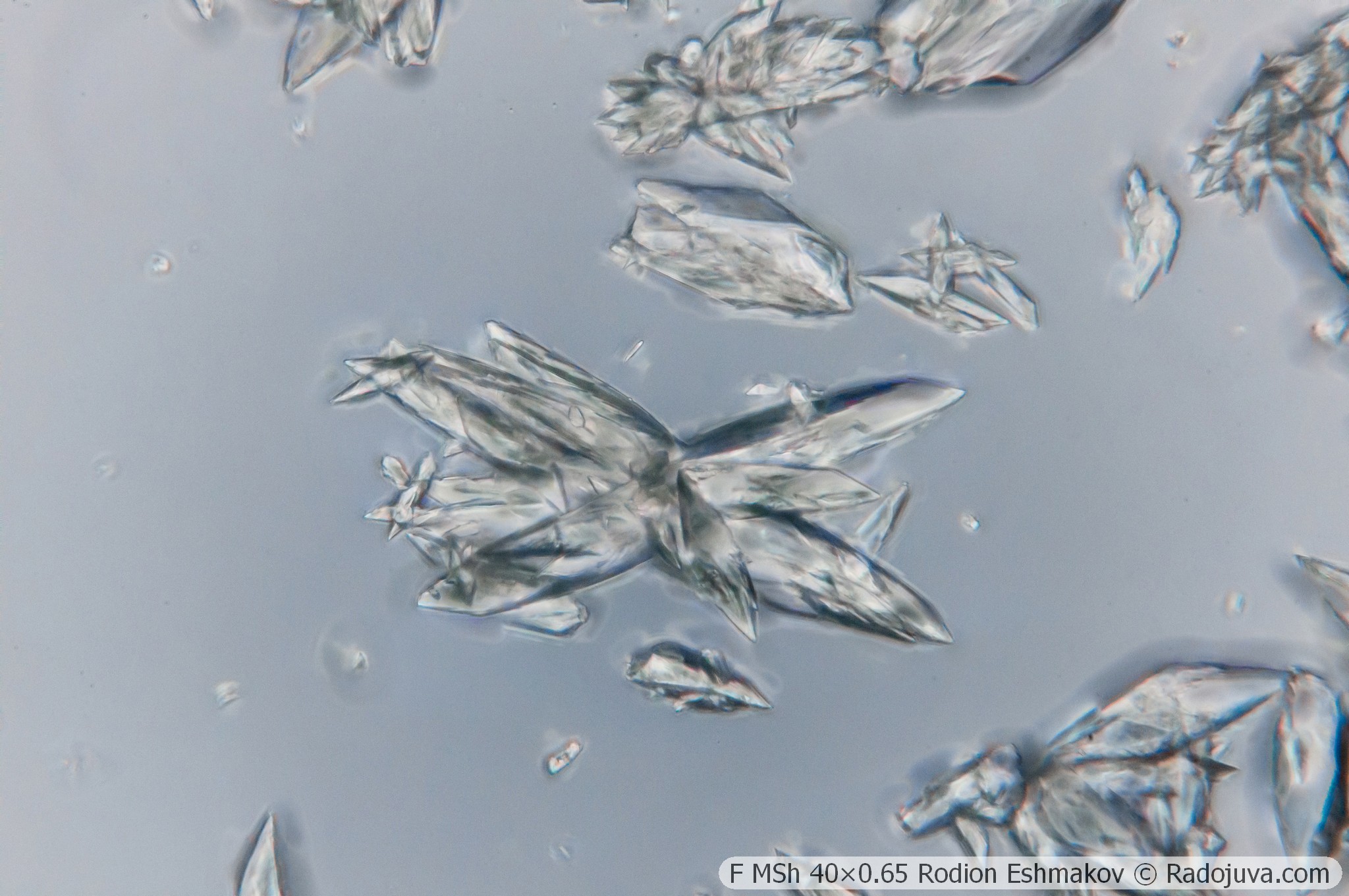
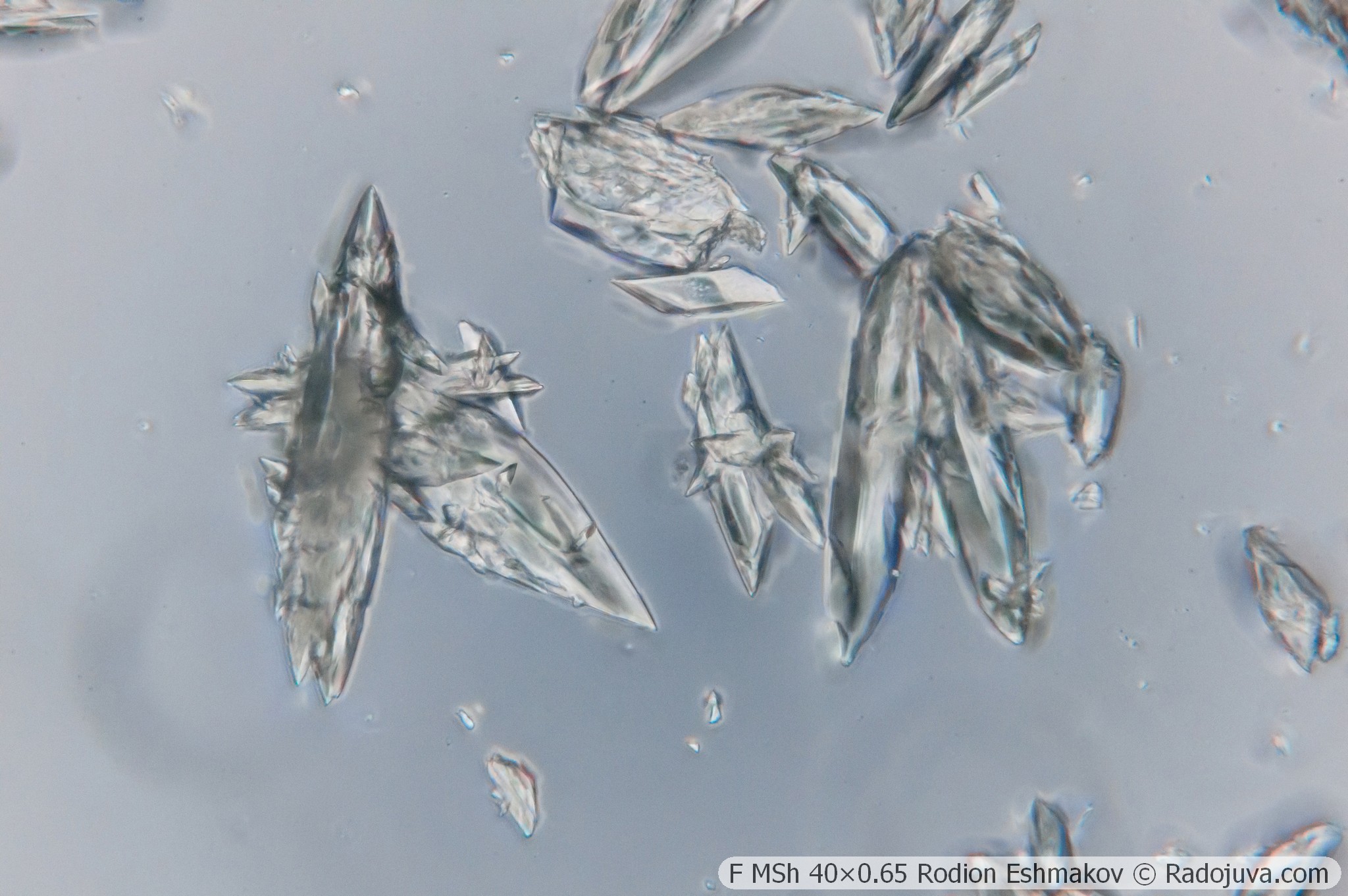
Below are examples of photos taken with the Sony NEX-6 and the F MS 40×0.65 lens in prime focus and using the relay from the K5× ML-2 eyepiece and the WitrueHD 75/2.8 10 Mpix lens.
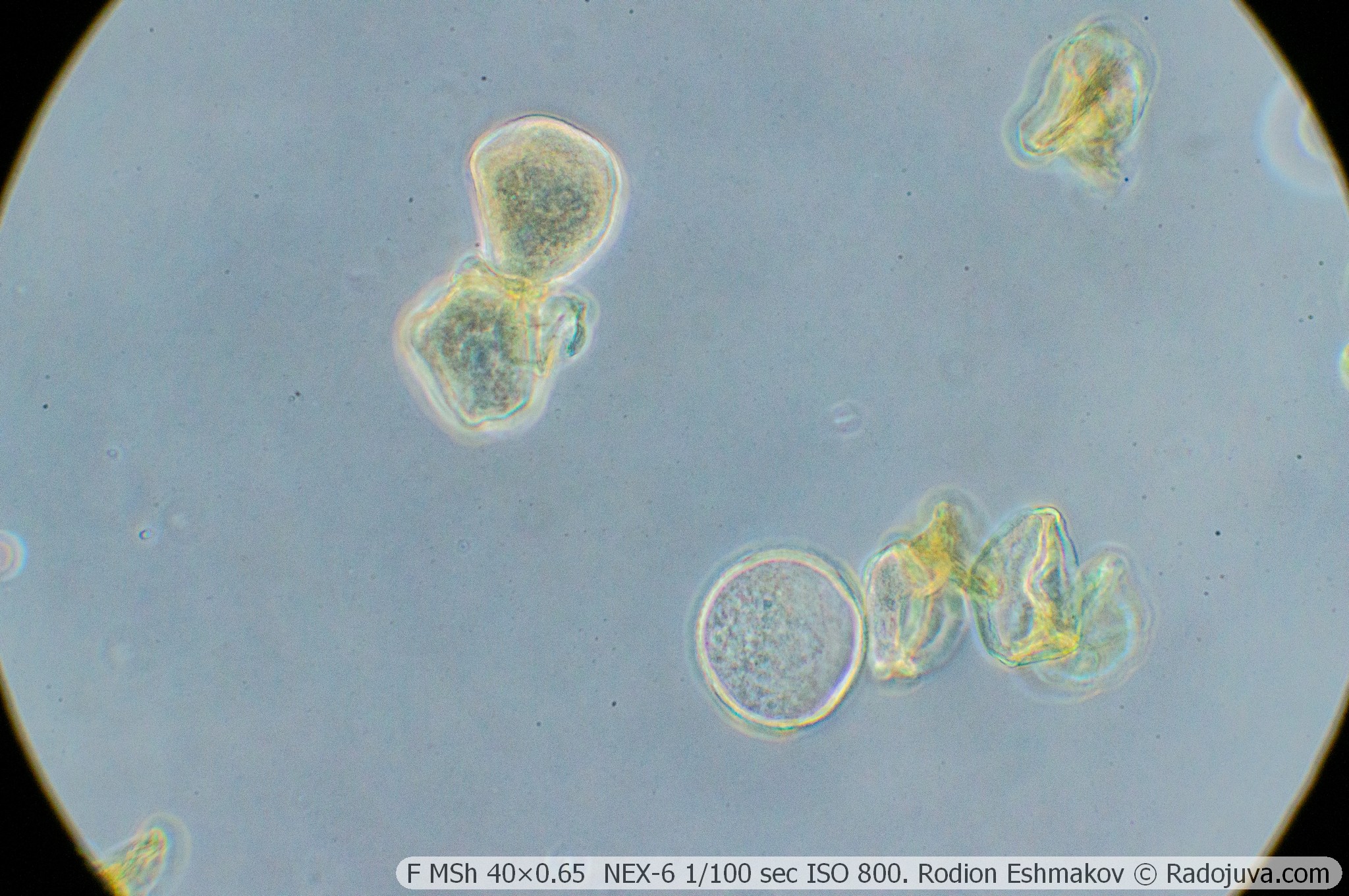
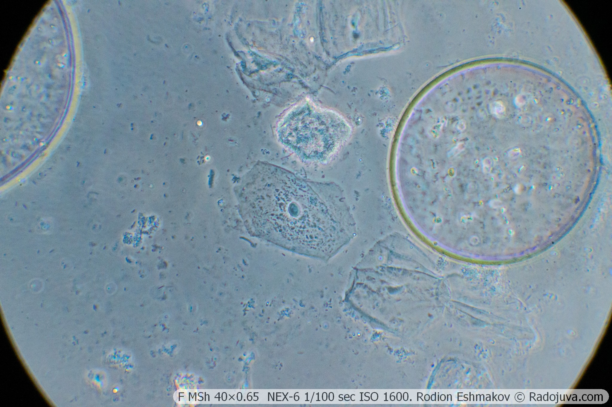
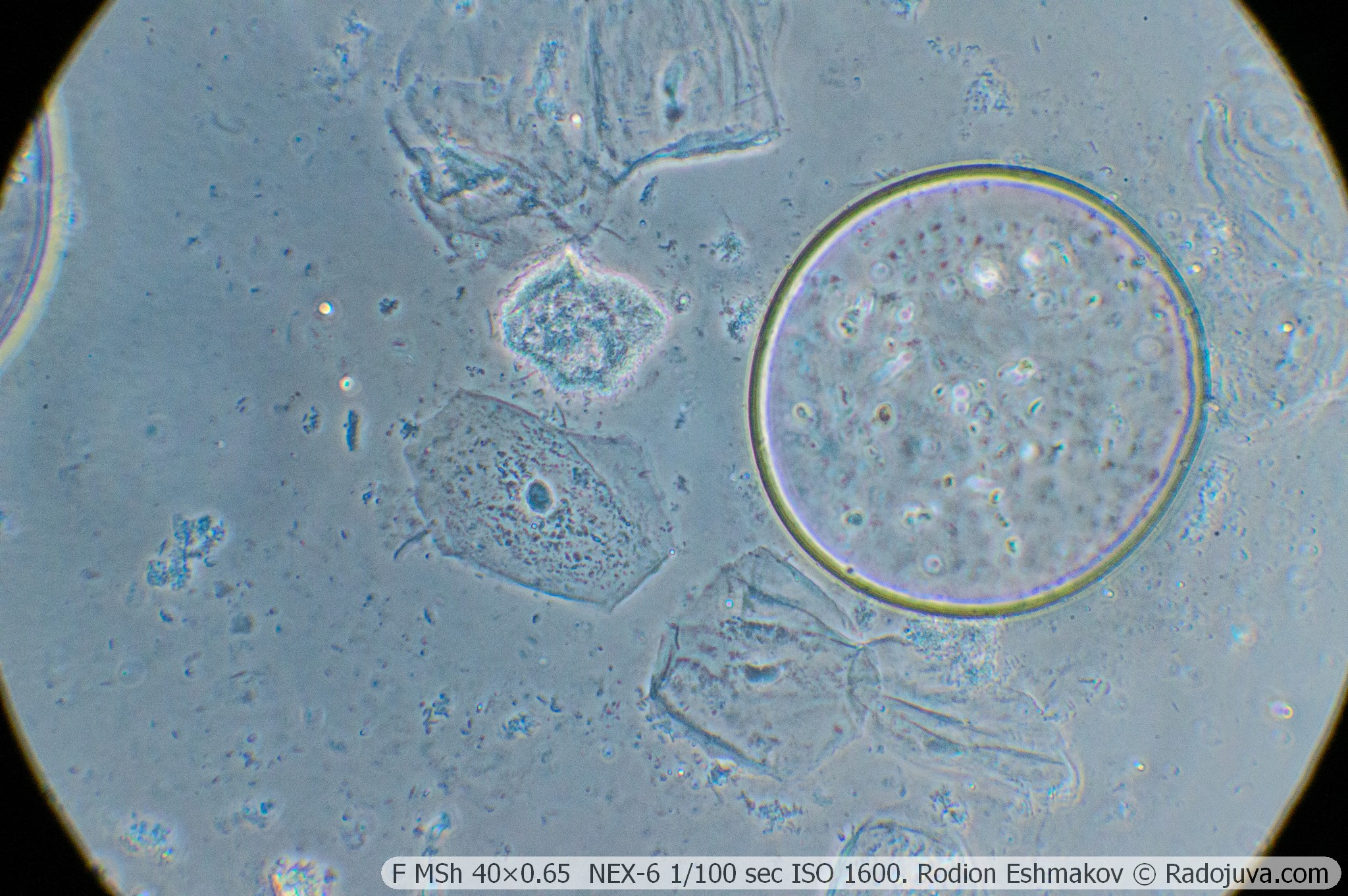
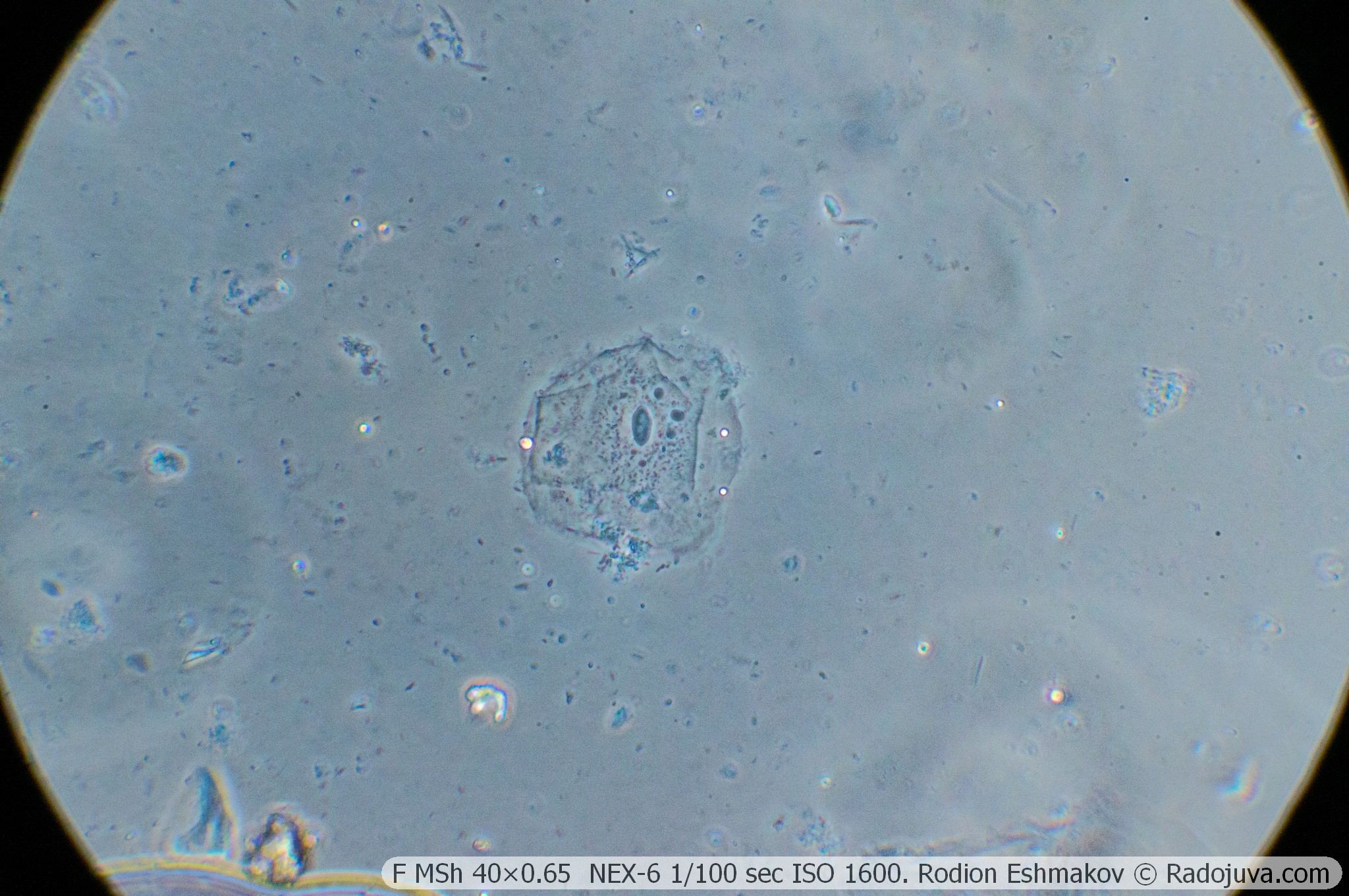
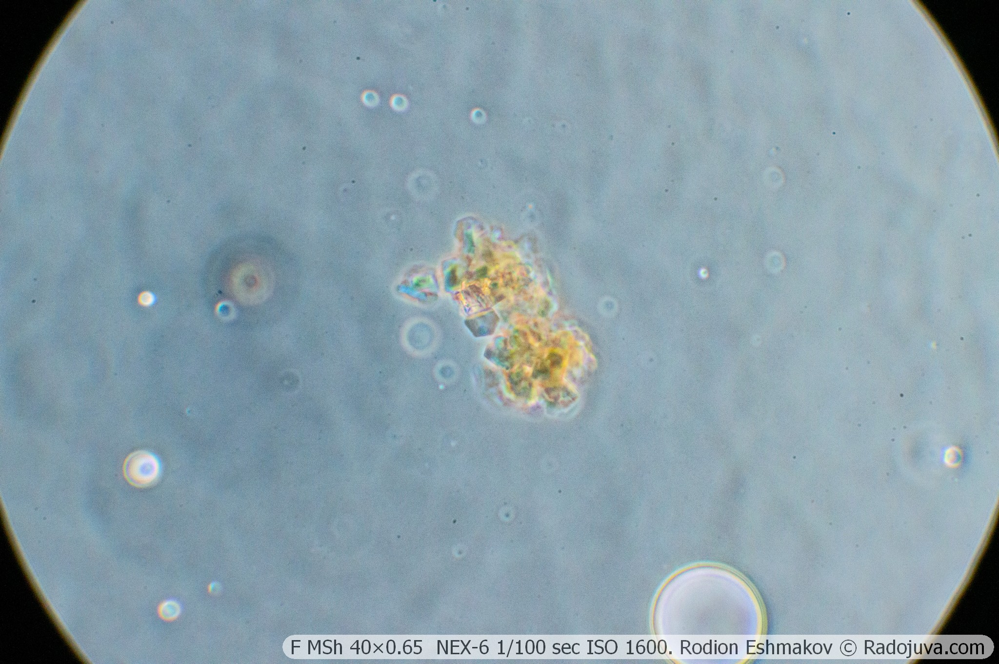
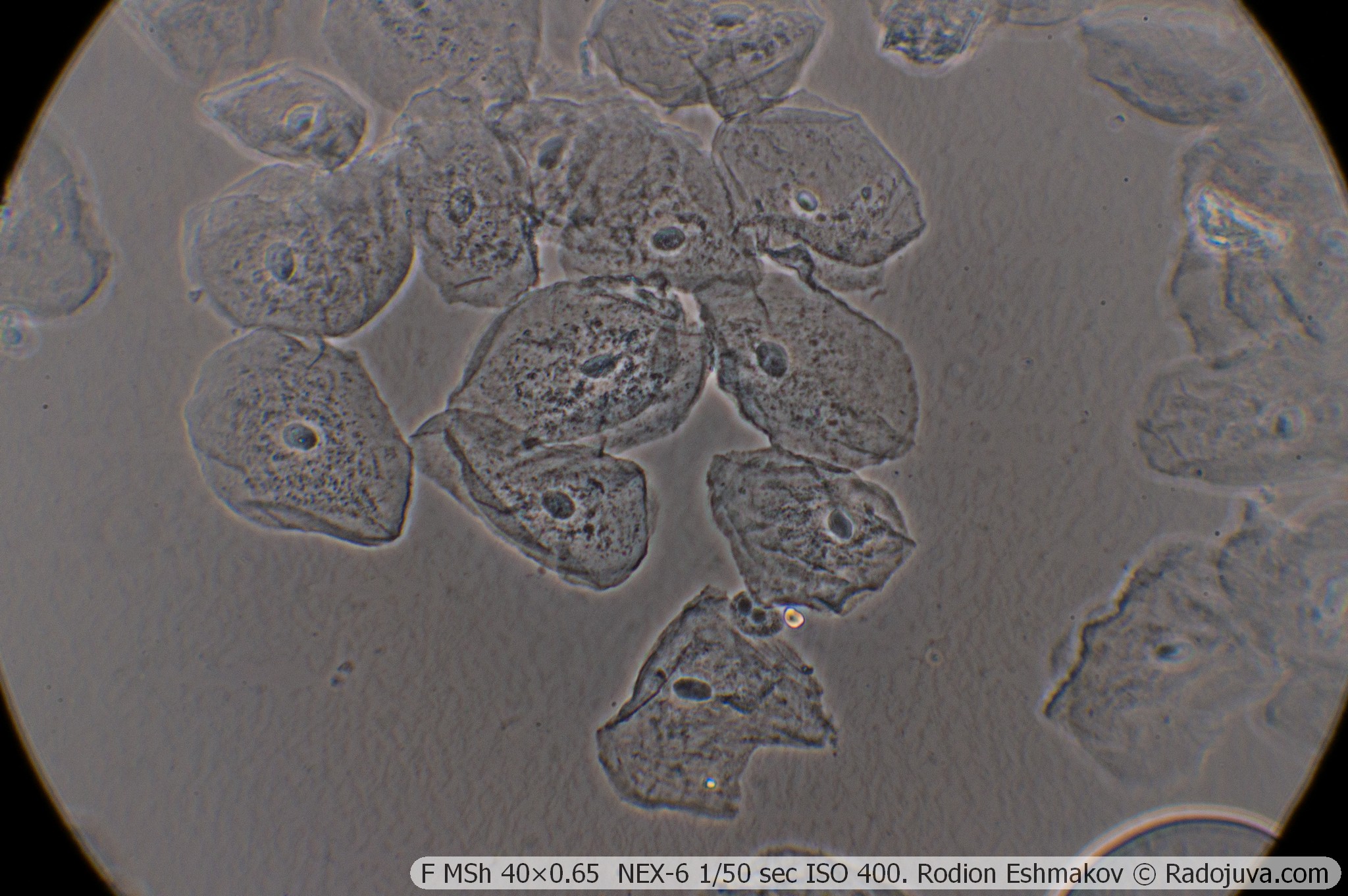
Below are examples of photos with stacking.
Lenses for the KF-1: 90×1.25 F MI OM-41
Optical design – 6 lenses in 4 groups, high-aperture aplanat;
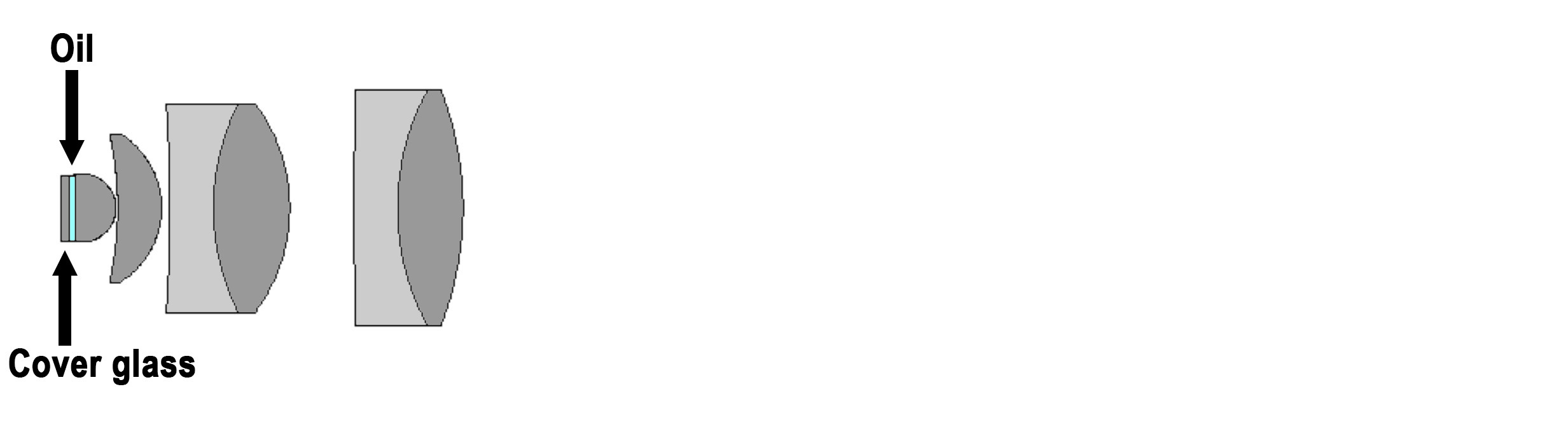
Drawing of the optical diagram of the F MI OM-41 90×1.25.
Type of correction – achromat;
Tube distance – 160 mm;
Parfocal distance – 32.7 mm;
Magnification: 90×;
Numerical aperture – 1.25 (with KF-1 0.8, phase ring 0.37);
Immersion required – yes (oil);
Working distance – 0.1 mm;
Cover glass thickness: 0-0.25 mm;
Mounting type – RMS standard (4/5” × 1/36” thread);
Lateral chromatic aberration – corrected with a compensating eyepiece (such as LOMO K5×);
Features - microscopic lens, does not have an iris diaphragm and a focusing mechanism.
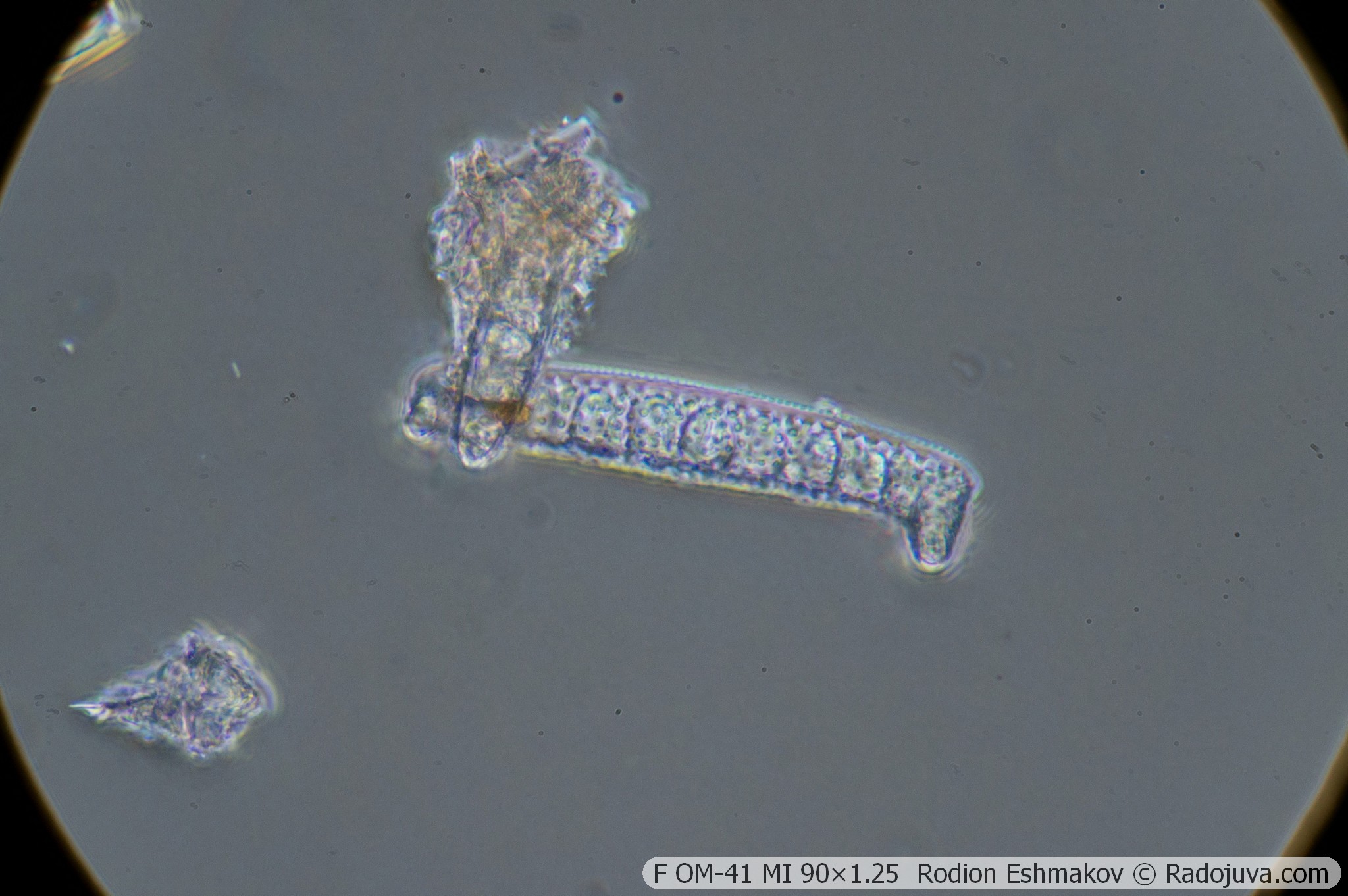
This lens, like any 100×1.25, performs very poorly in brightfield photography, too. The phase-contrast version actually operates at a very low numerical aperture, making the image resolution unacceptably low for photography. Nevertheless, it is the highest-aperture lens in the KF-1 kit. Only this lens allows, for example, the visualization of such small objects as bacteria. Of course, the quality is not great, but it is possible to distinguish rods from cocci—and all without staining the specimen. You can even observe the movements of living bacteria. However, it's difficult to find aesthetically pleasing in this.
The lens exhibits incredibly high aberrations. Lateral chromatic aberrations are compensated for using a K5× ML-2 eyepiece.
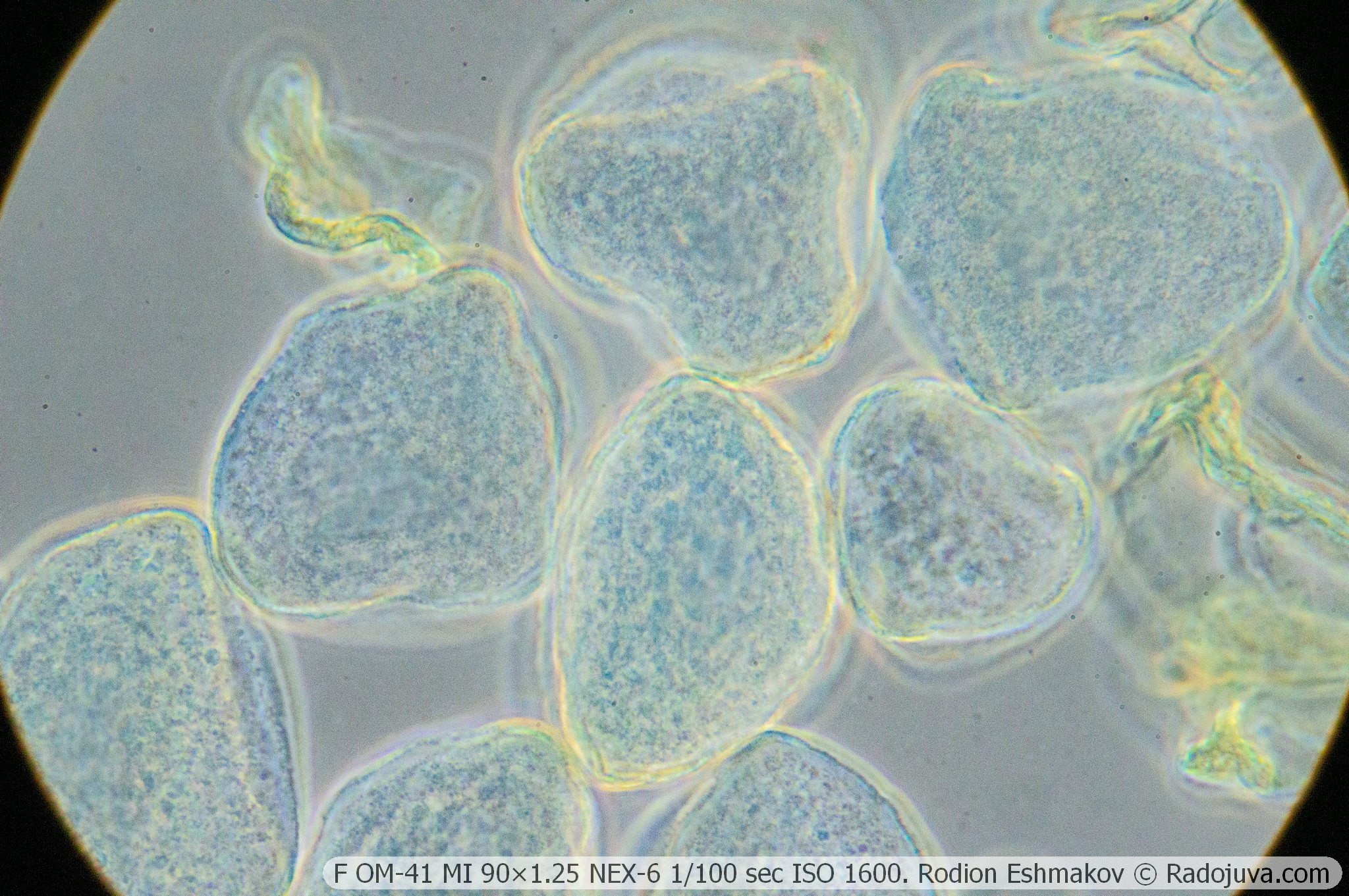
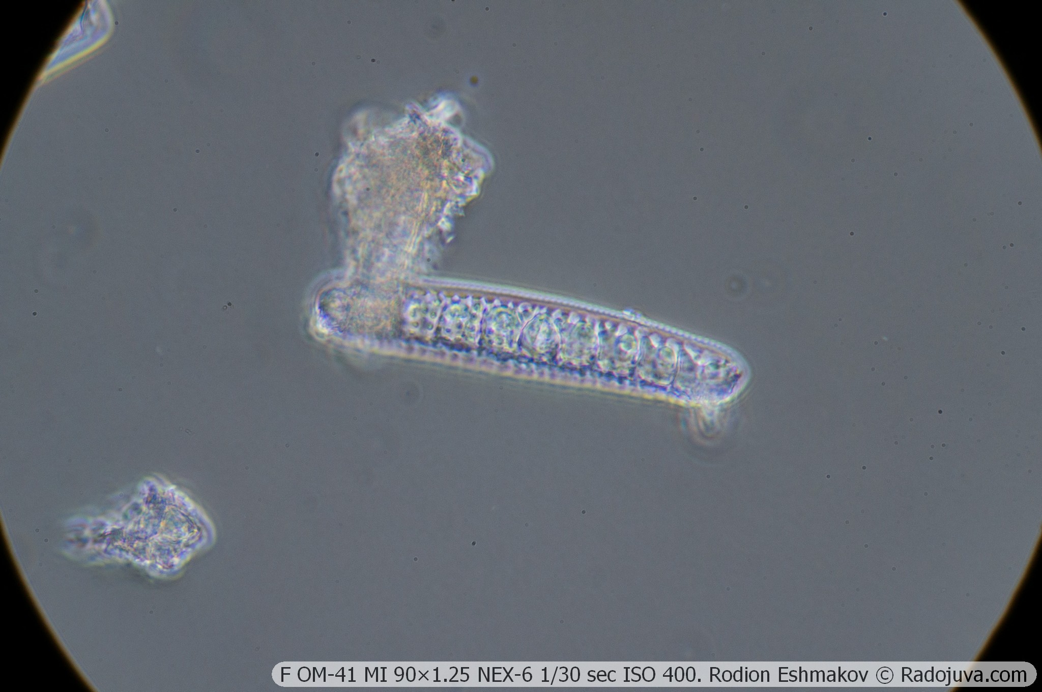
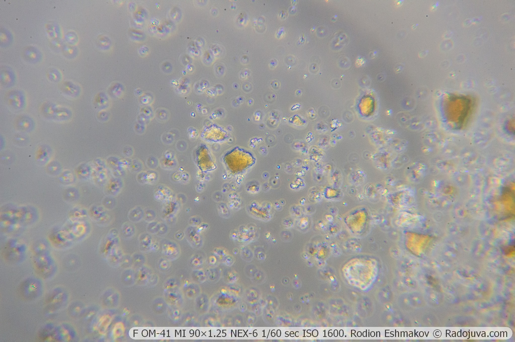
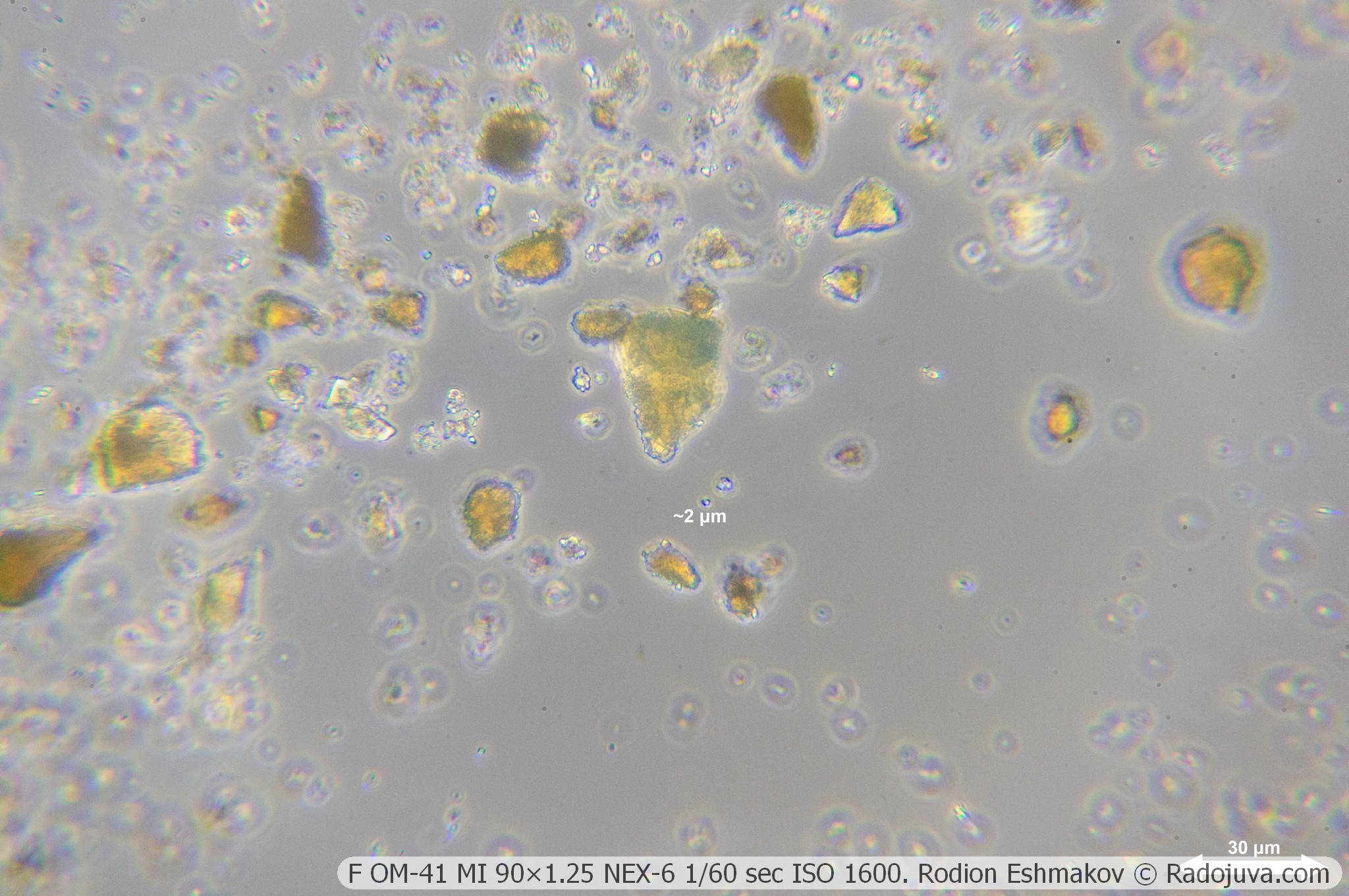
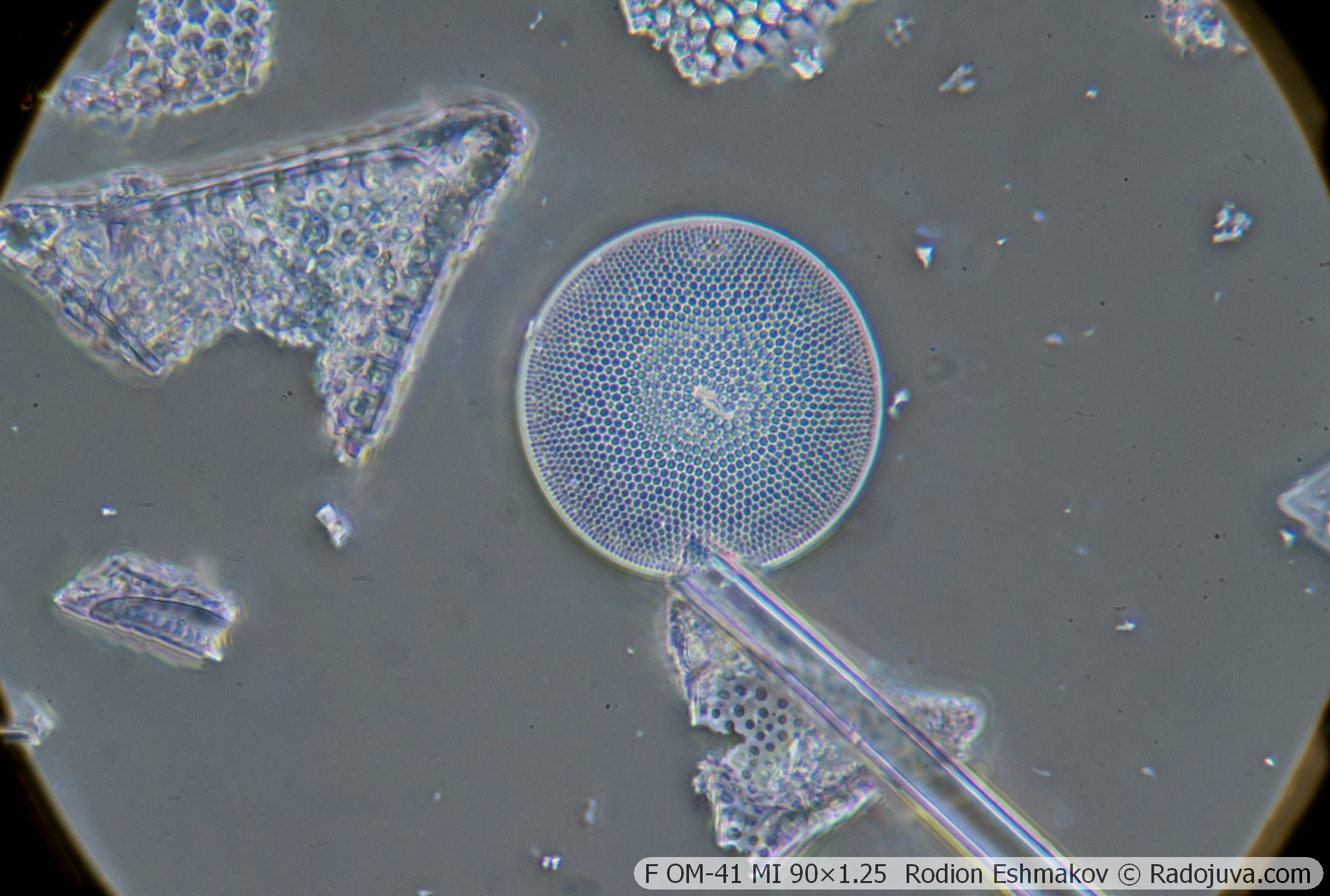
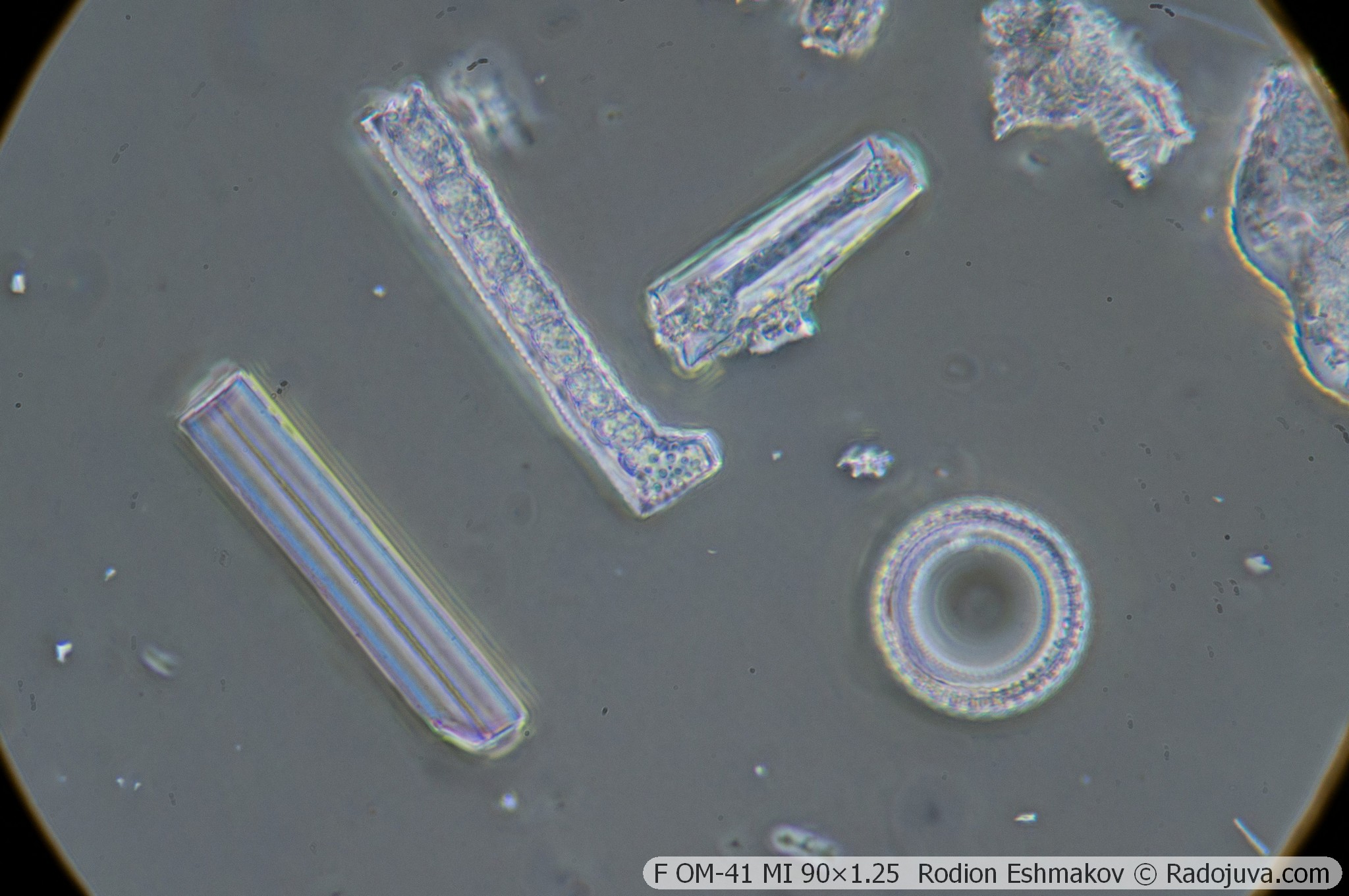
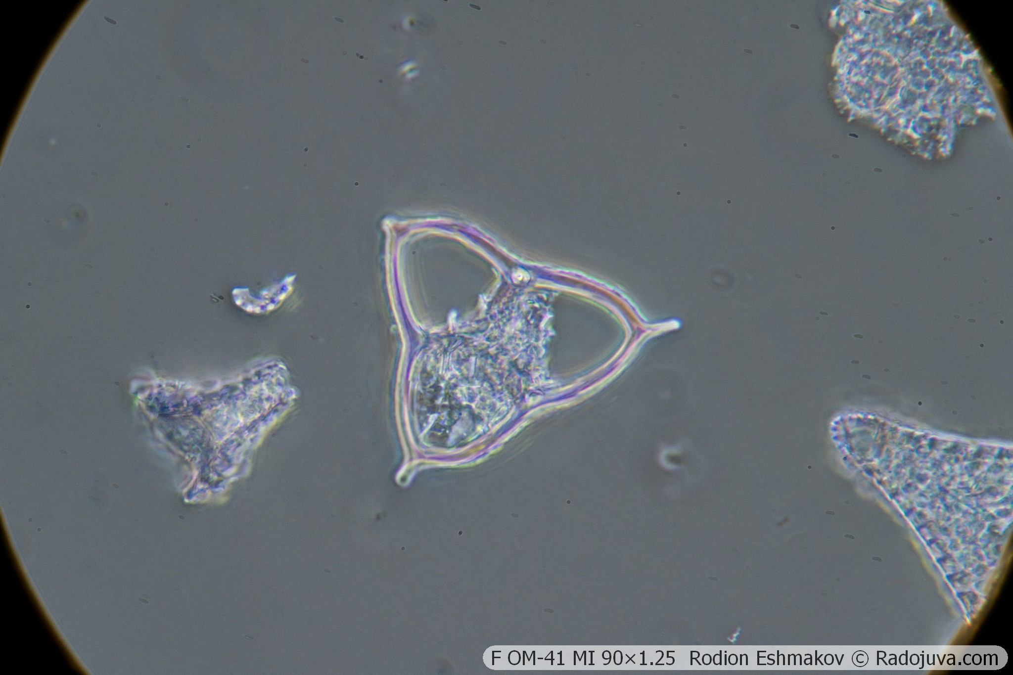
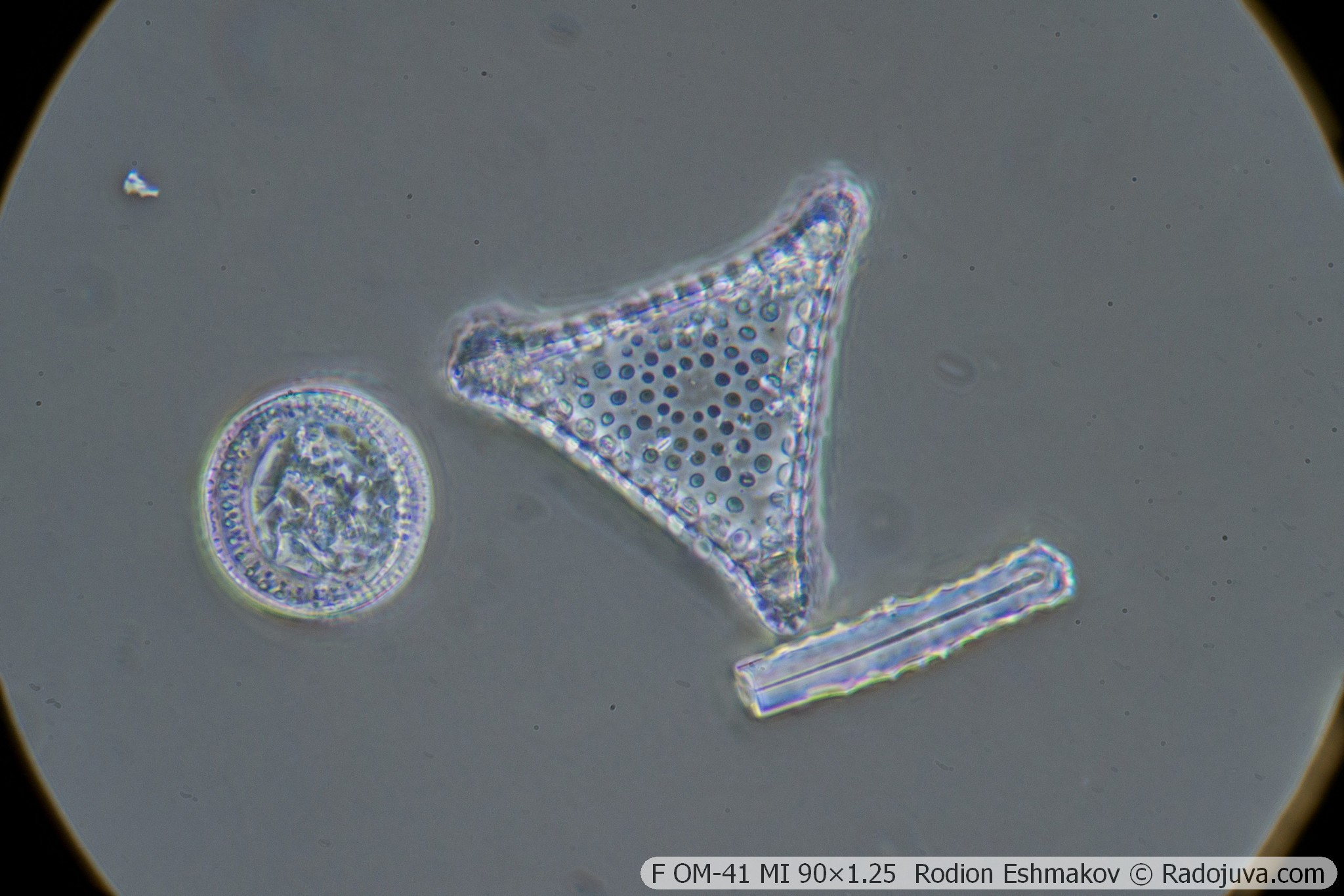
Below are some sample photos taken with the Sony NEX-6 and the F OM-41 MI 90×1.25 lens in prime focus and using relay from the K5× ML-2 eyepiece and objective WitrueHD 75/2.8 10 Mpix.
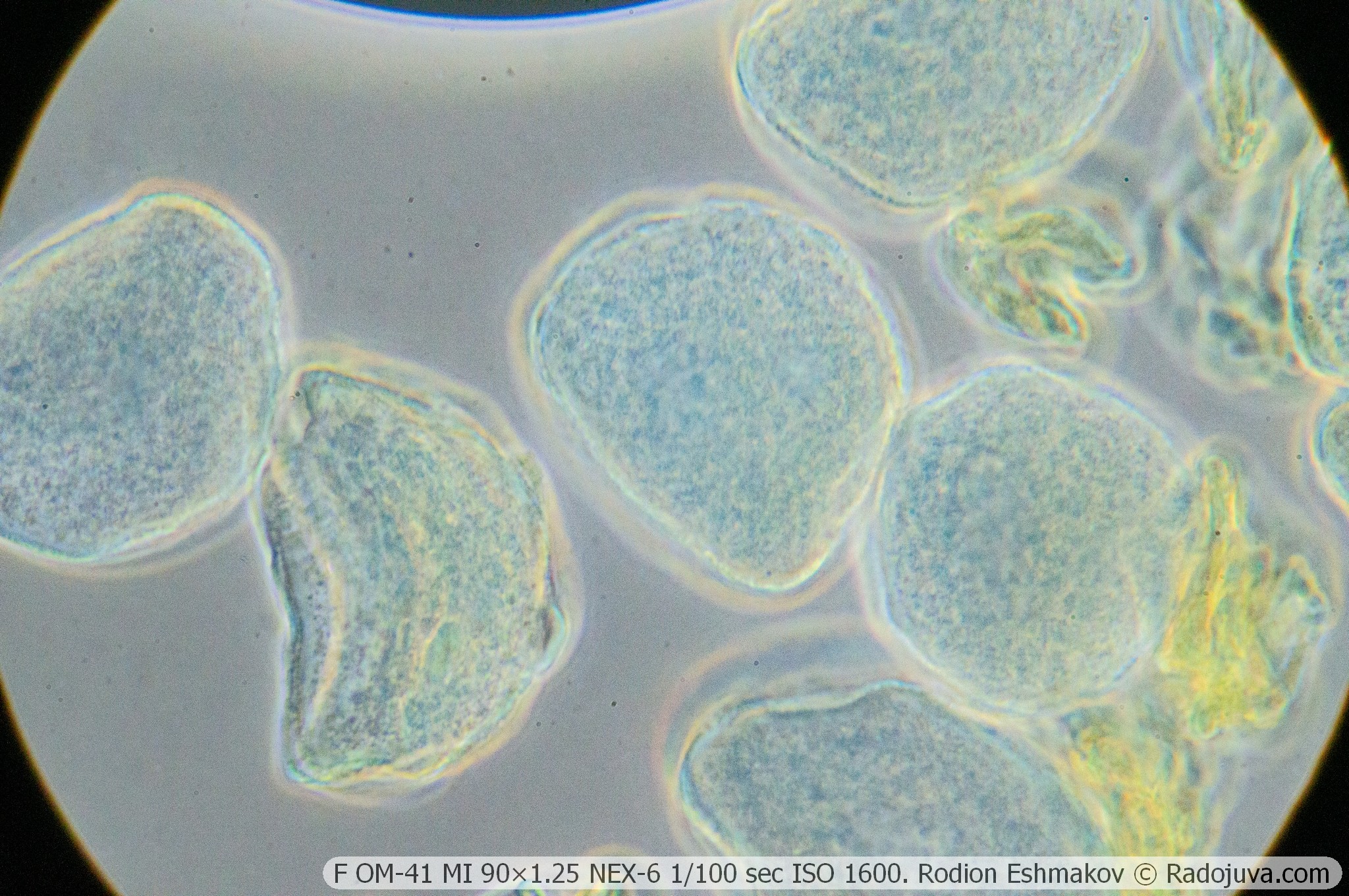
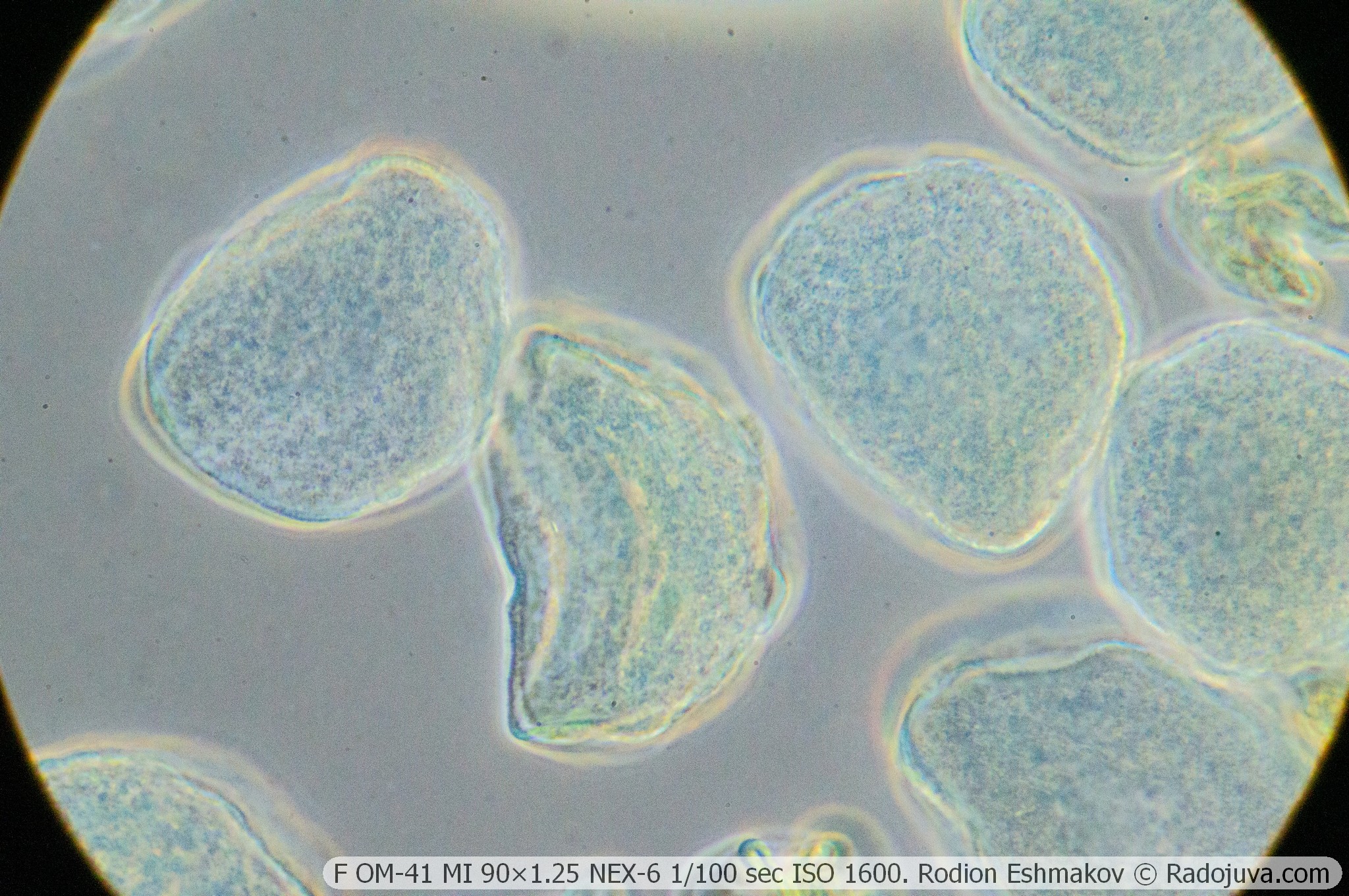
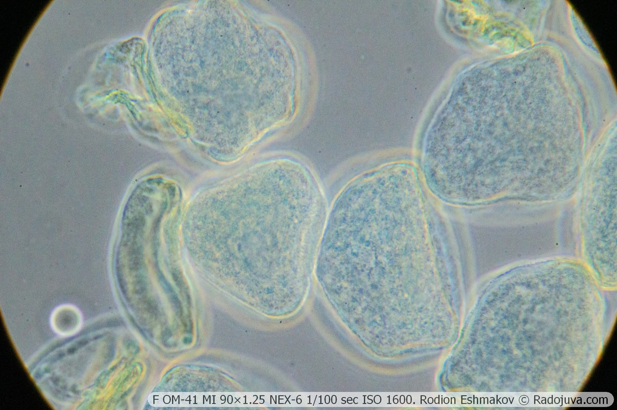
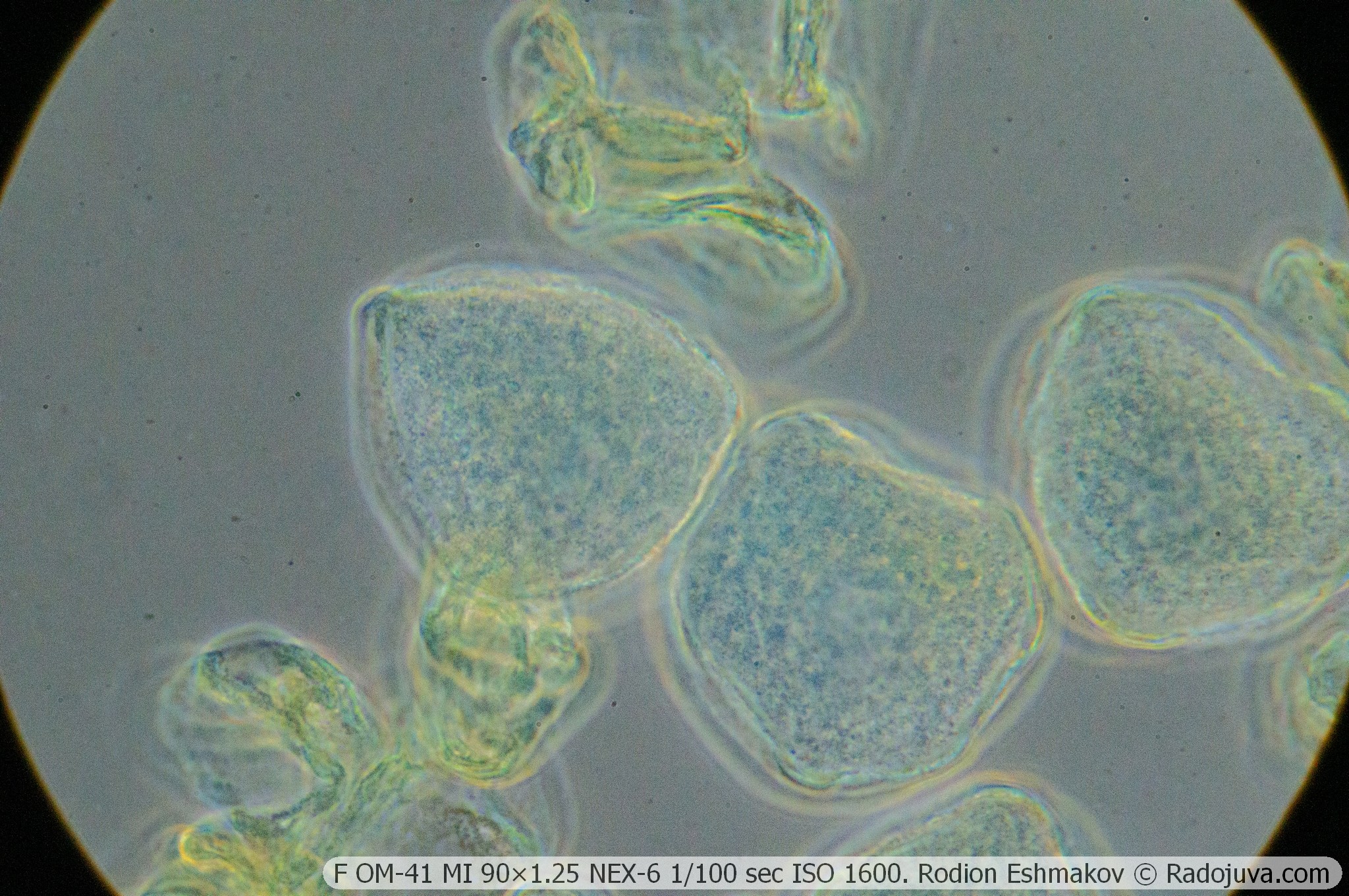
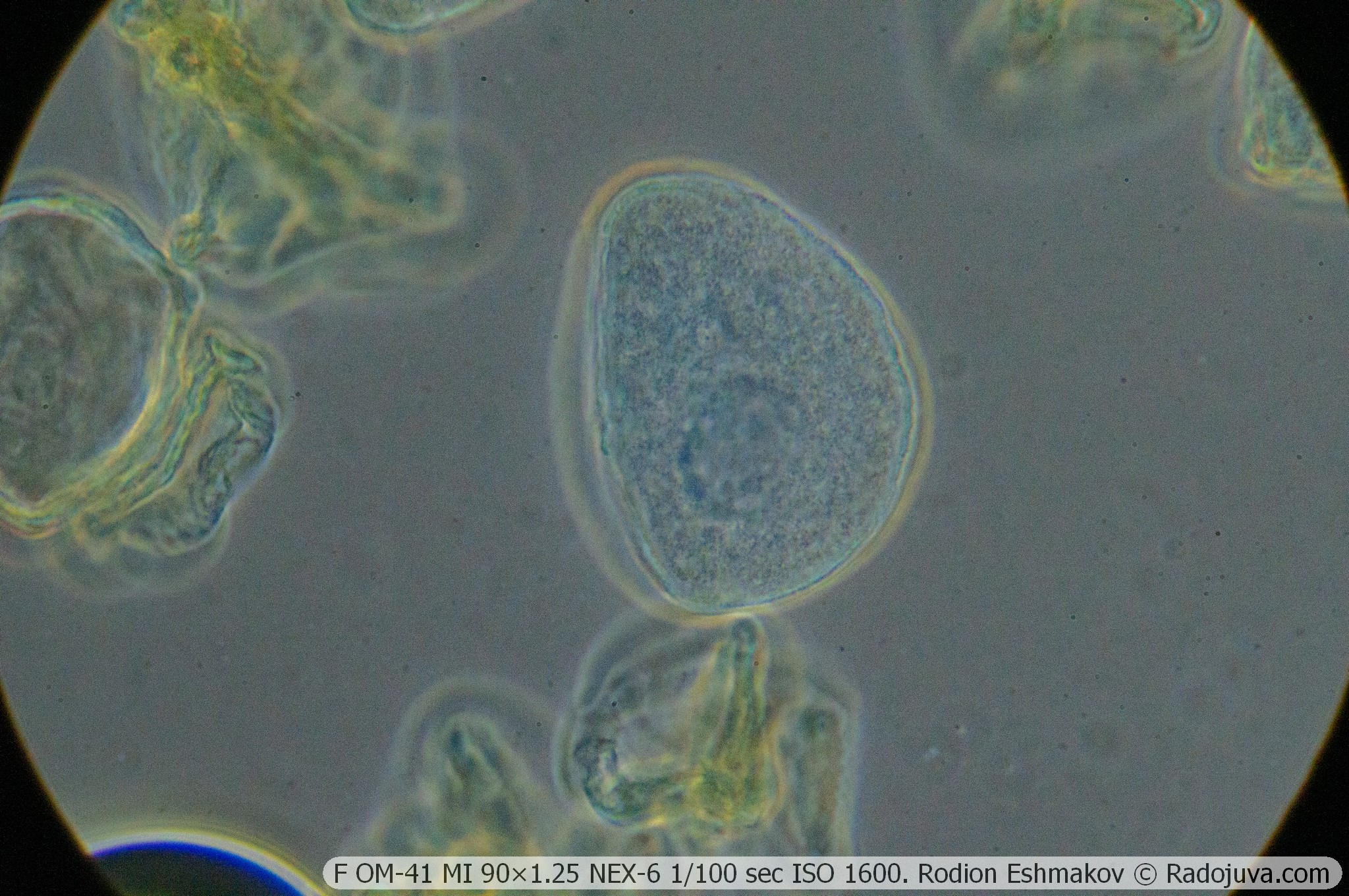
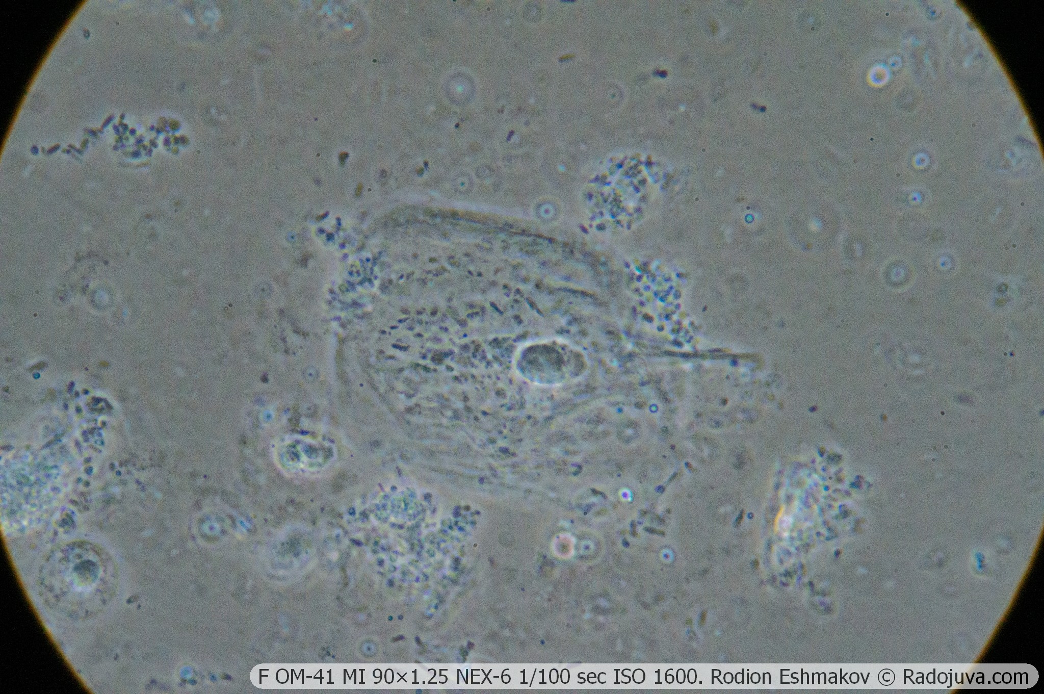
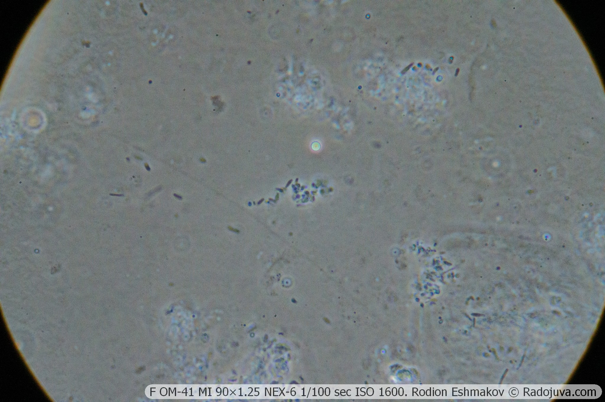
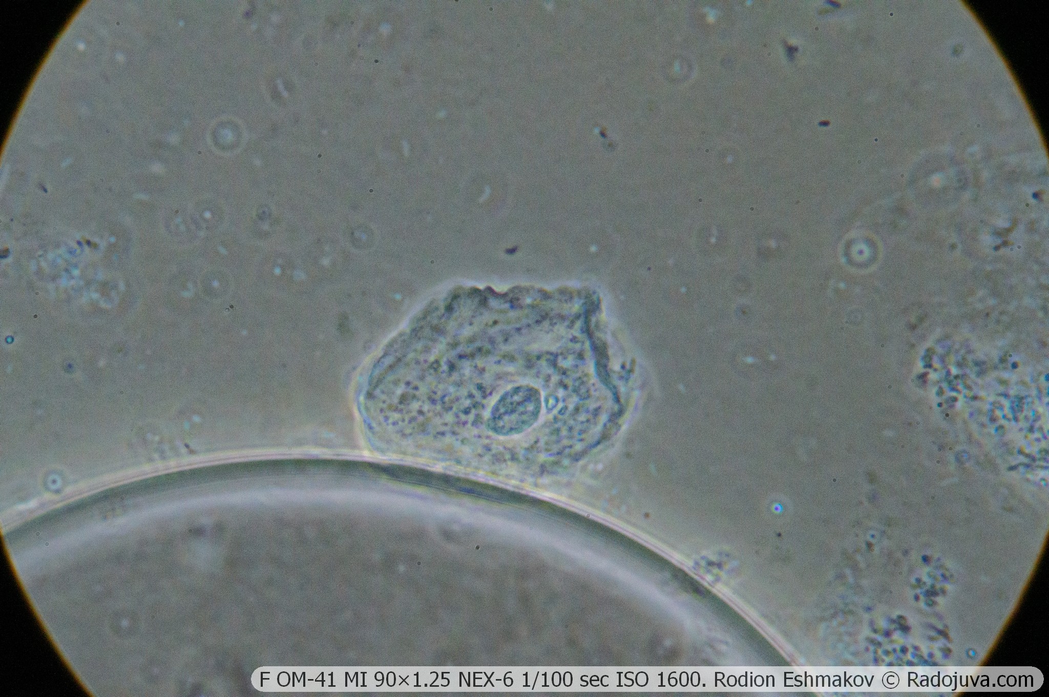
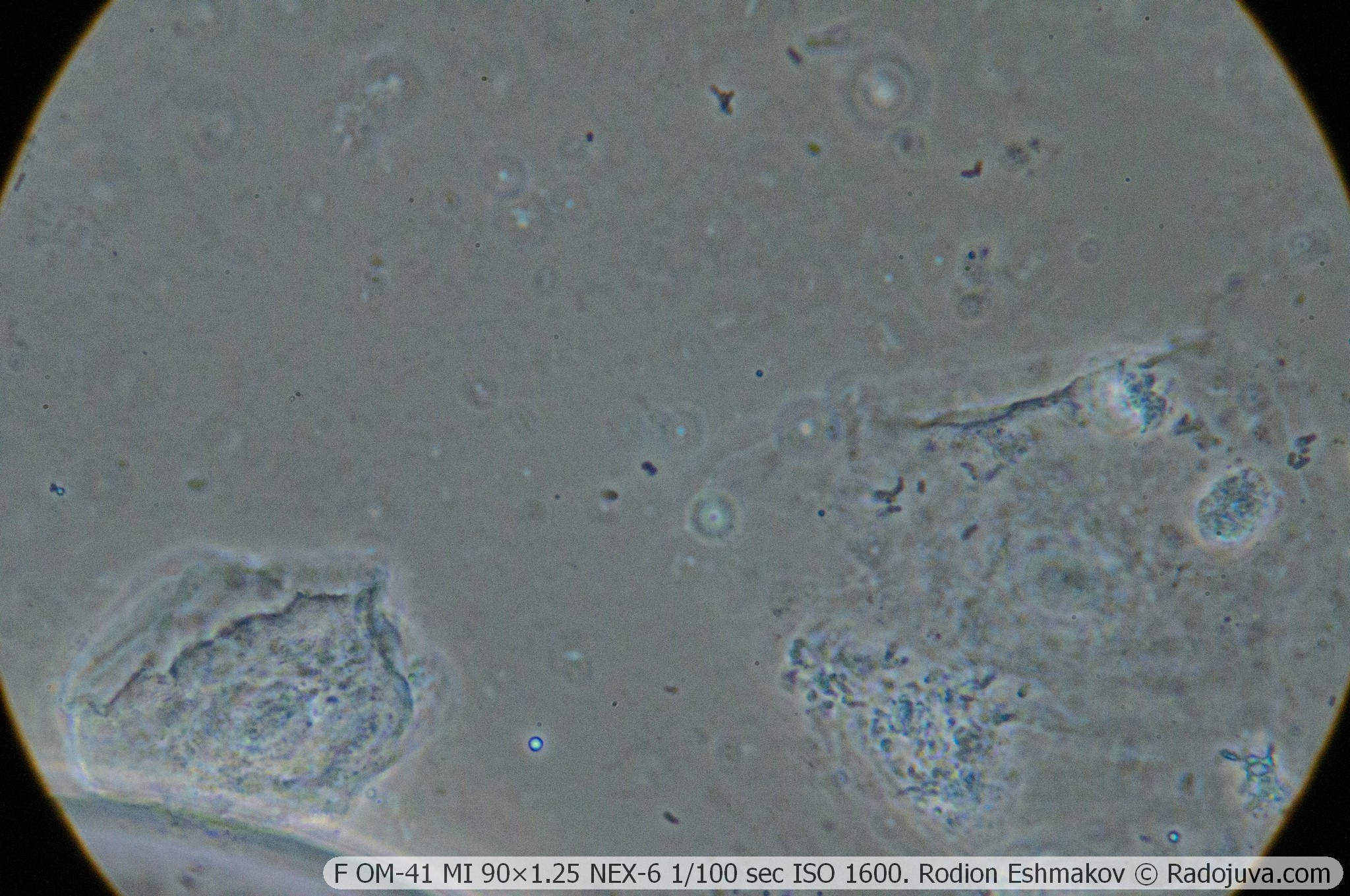
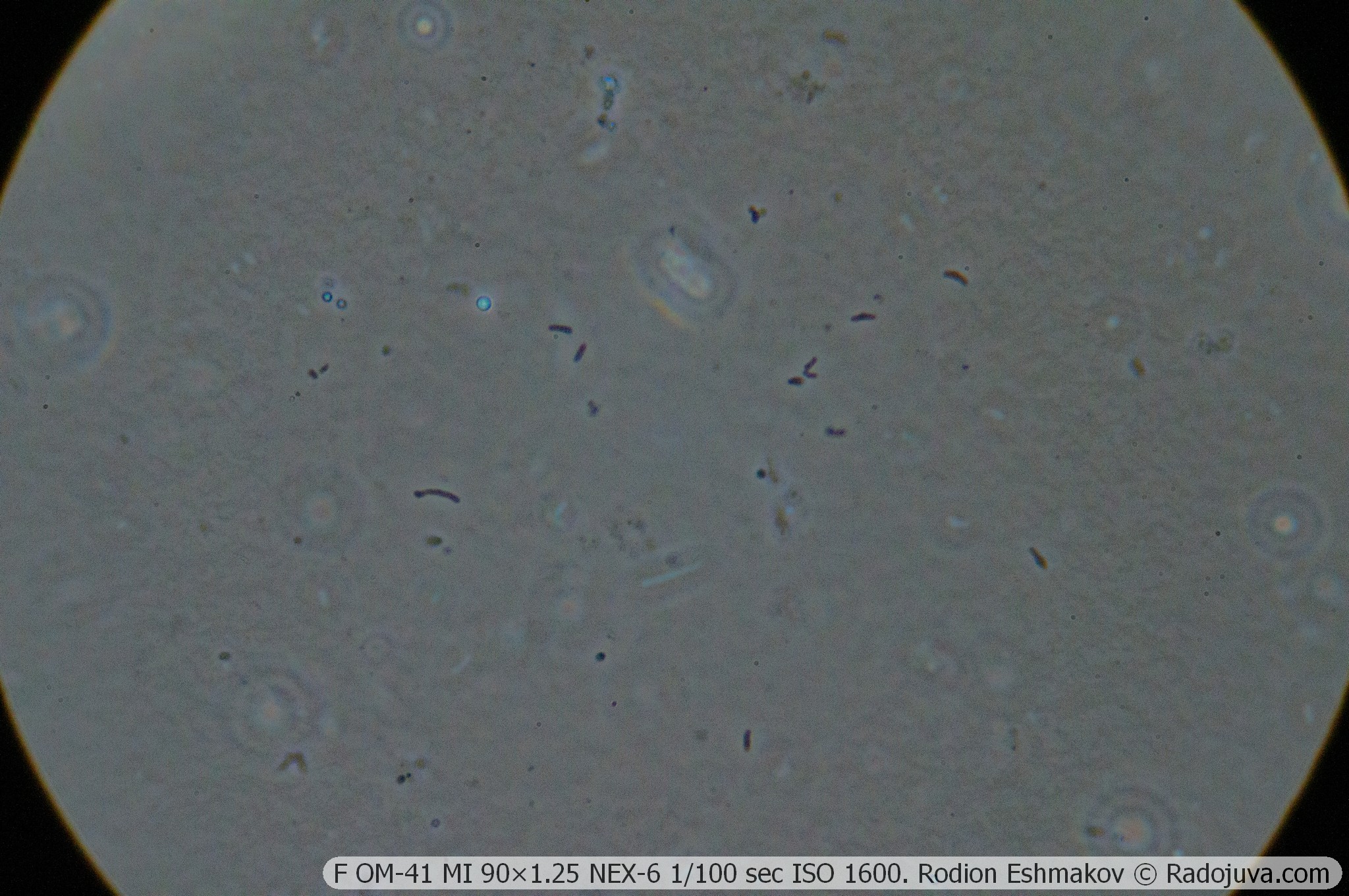
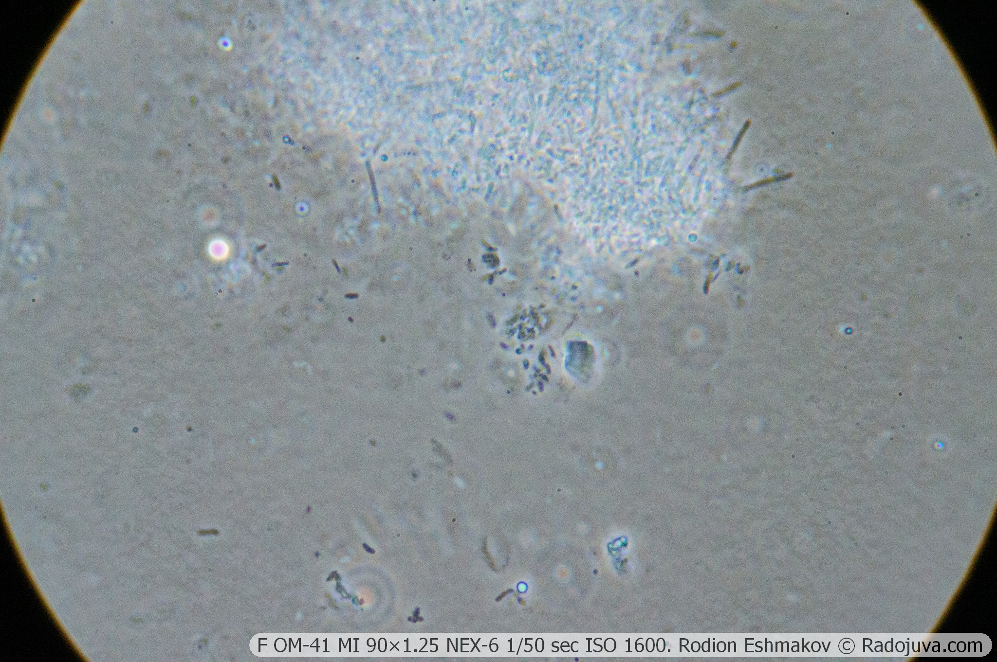
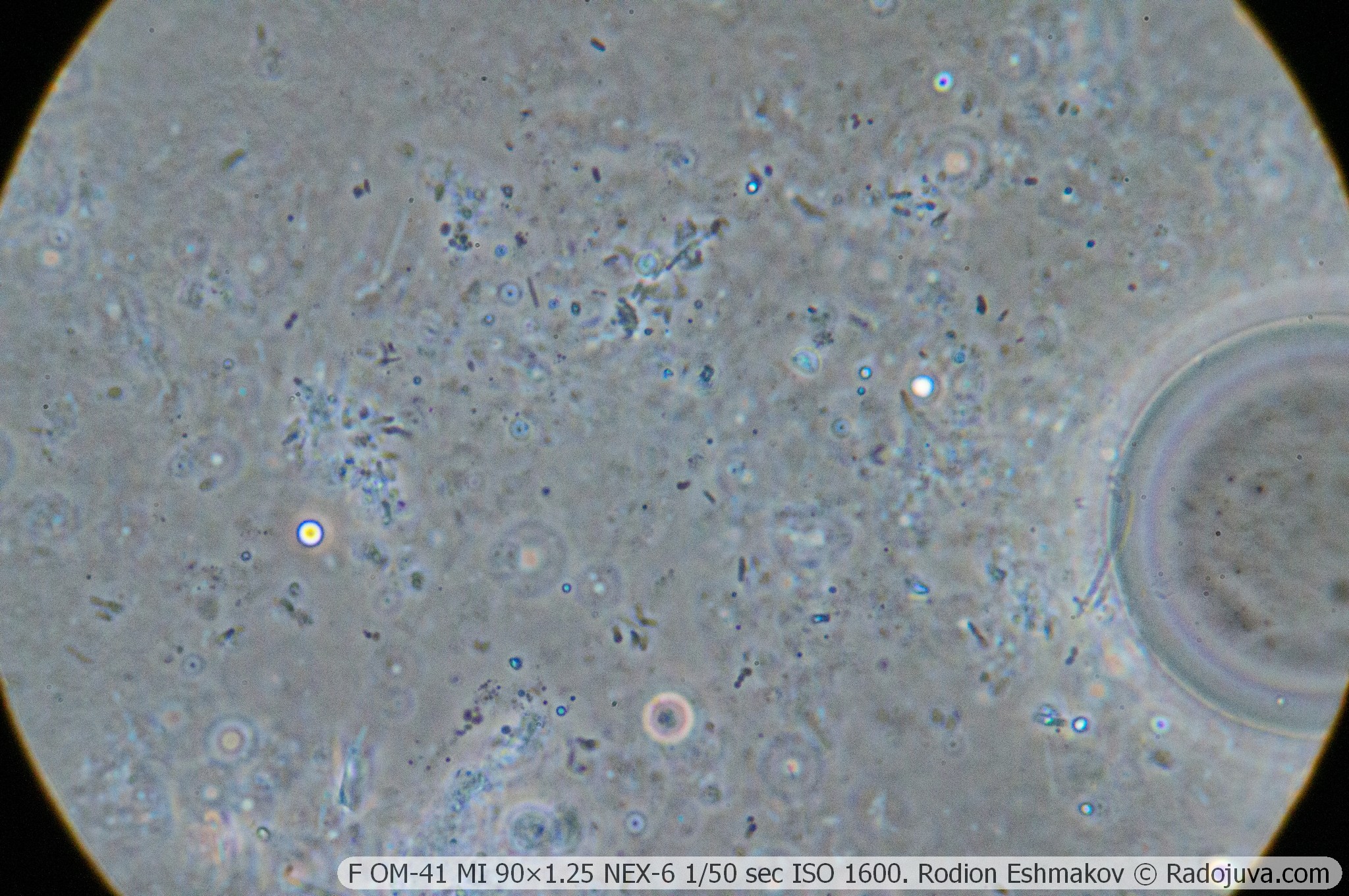
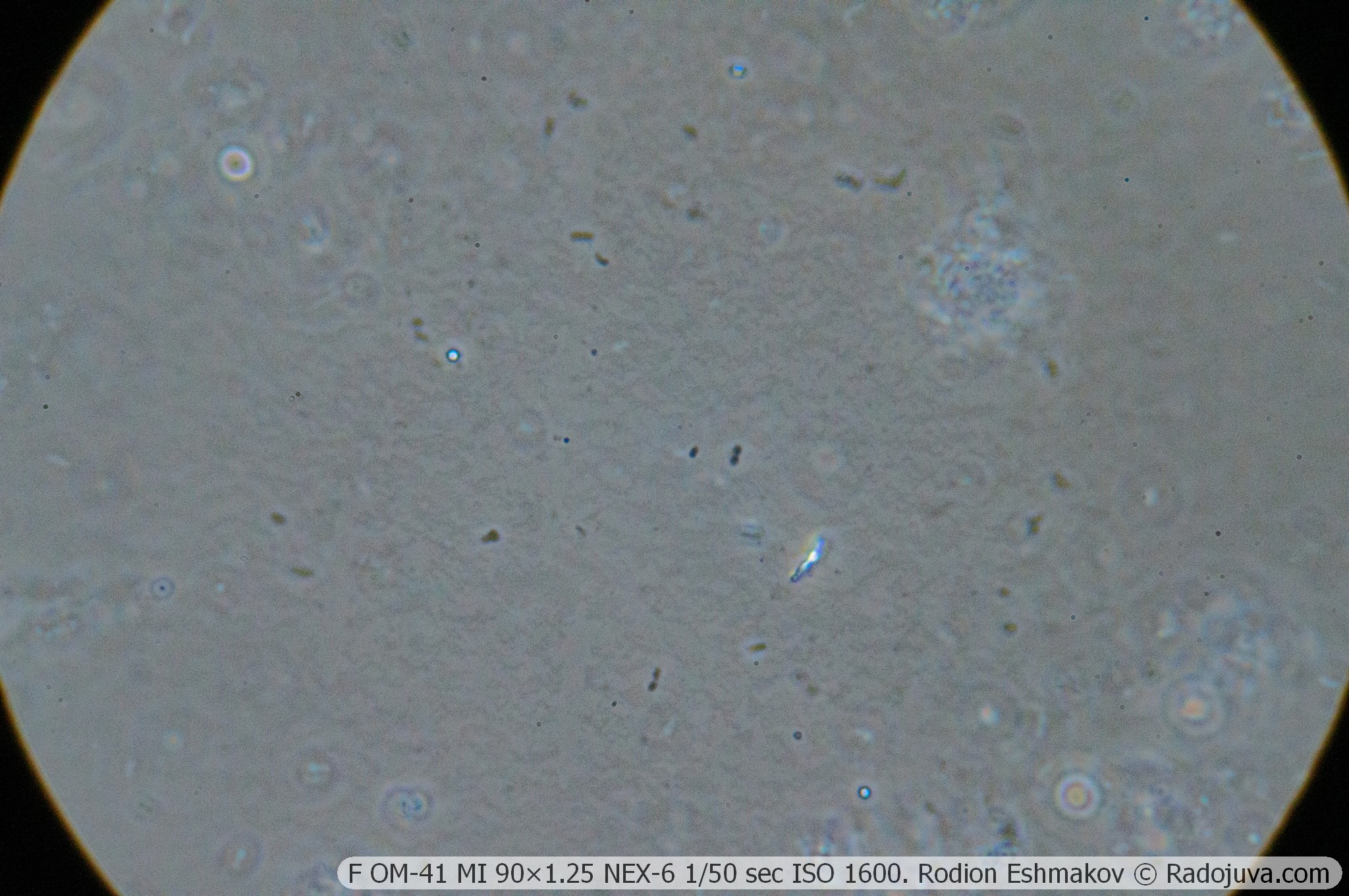
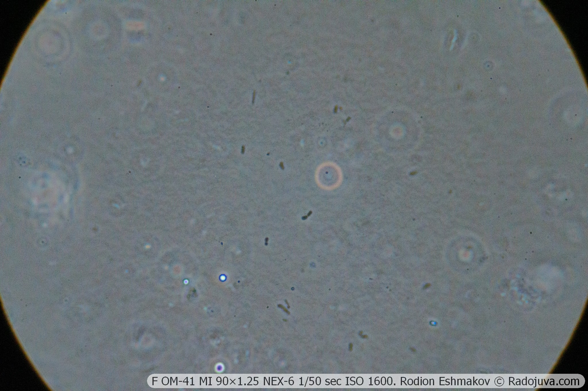
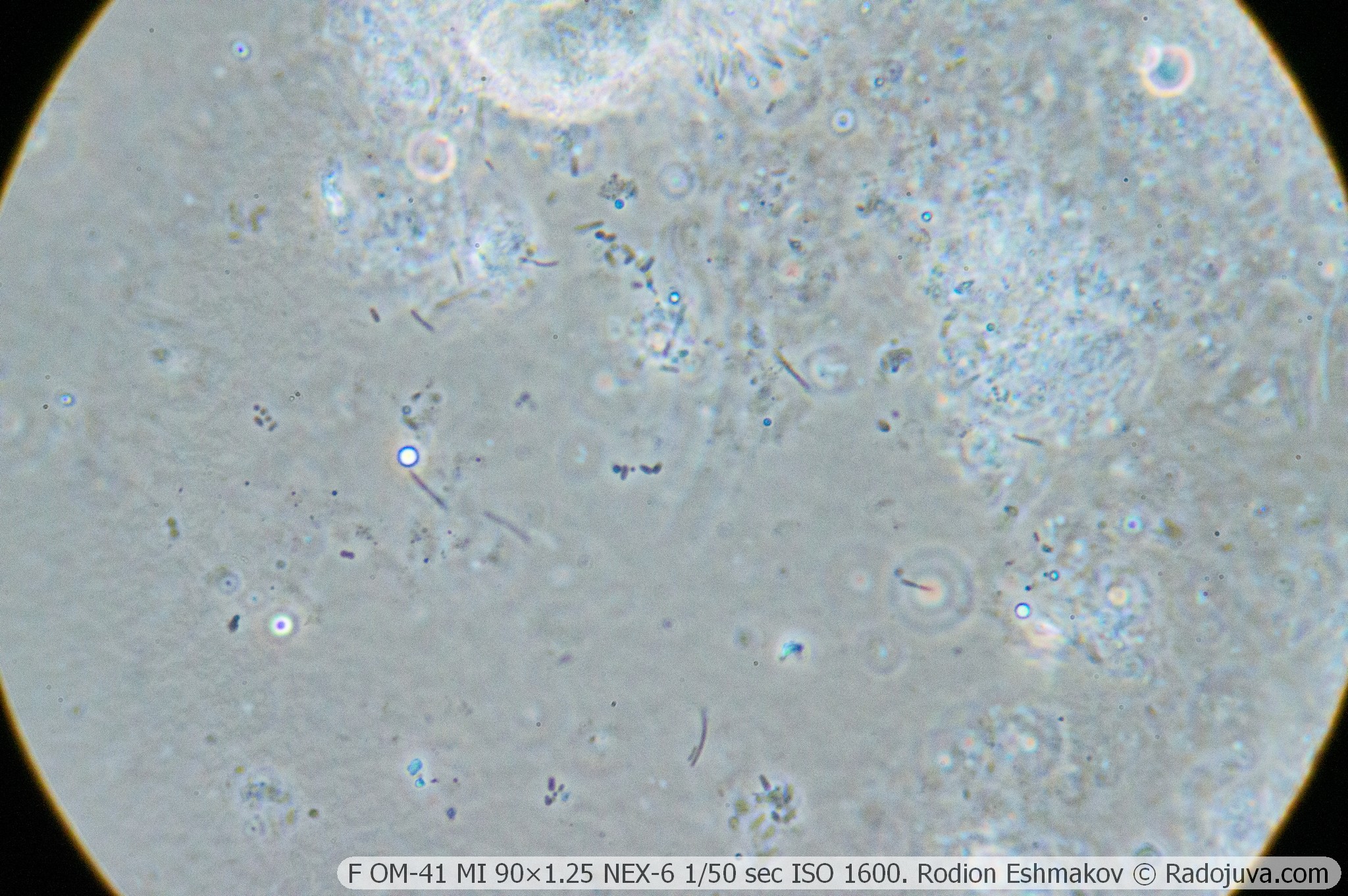
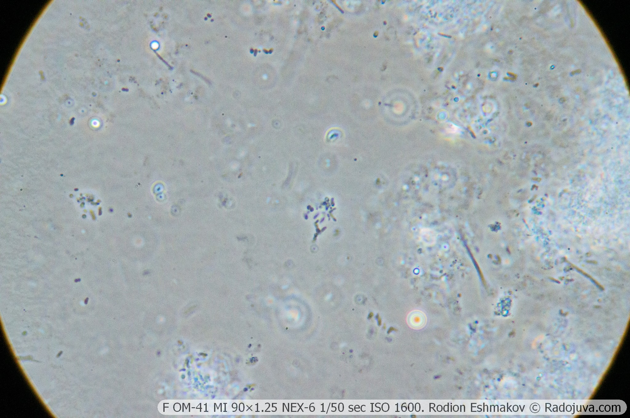
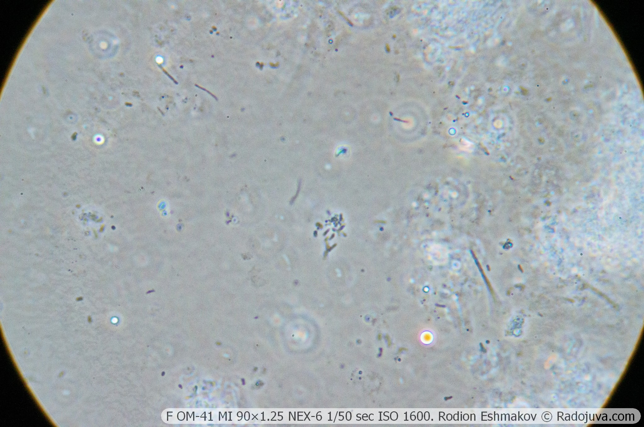
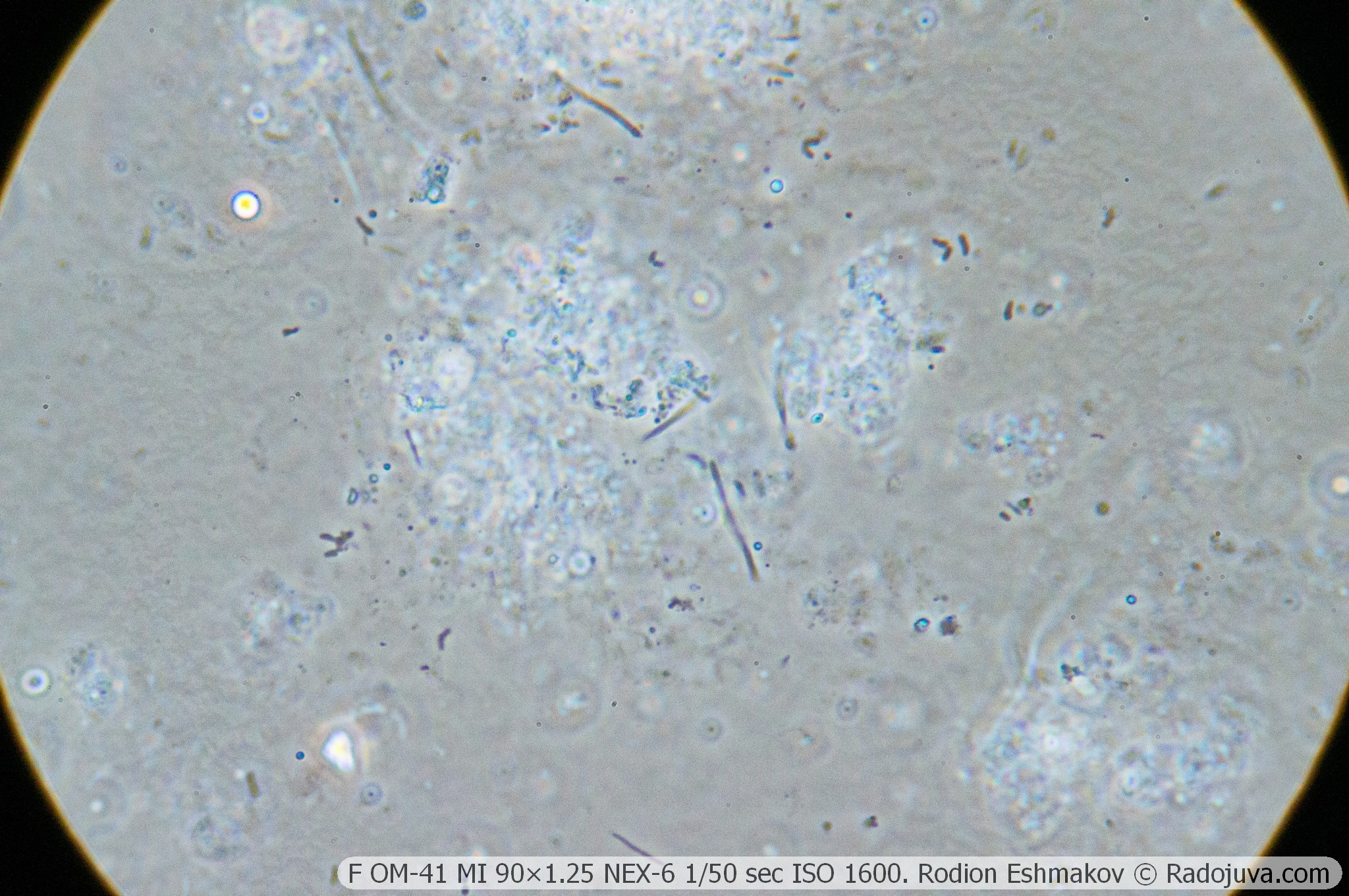
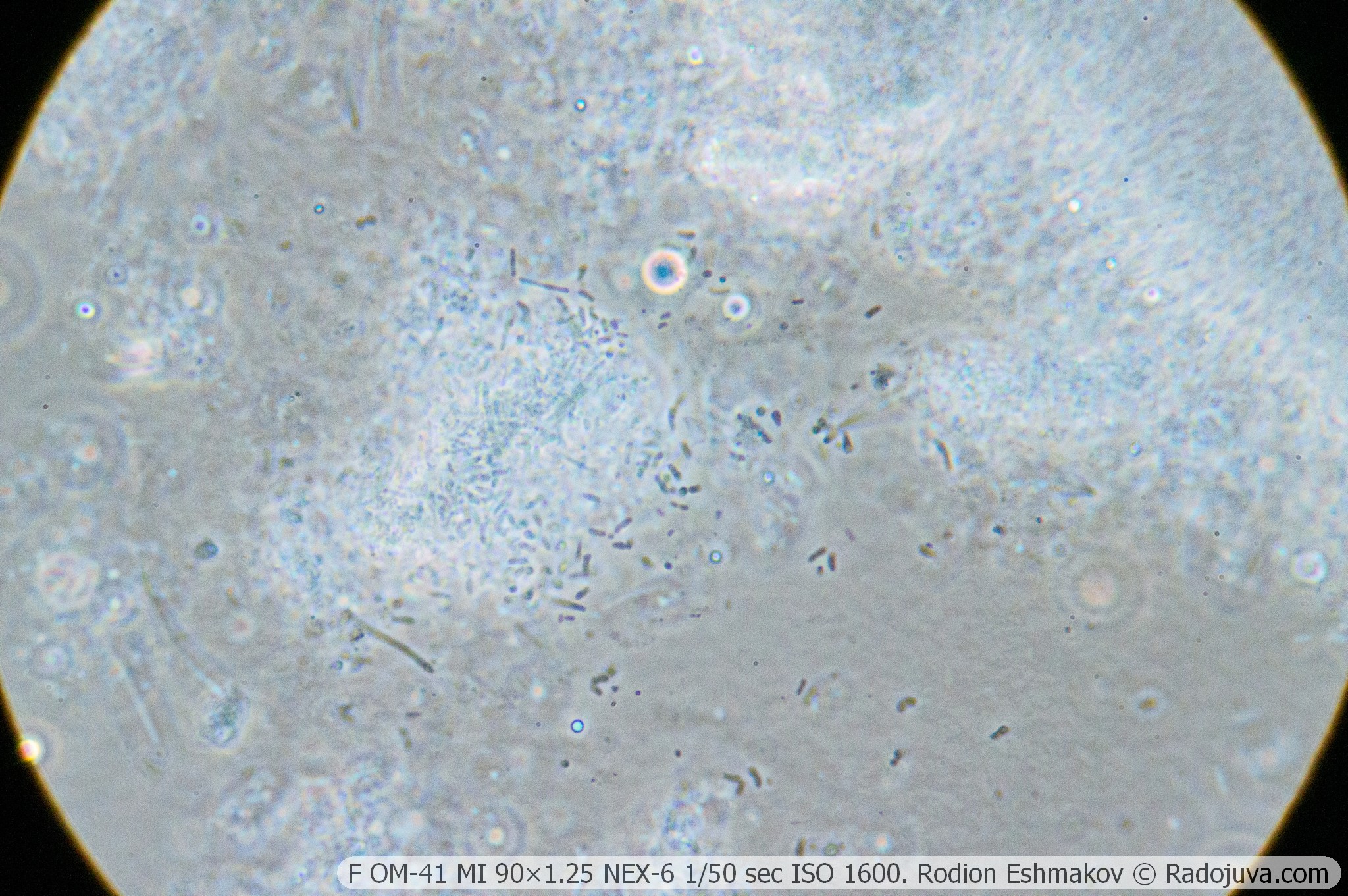
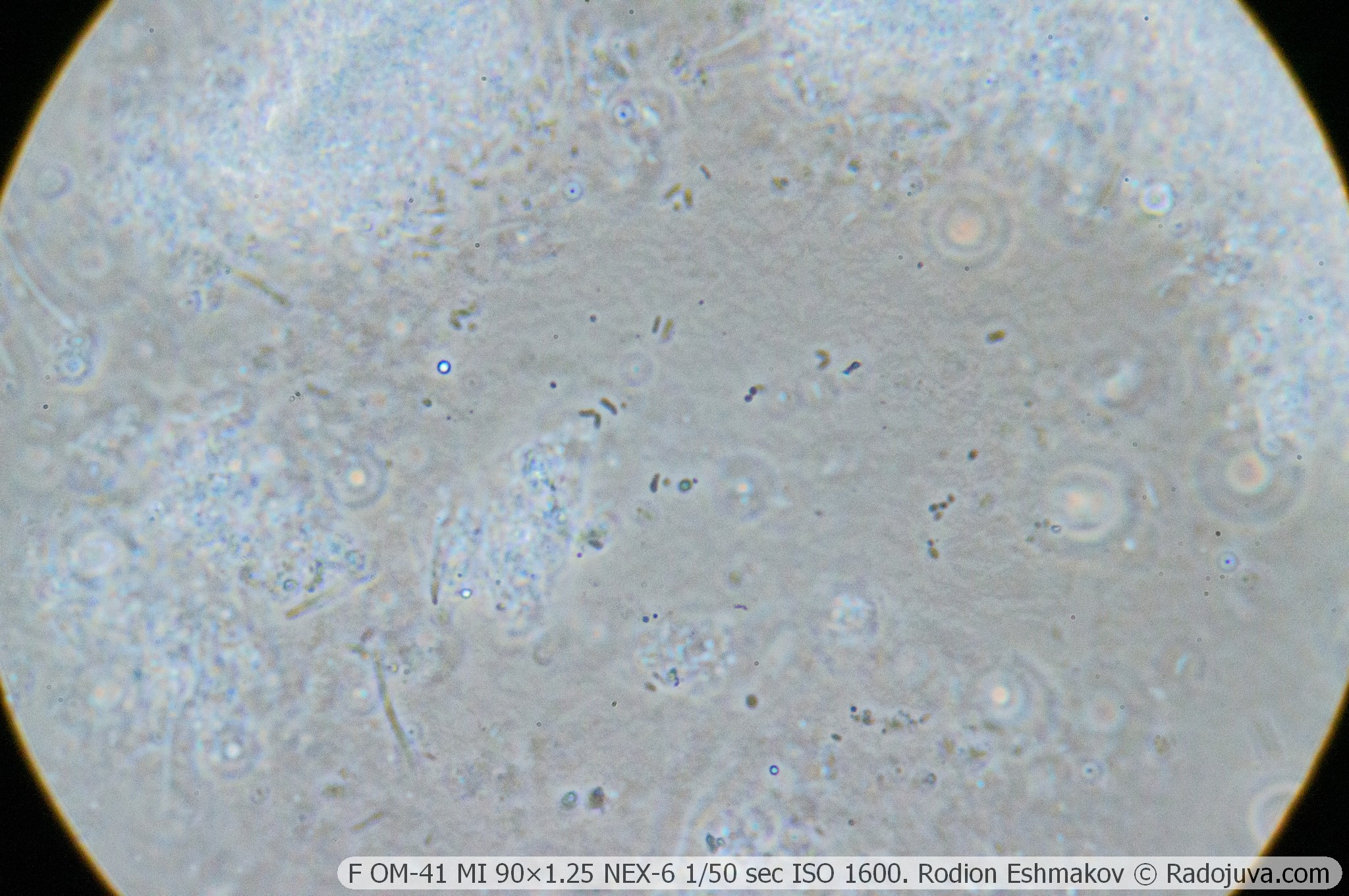
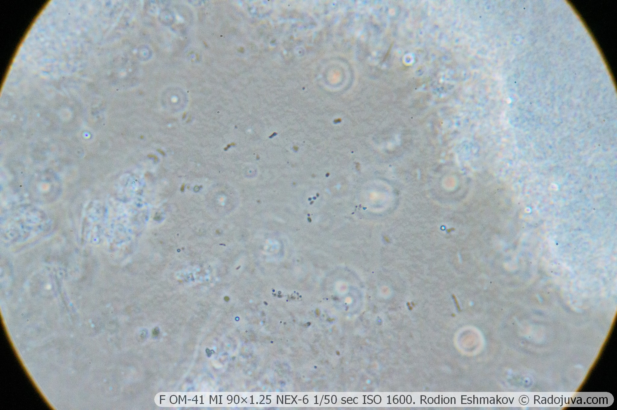
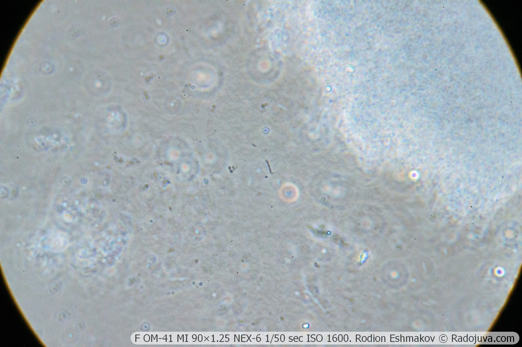
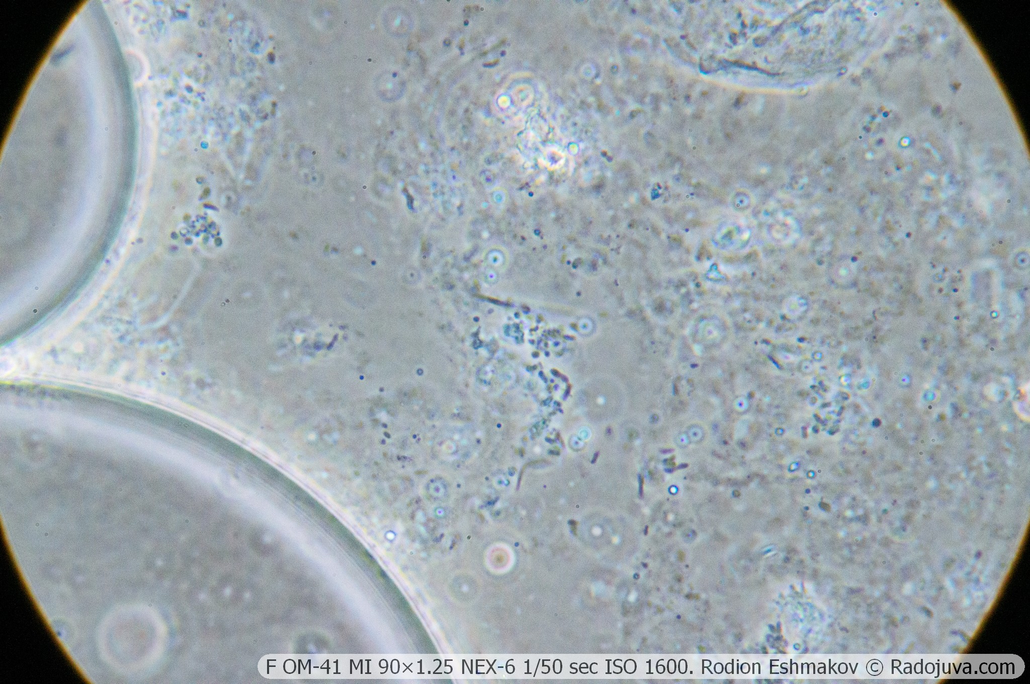
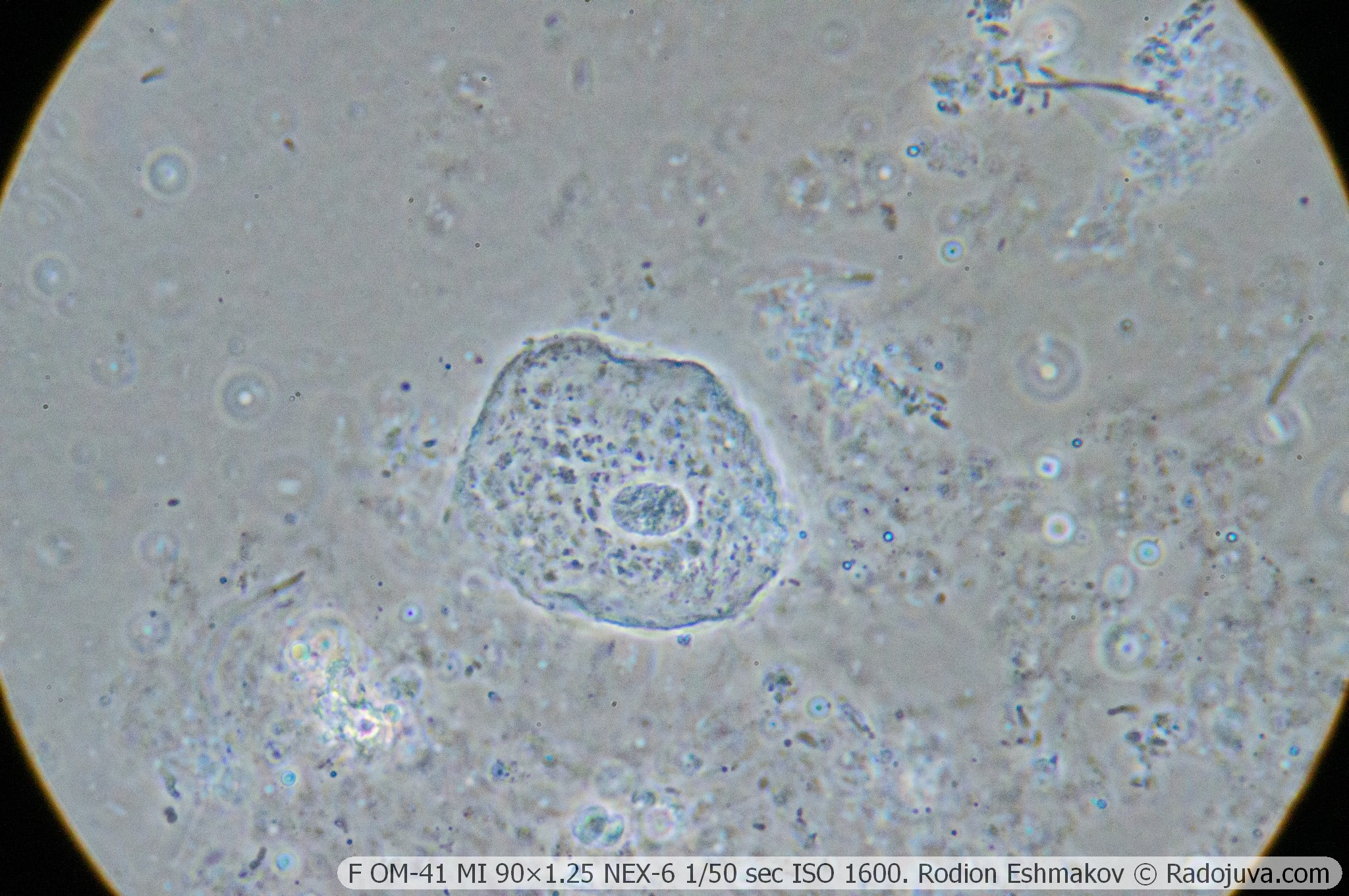
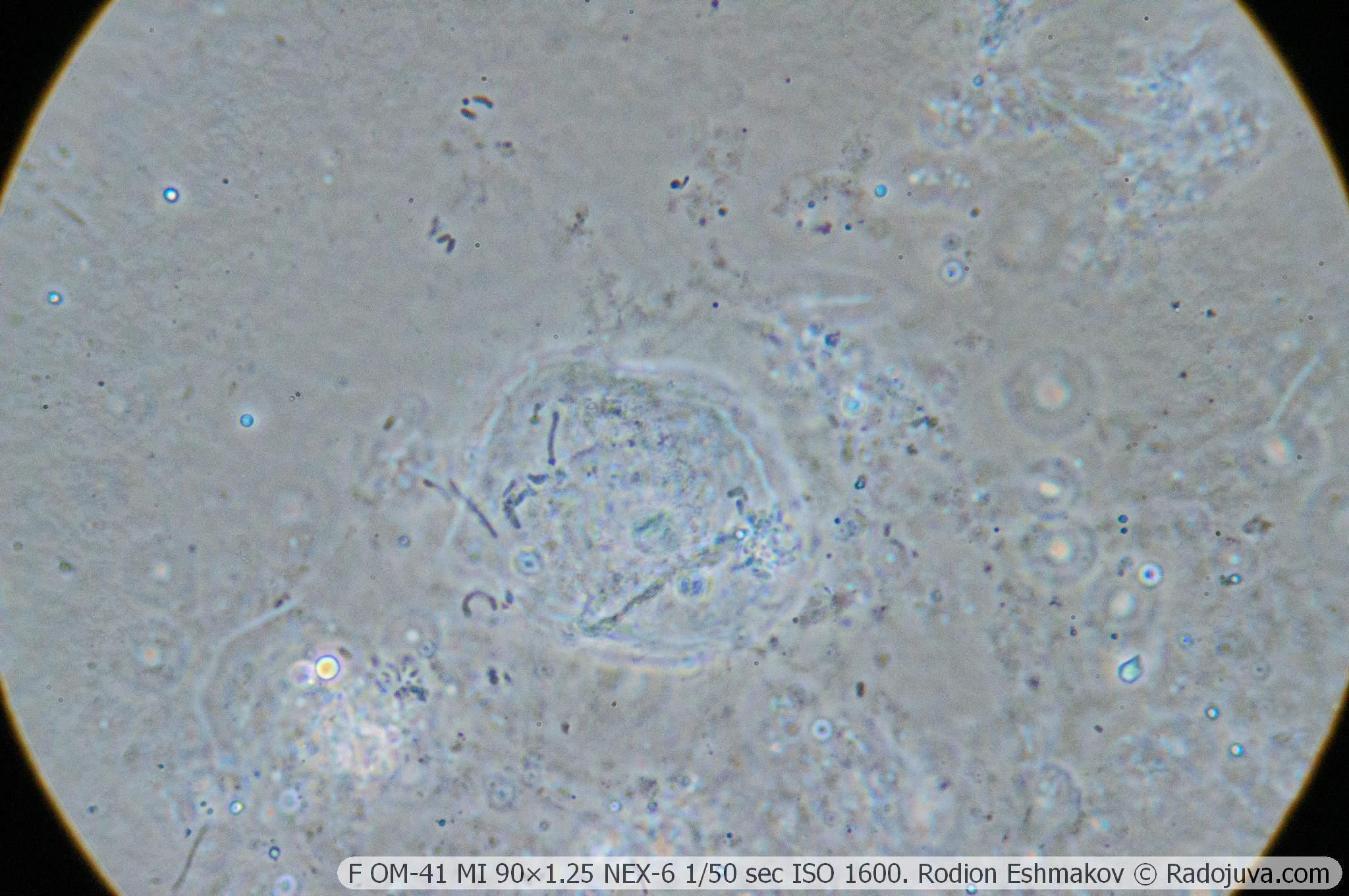
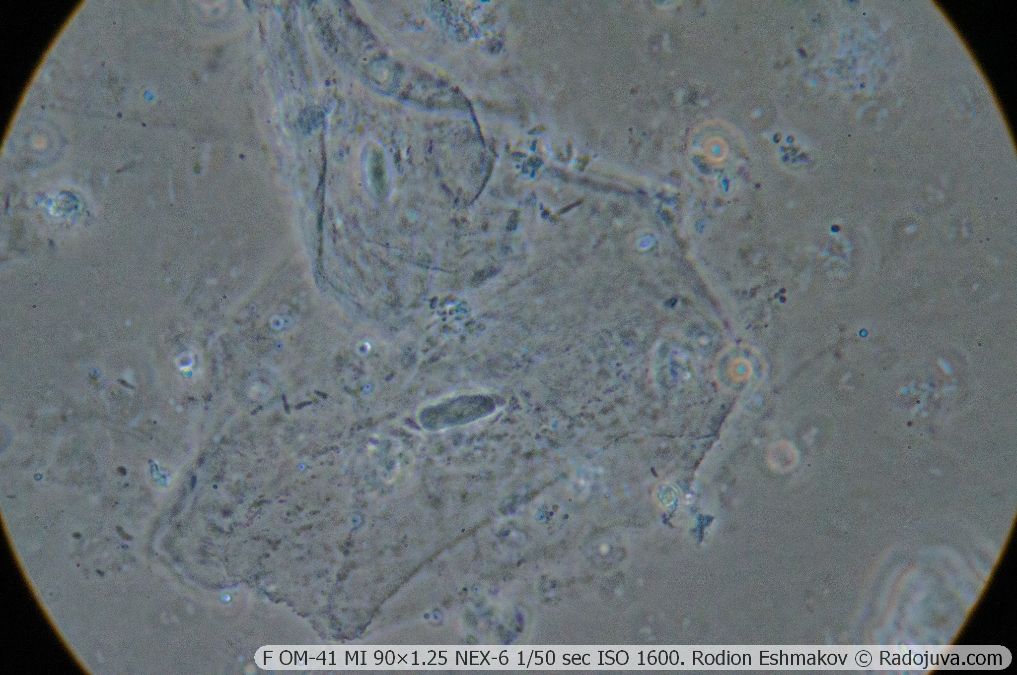
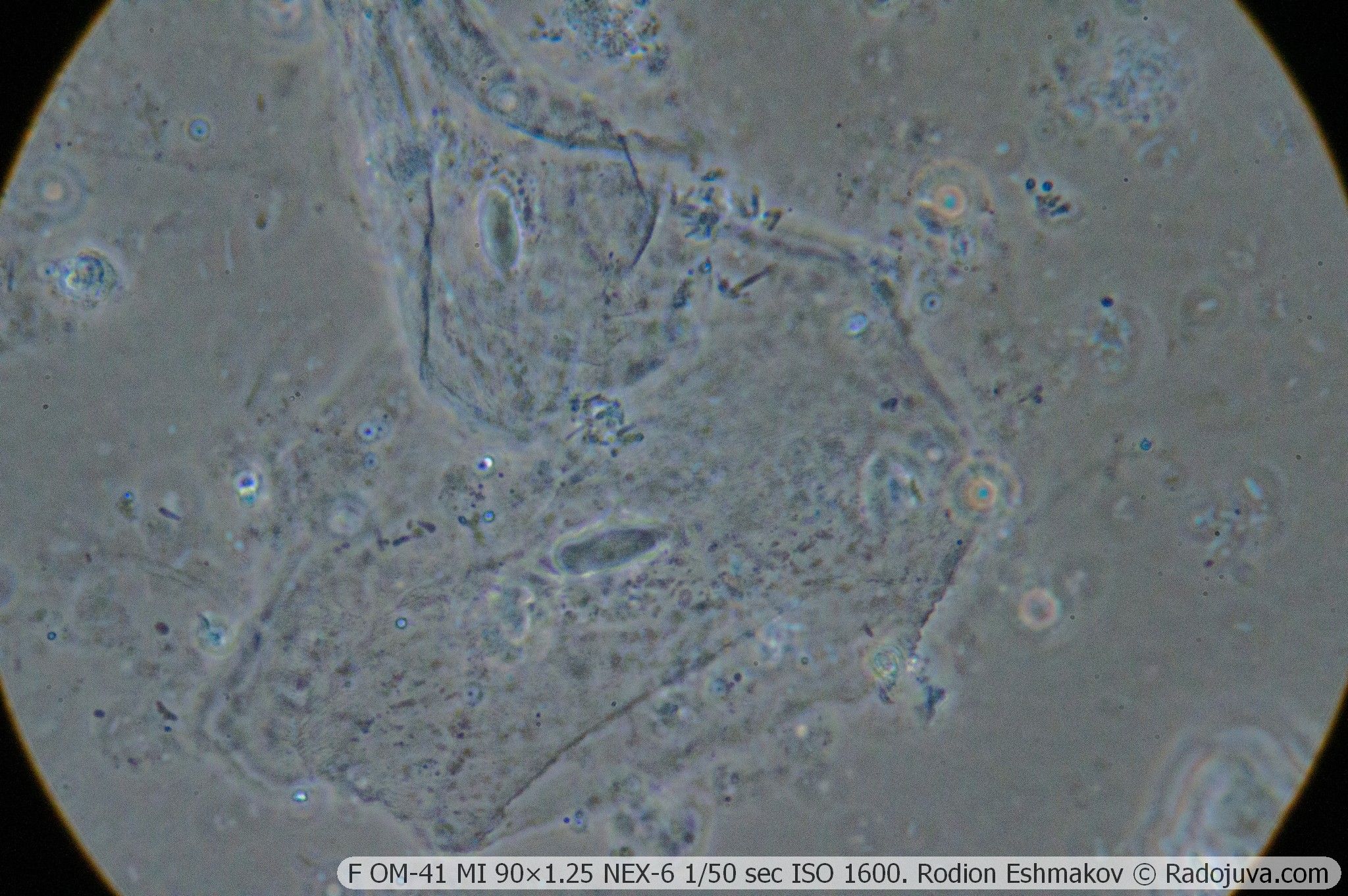
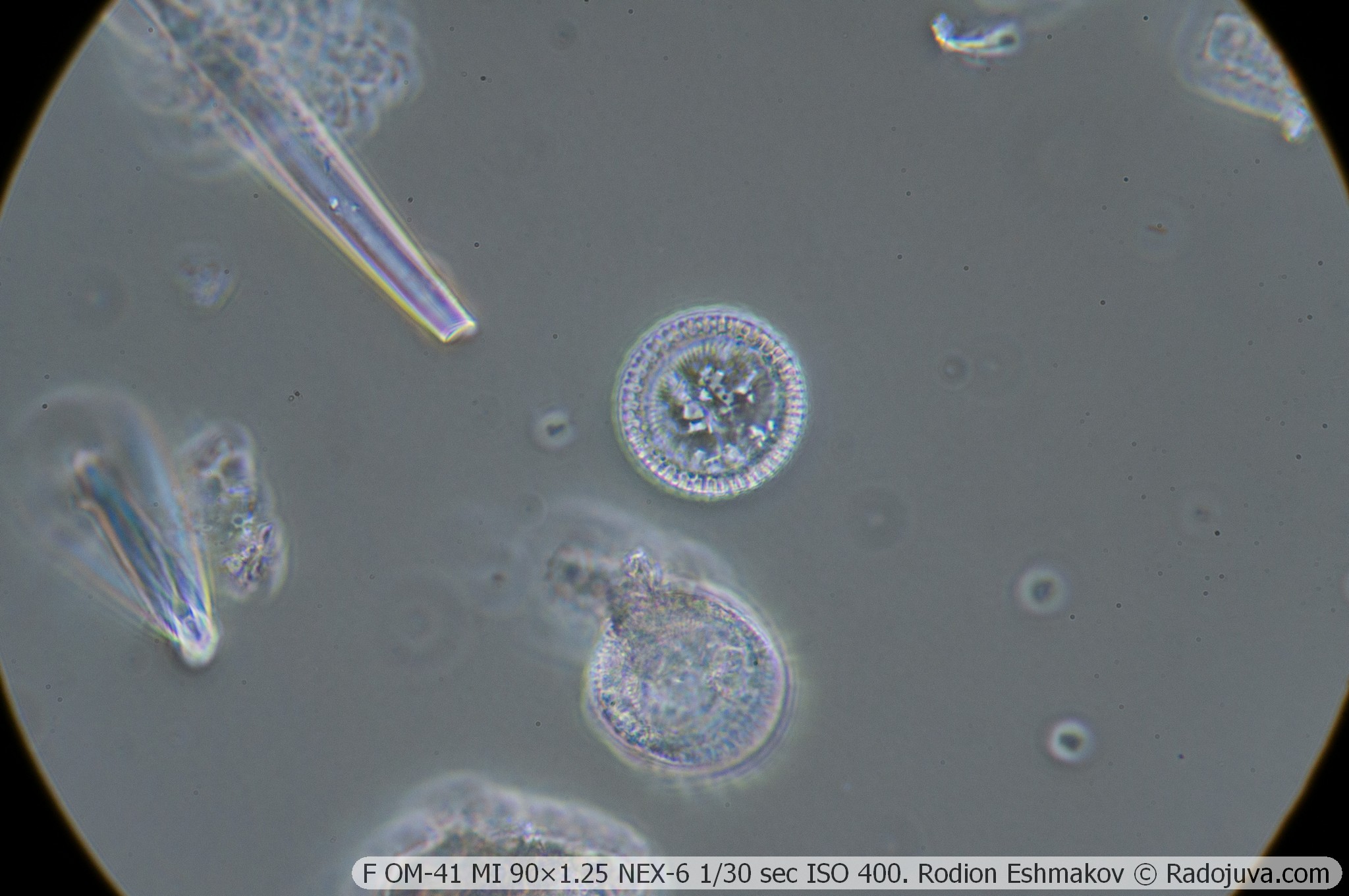
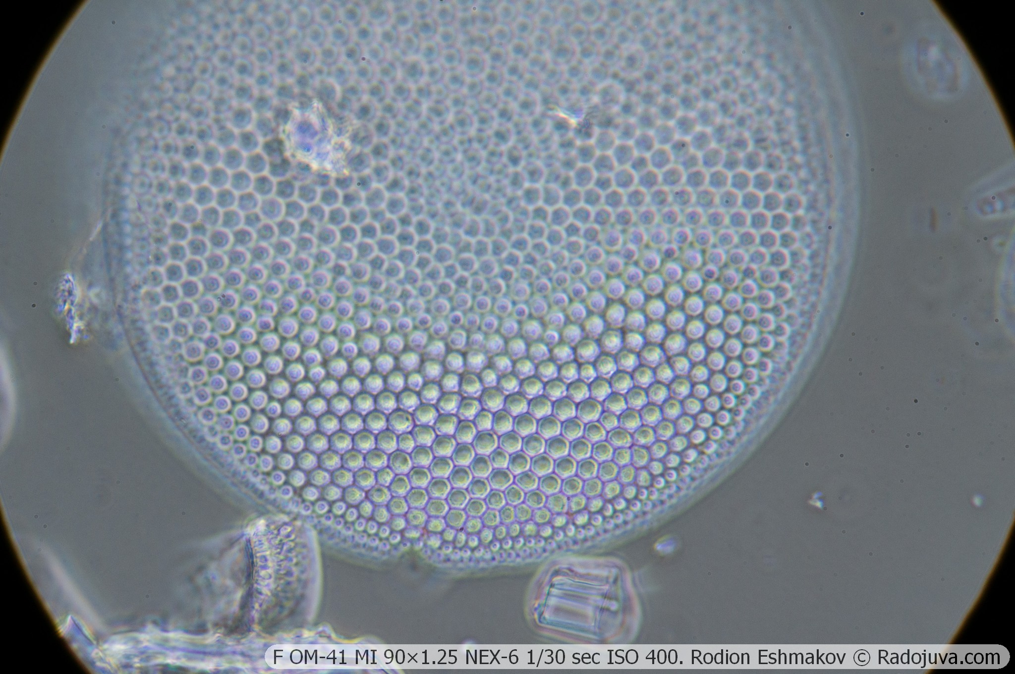
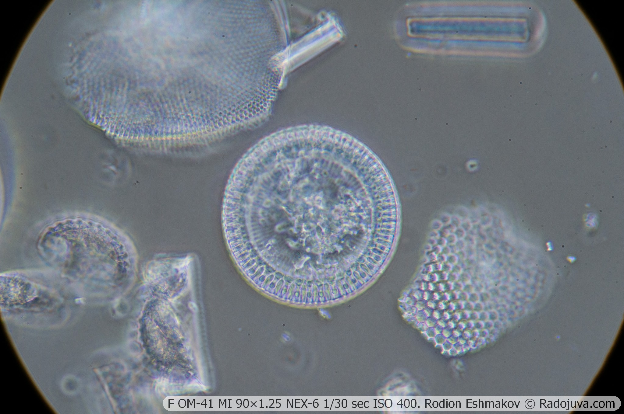
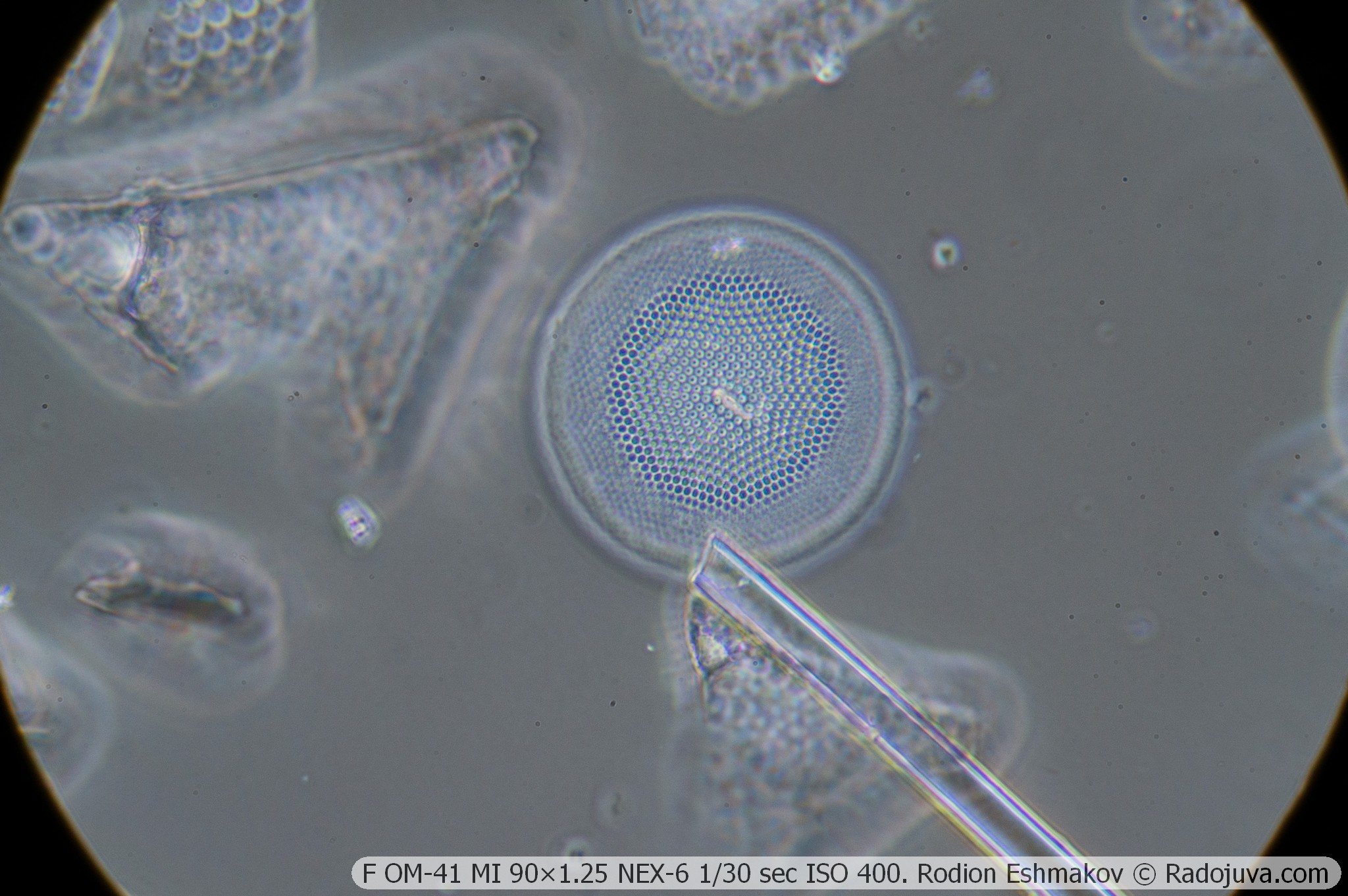
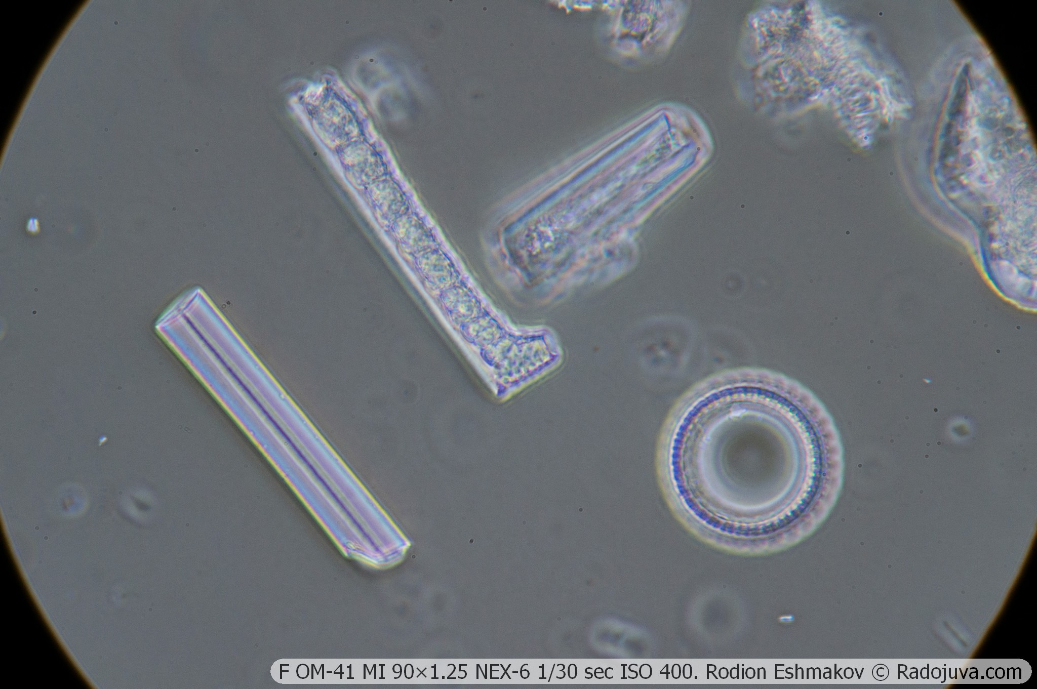
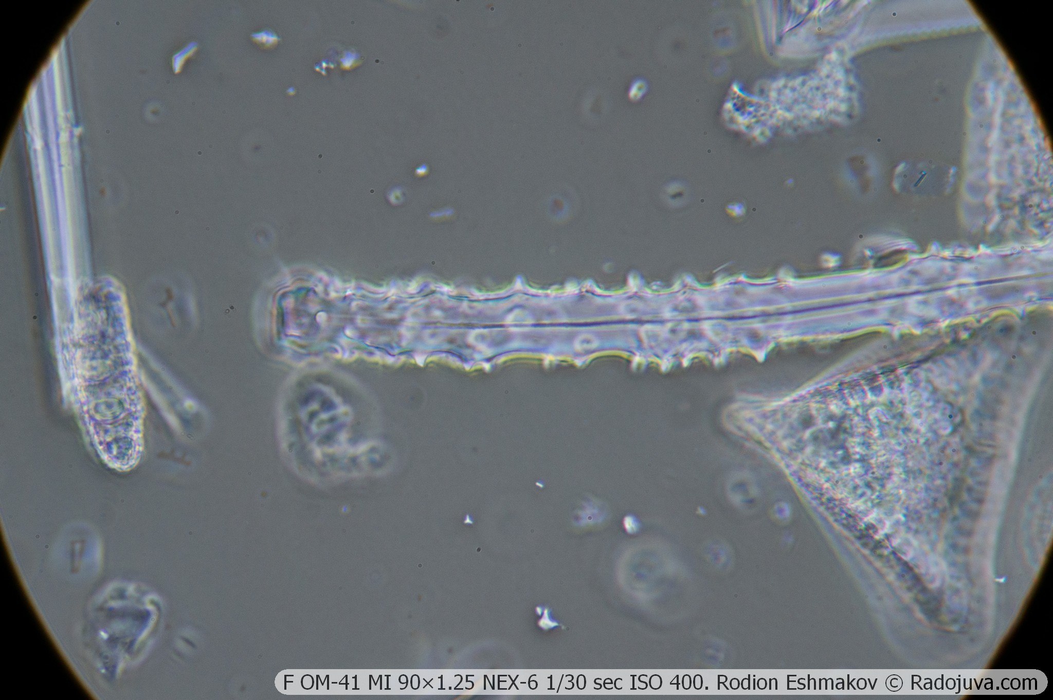
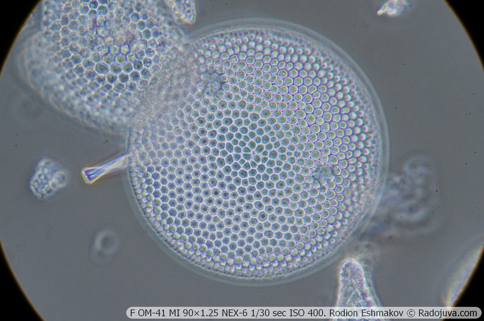
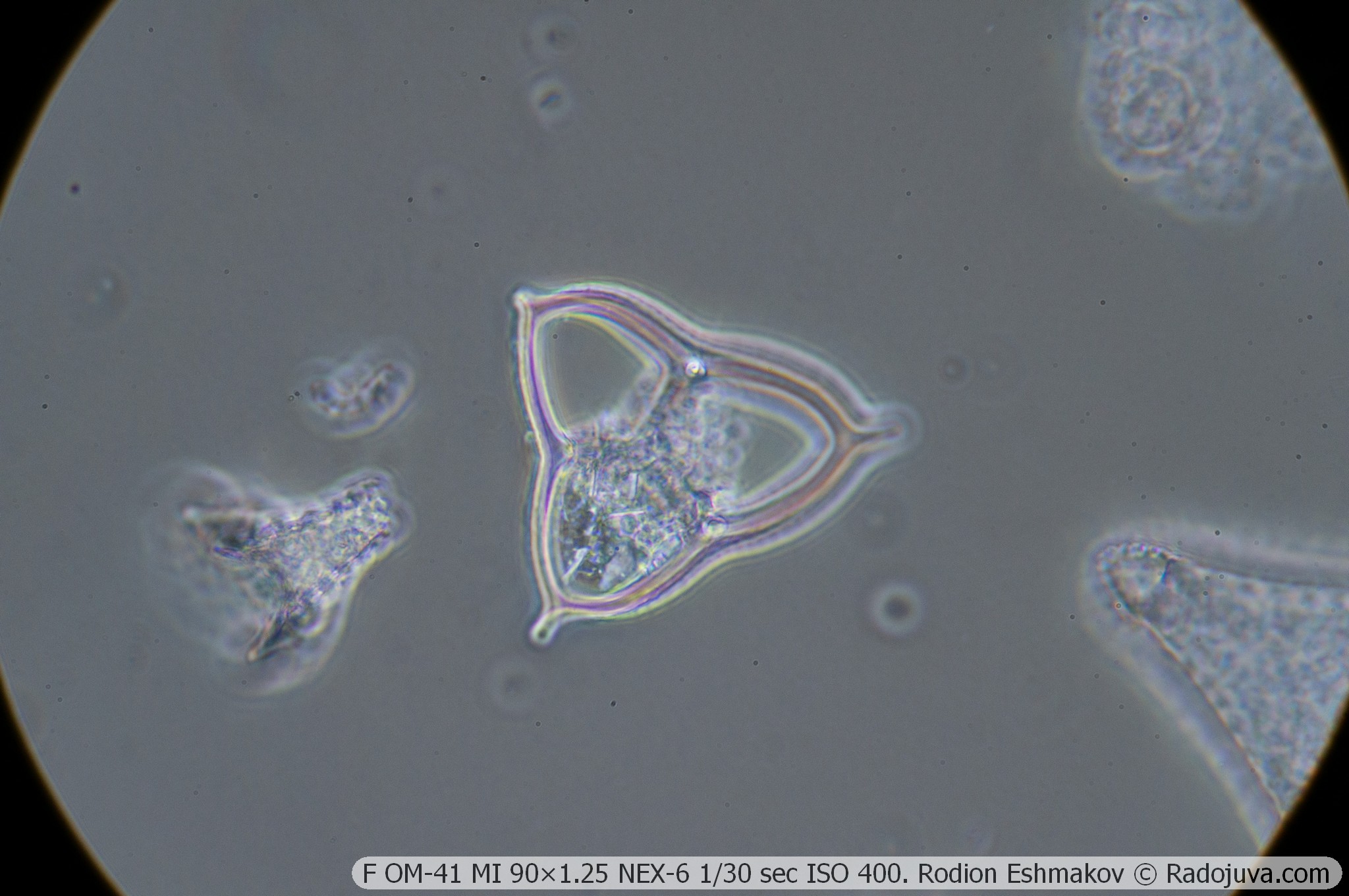
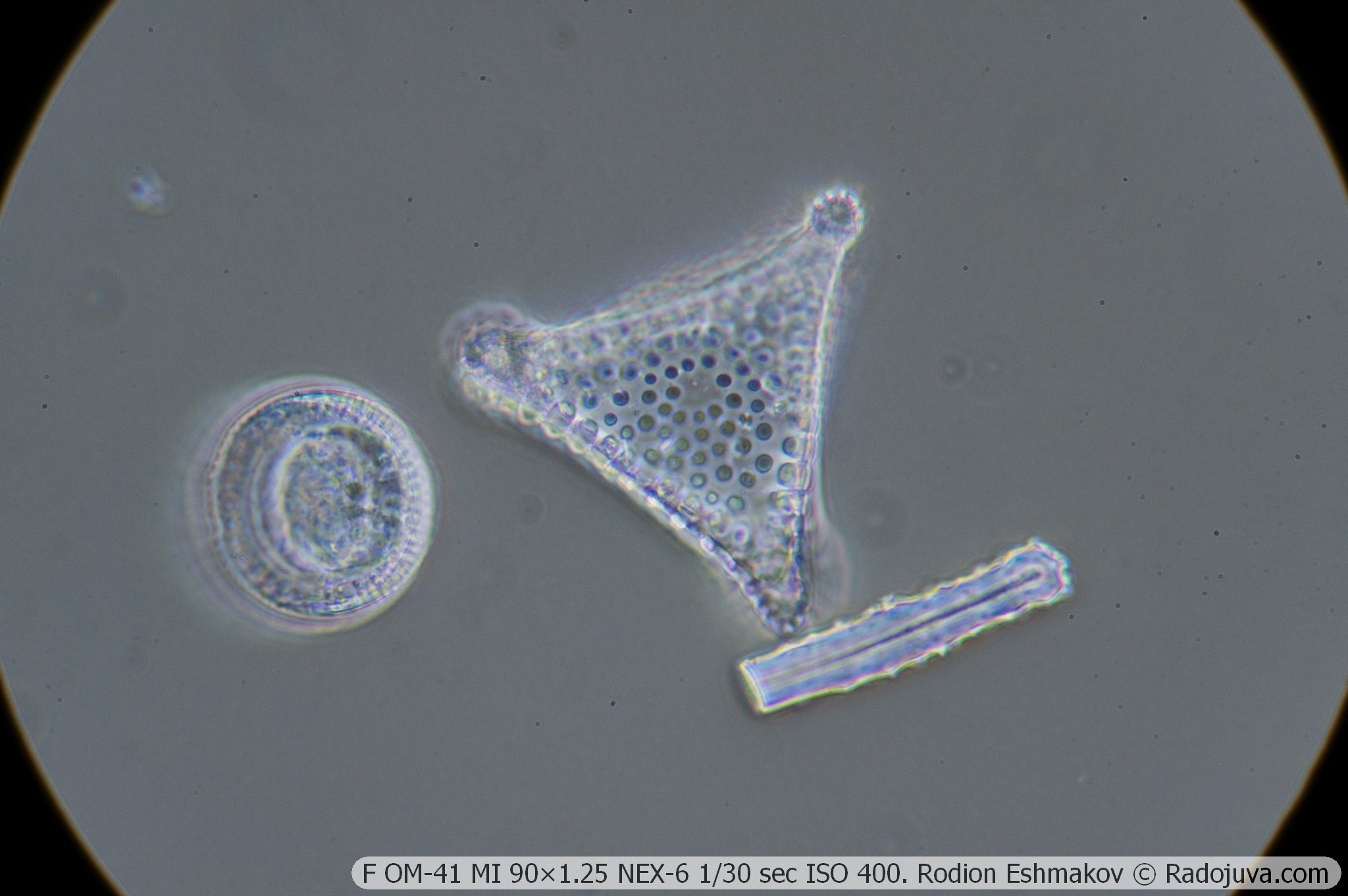
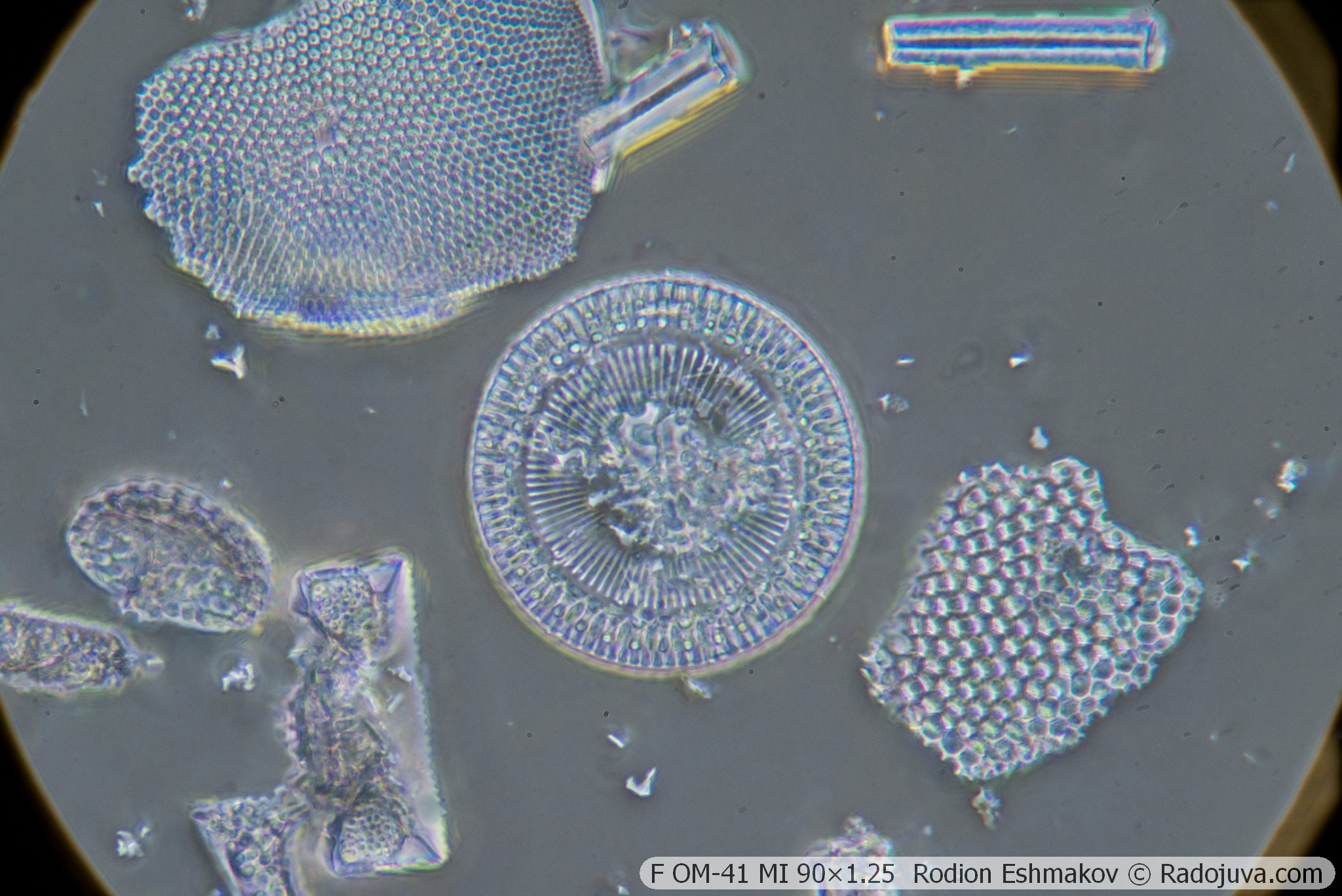
Below are examples of photos with stacking.
Lenses for the KF-1: 40×0.75 F VI OM-23
Optical design – 6 lenses in 4 groups, high-aperture aplanat;
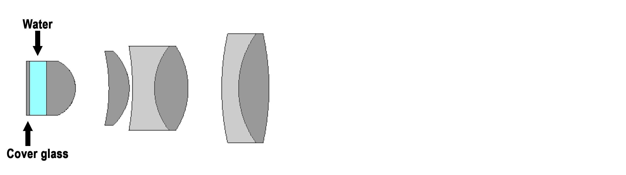
Drawing of the optical diagram of the F VI OM-23 40×0.75.
Type of correction – achromat;
Tube distance – 160 mm;
Parfocal distance – 32.7 mm;
Magnification: 40×;
Numerical aperture – 0.75 (phase ring 0.26);
Immersion required – yes (water);
Working distance – 1.8 mm;
Cover glass thickness – 0.17 mm;
Mounting type – RMS standard (4/5” × 1/36” thread);
Lateral chromatic aberration – corrected with a compensating eyepiece (such as LOMO K5×);
Features - microscopic lens, does not have an iris diaphragm and a focusing mechanism.
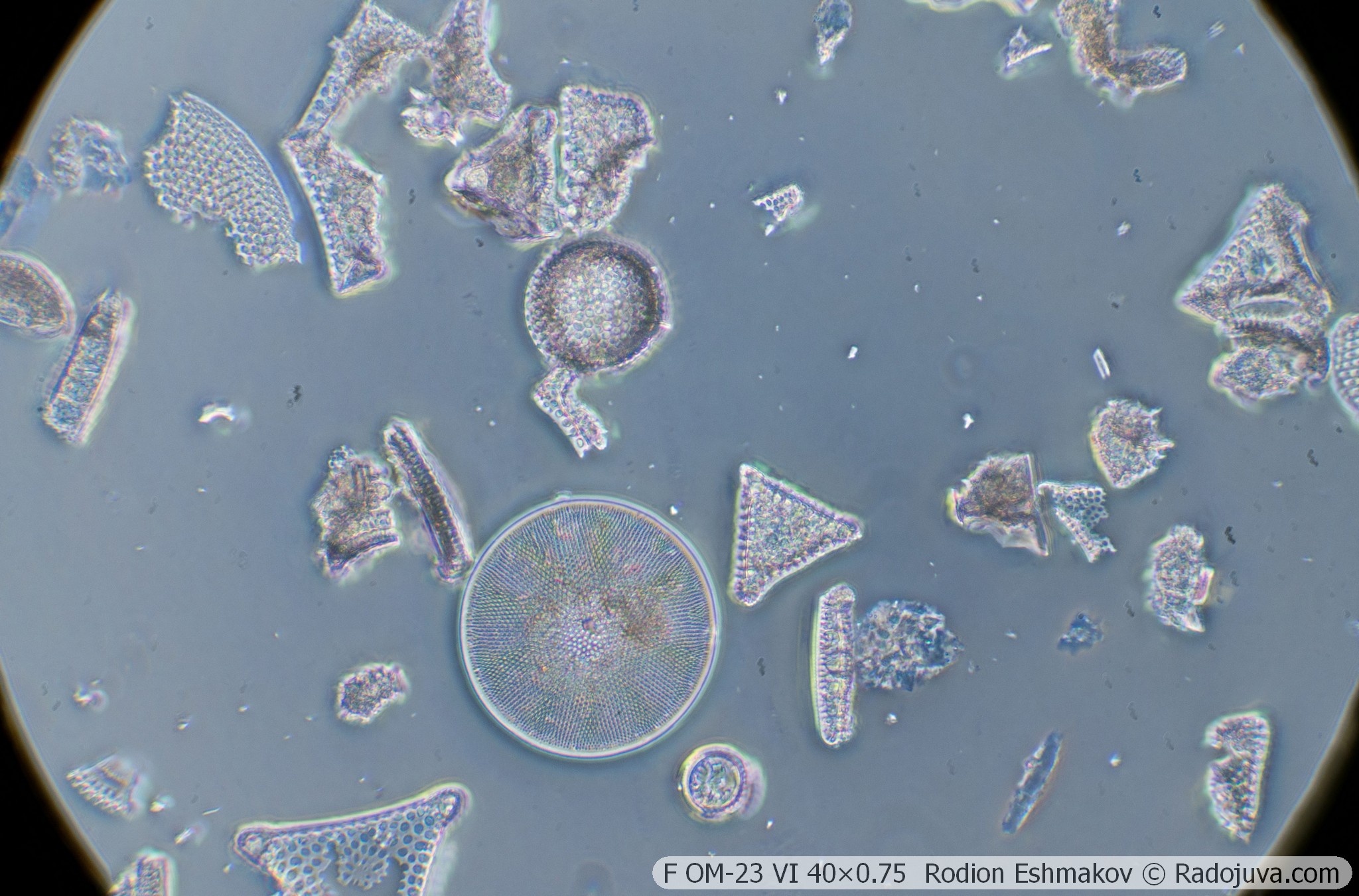
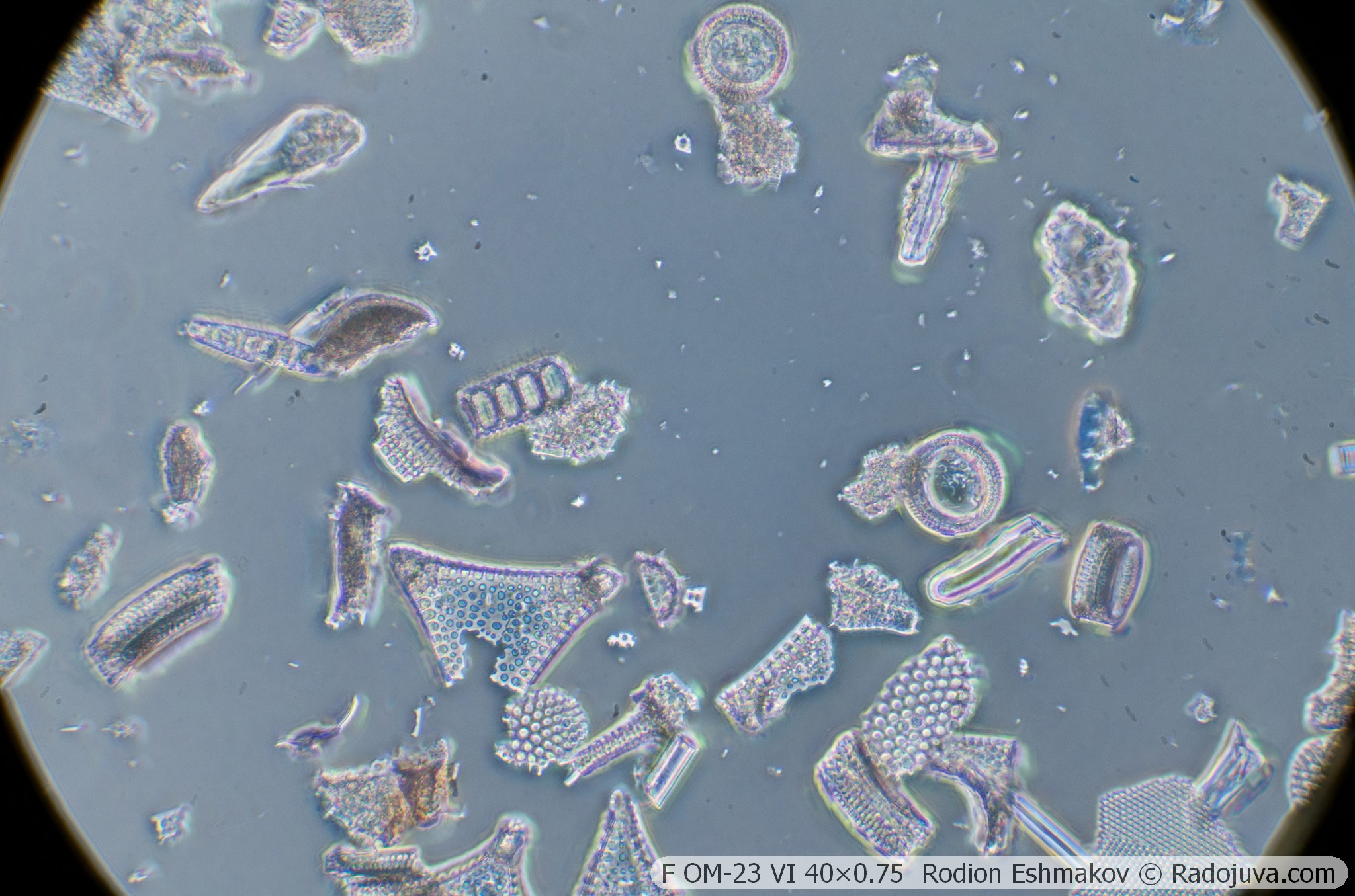
This objective can be considered an alternative to the F MS 40×0.65. Due to its more complex optical design, its image quality is somewhat higher both in the center and across the field. Lateral chromatic aberrations of this objective can be compensated for with K-5× ML-2 eyepieces. Its main drawback is the need for water immersion.
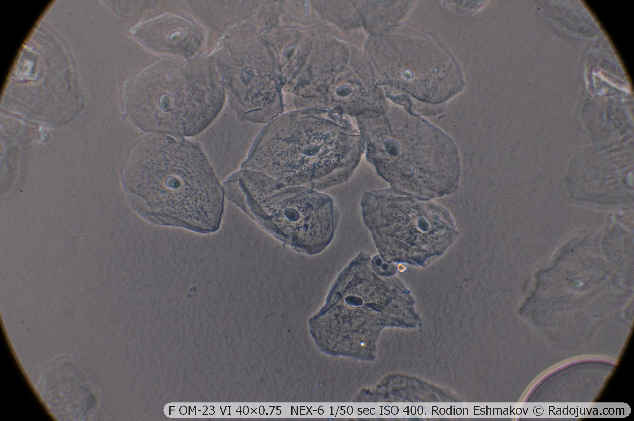
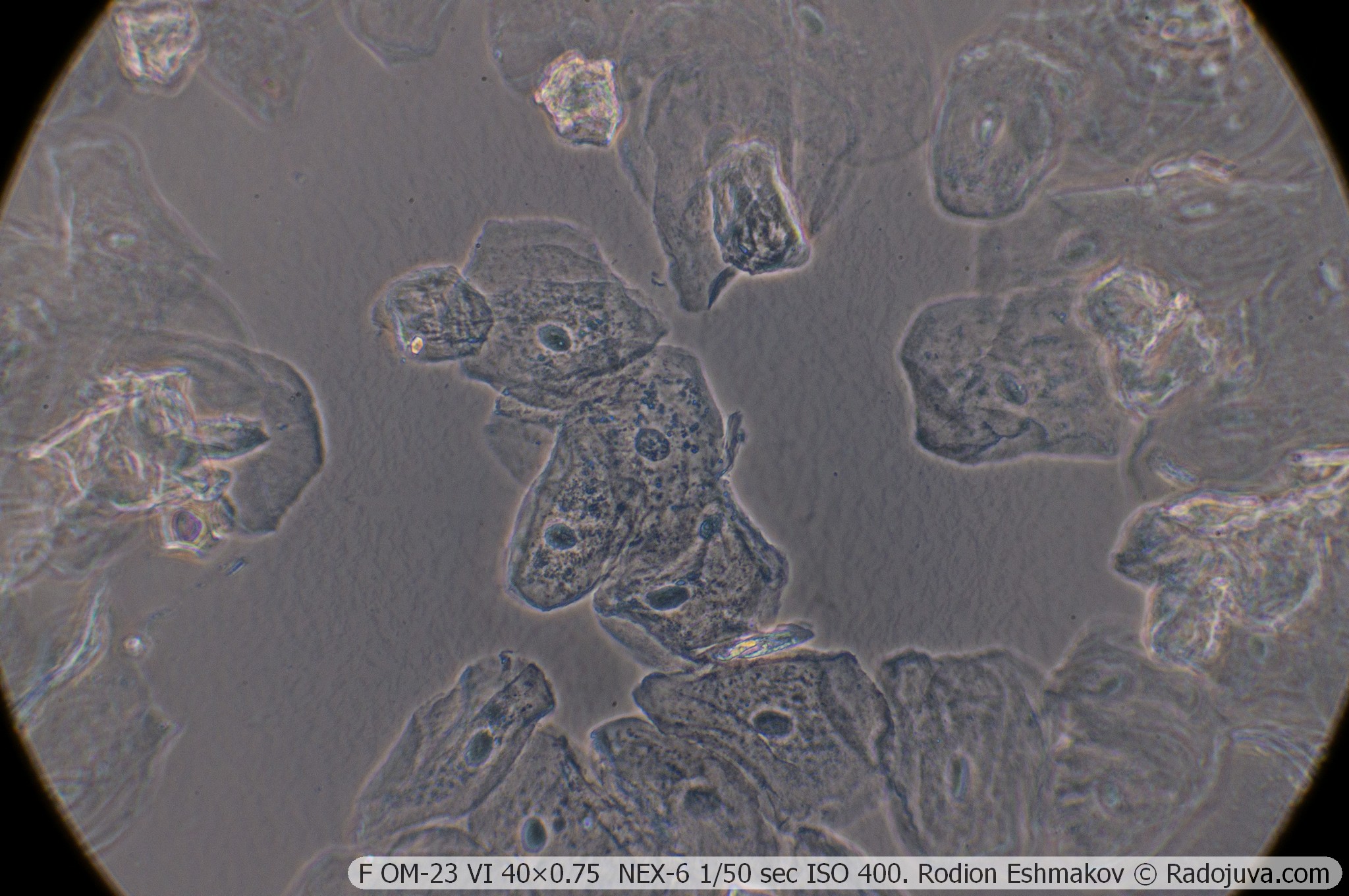
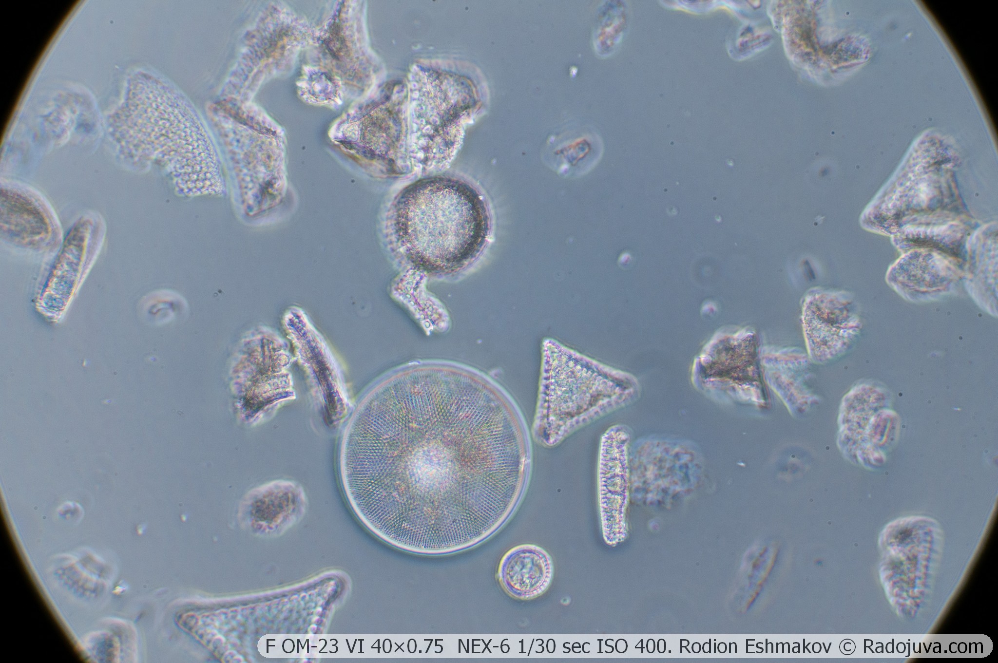
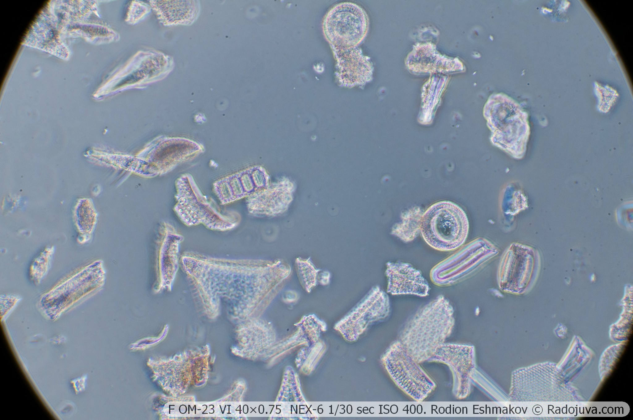
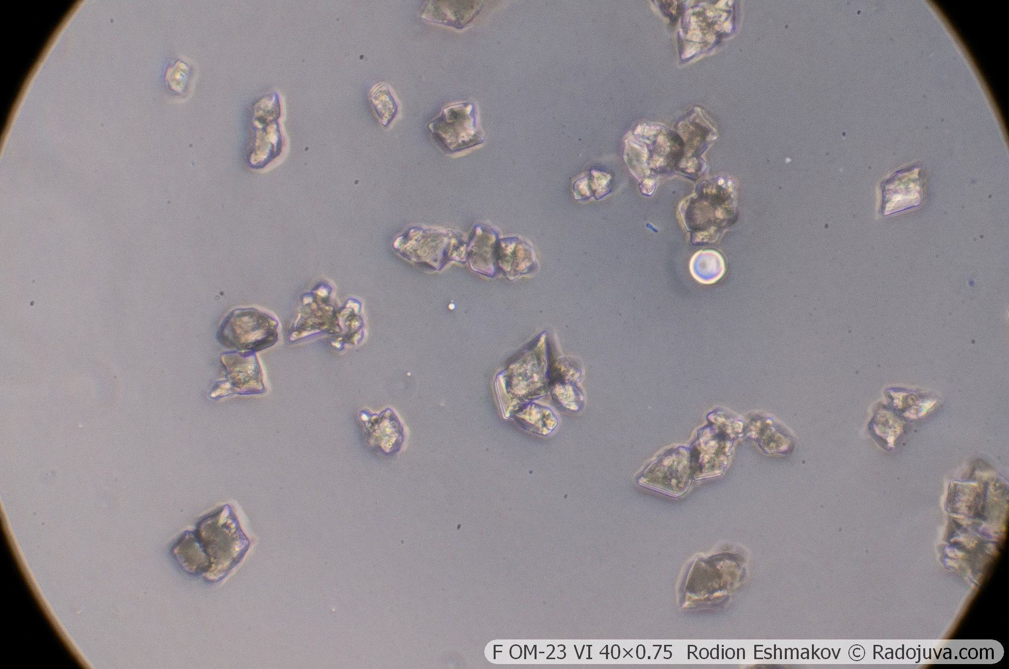
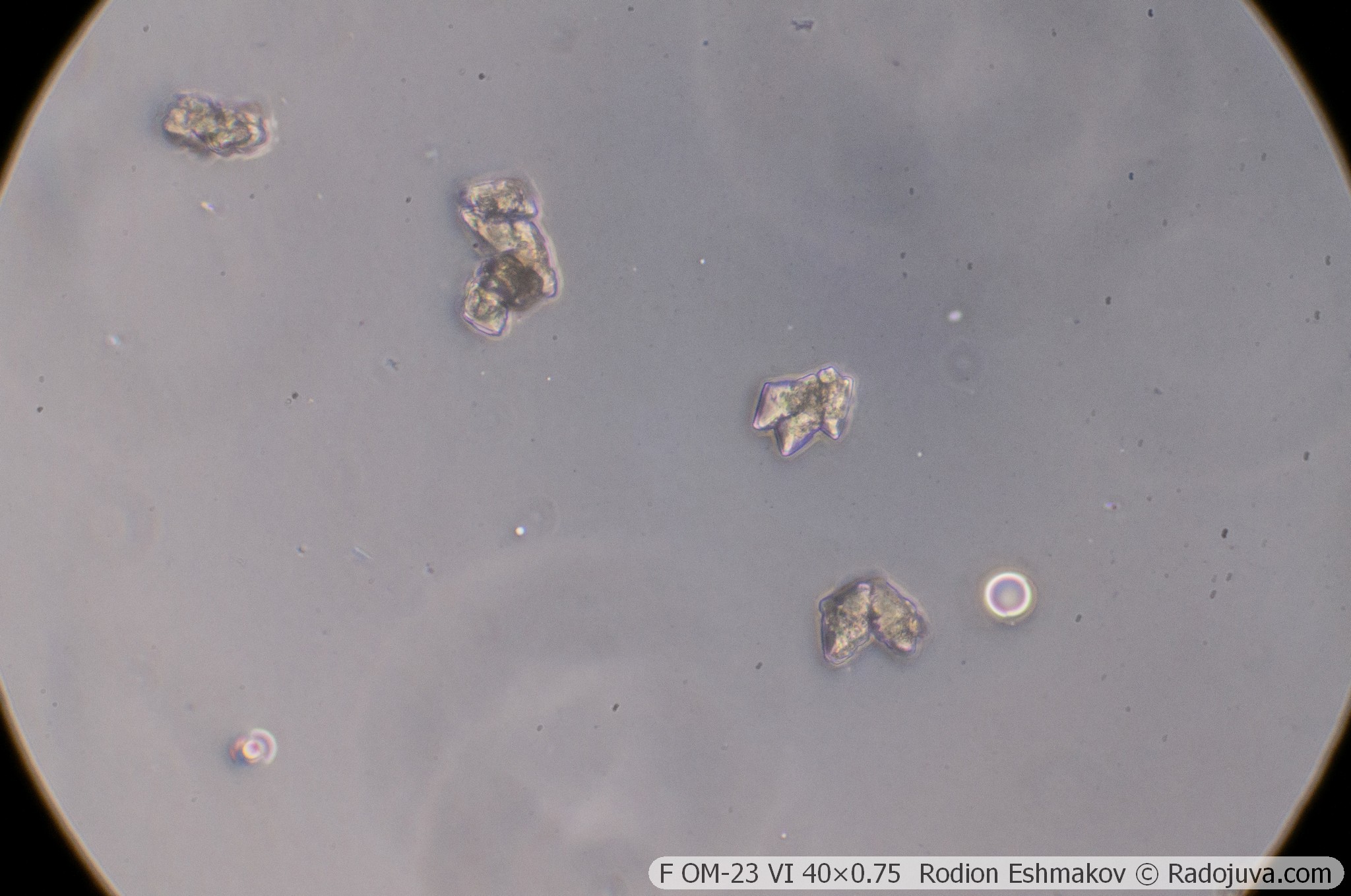
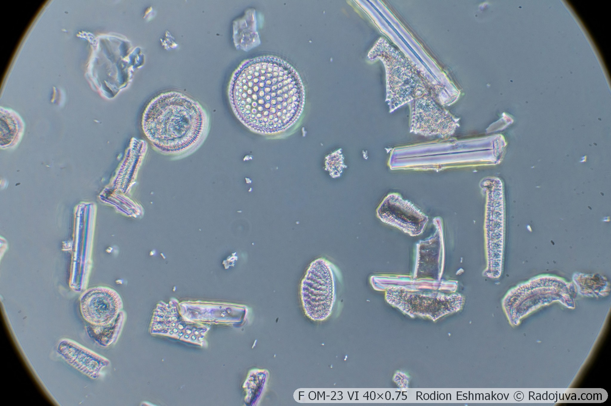
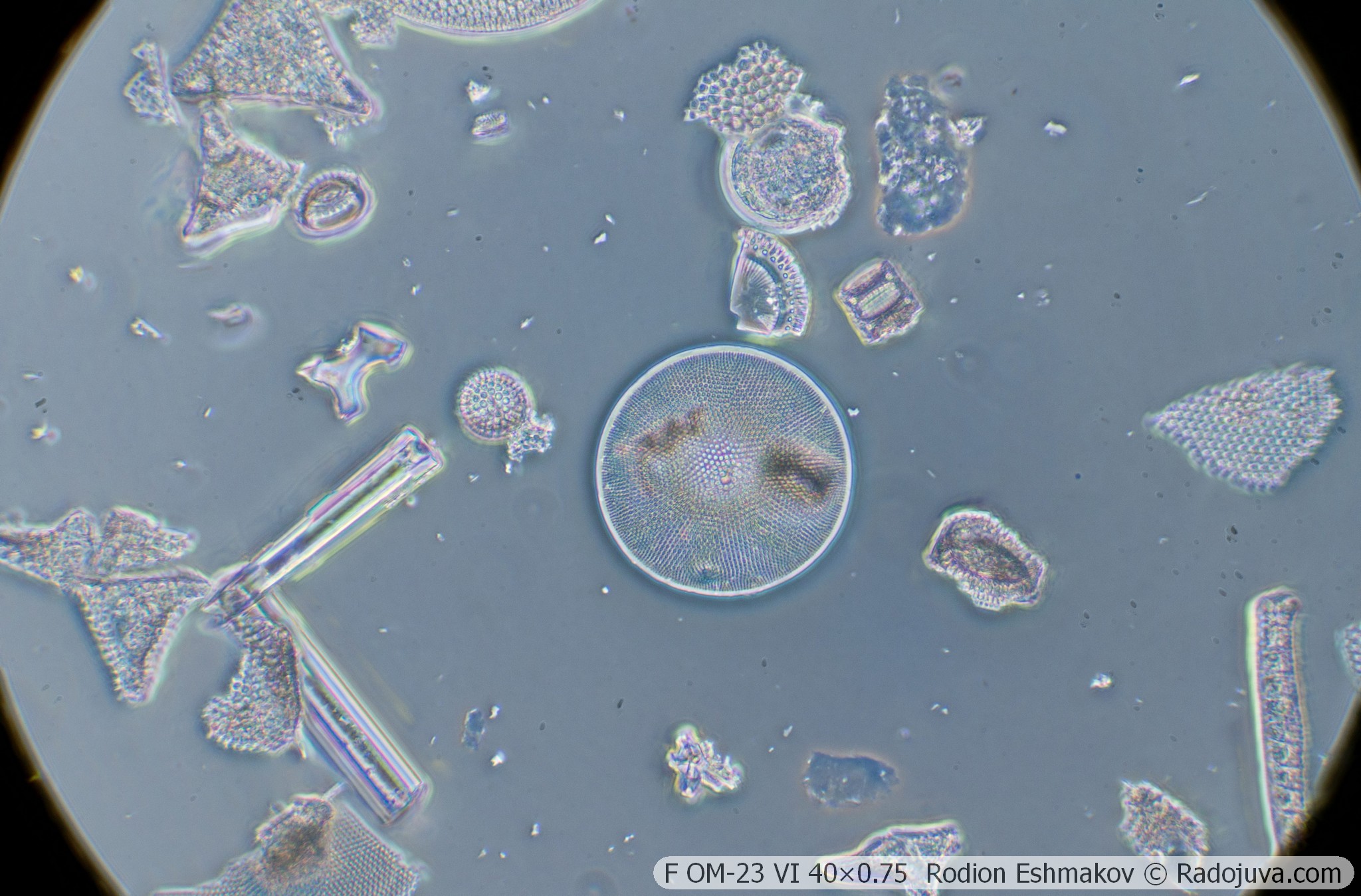
Below are examples of photos taken on a Sony NEX-6 and a F OM-23 VI 40×0.75 lens using a relay from a K5× ML-2 eyepiece and a WitrueHD 75/2.8 10 Mpix lens.
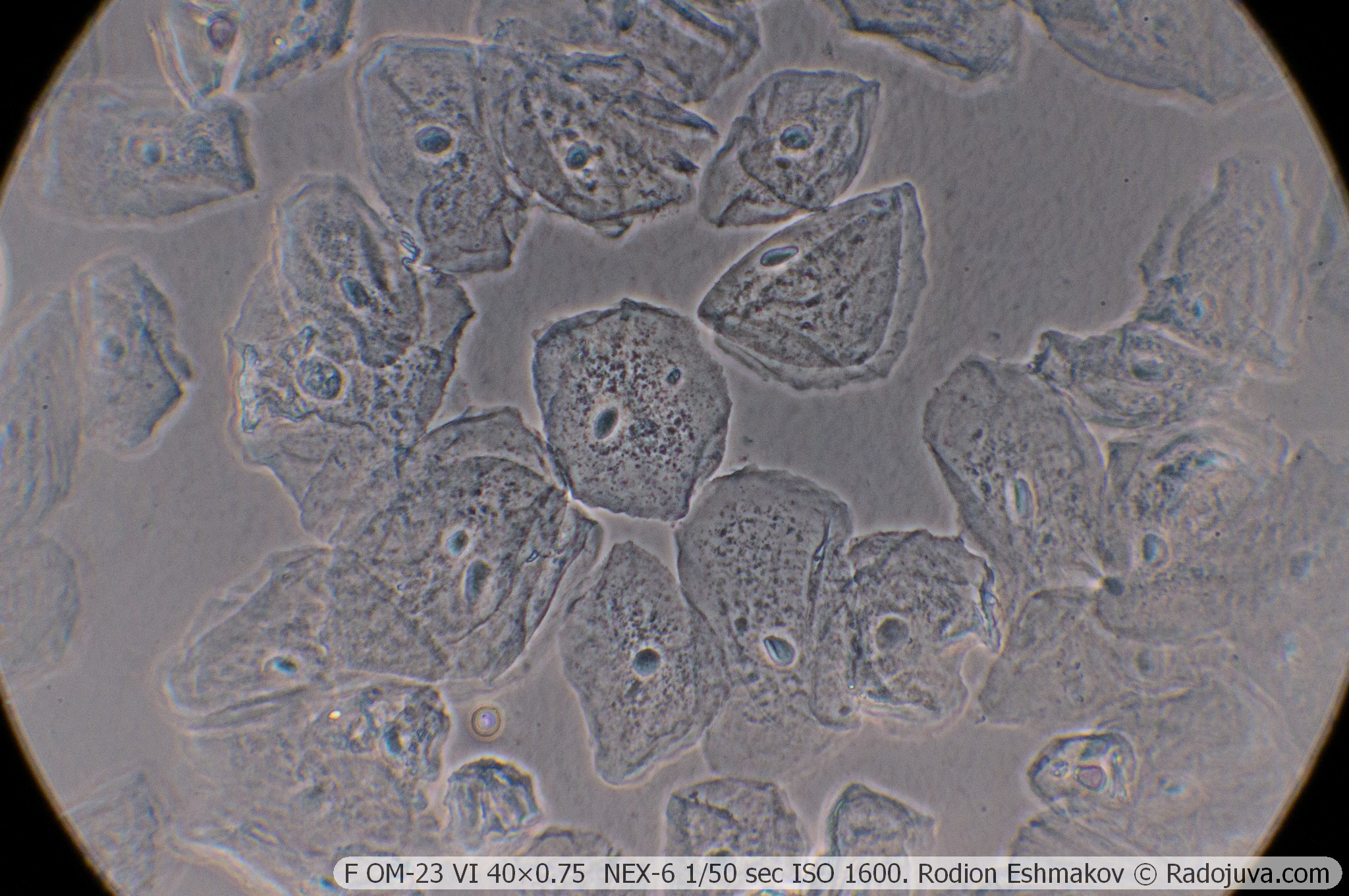
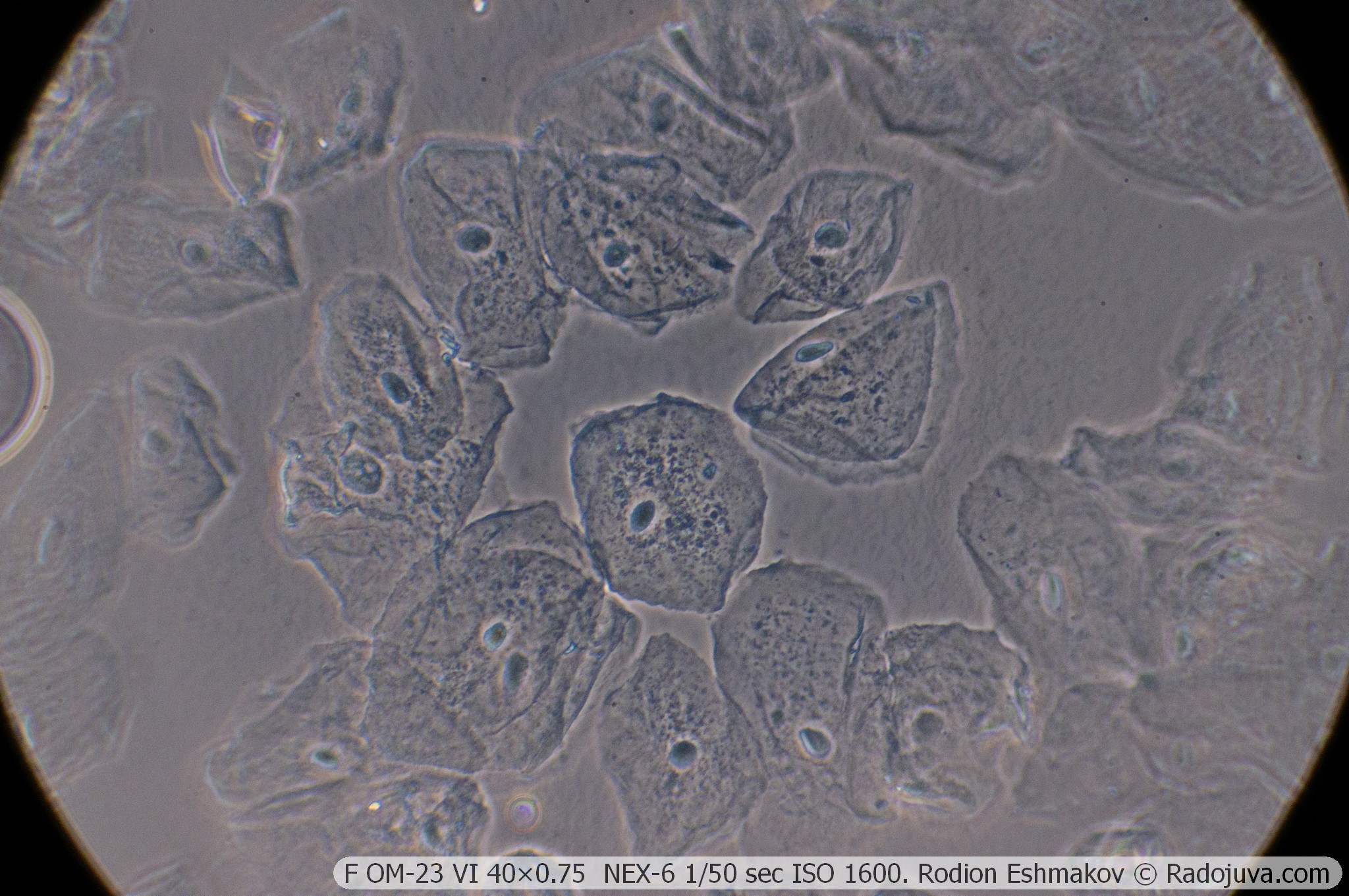
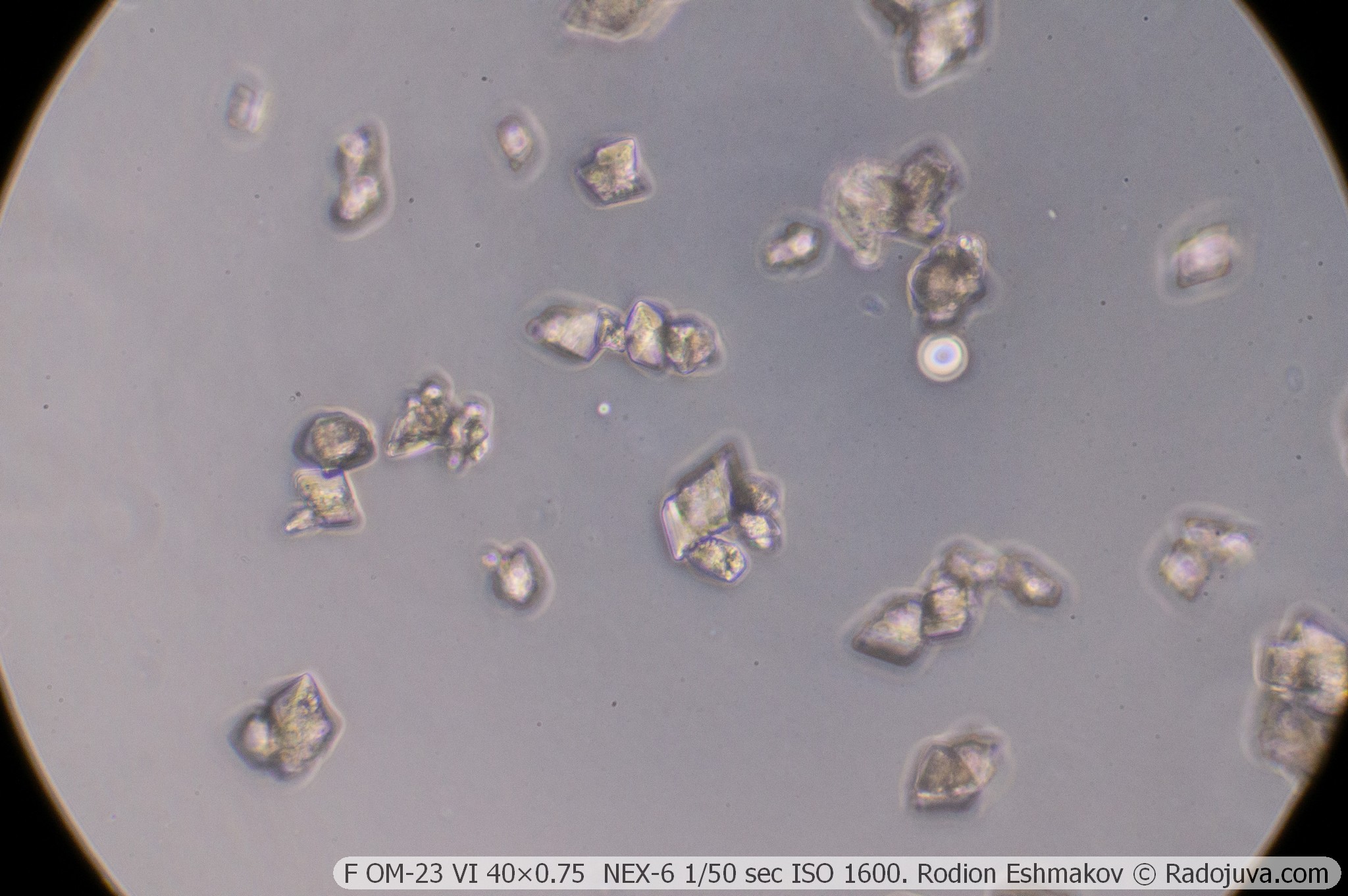
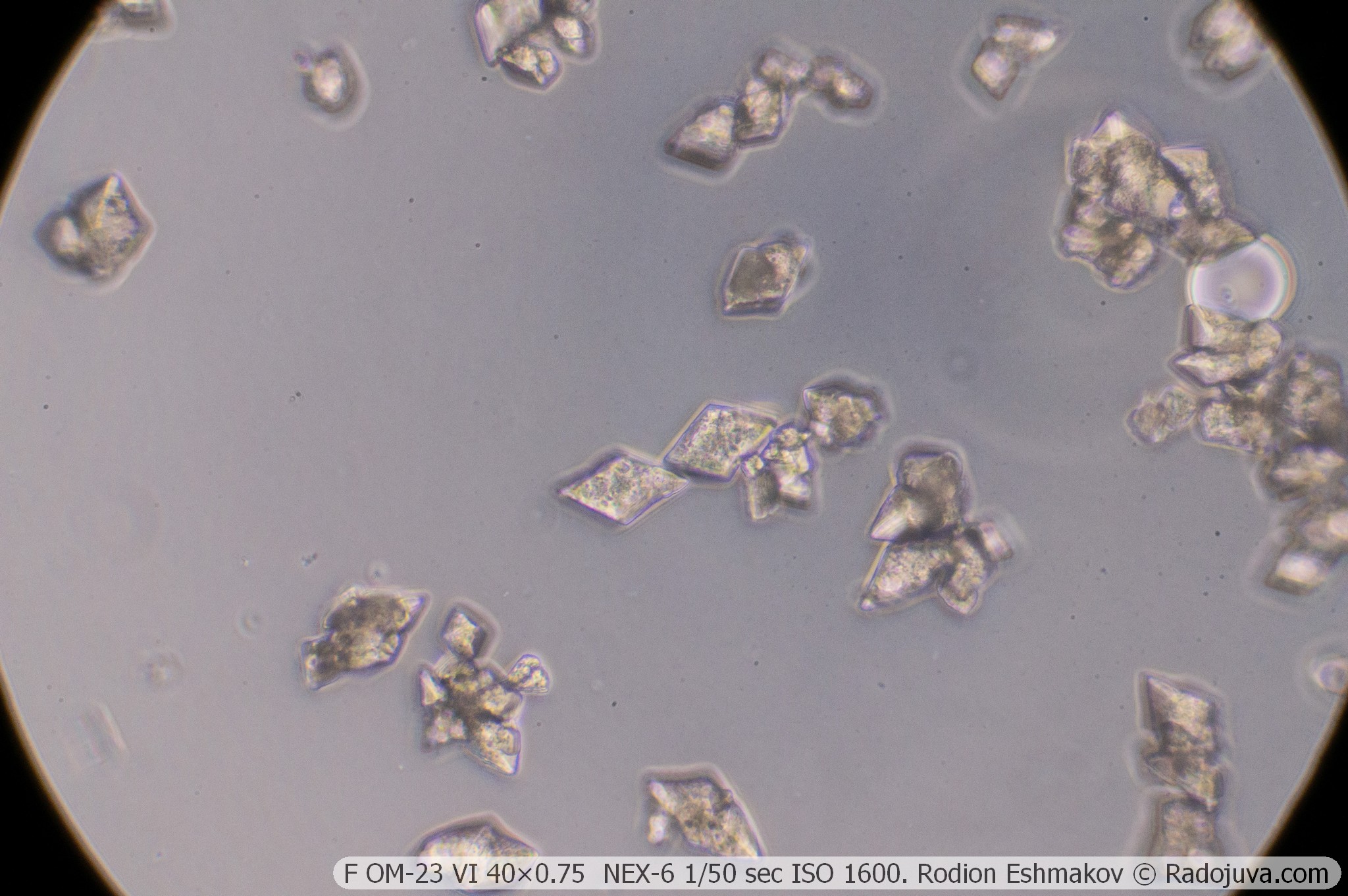
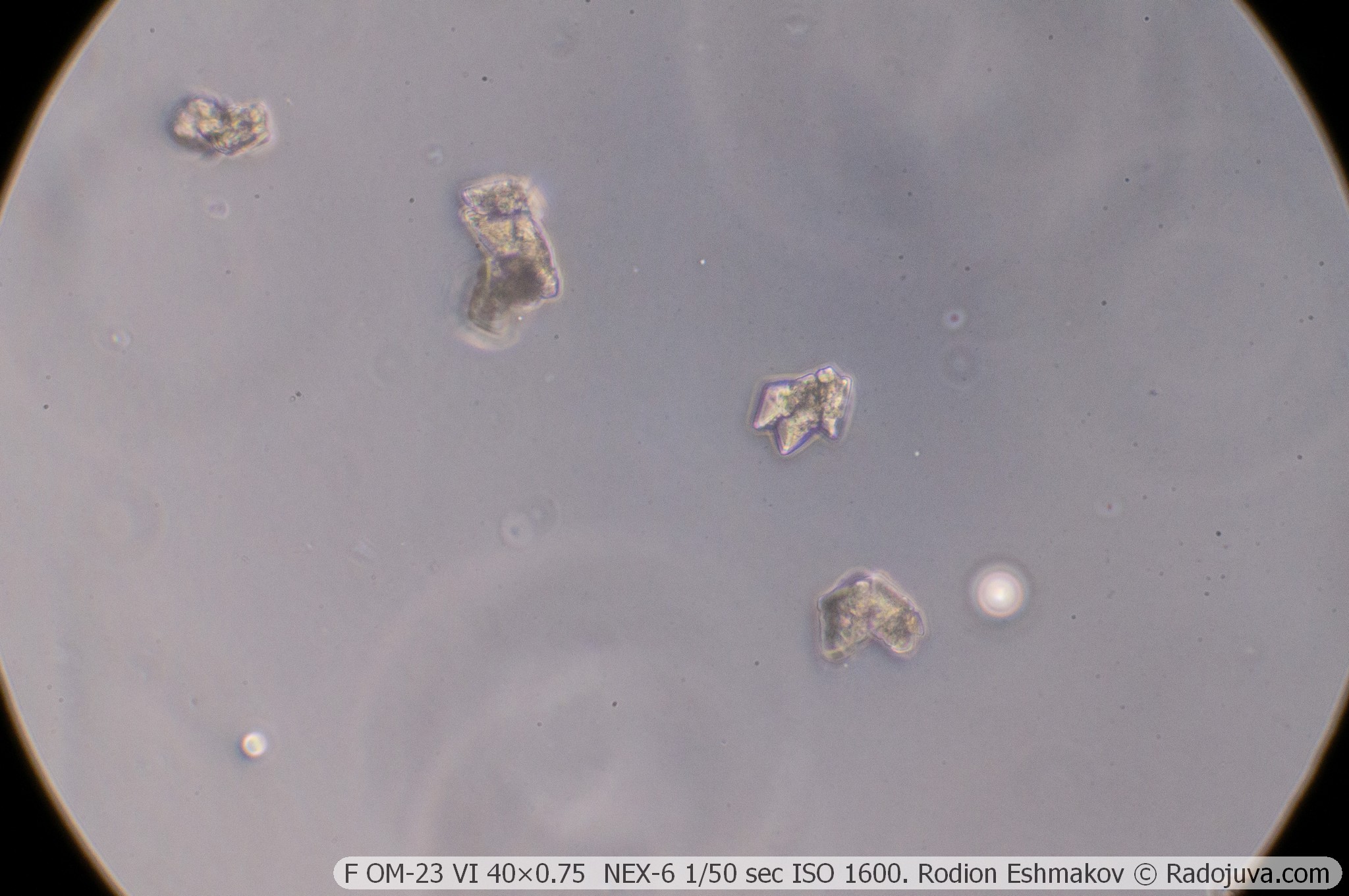
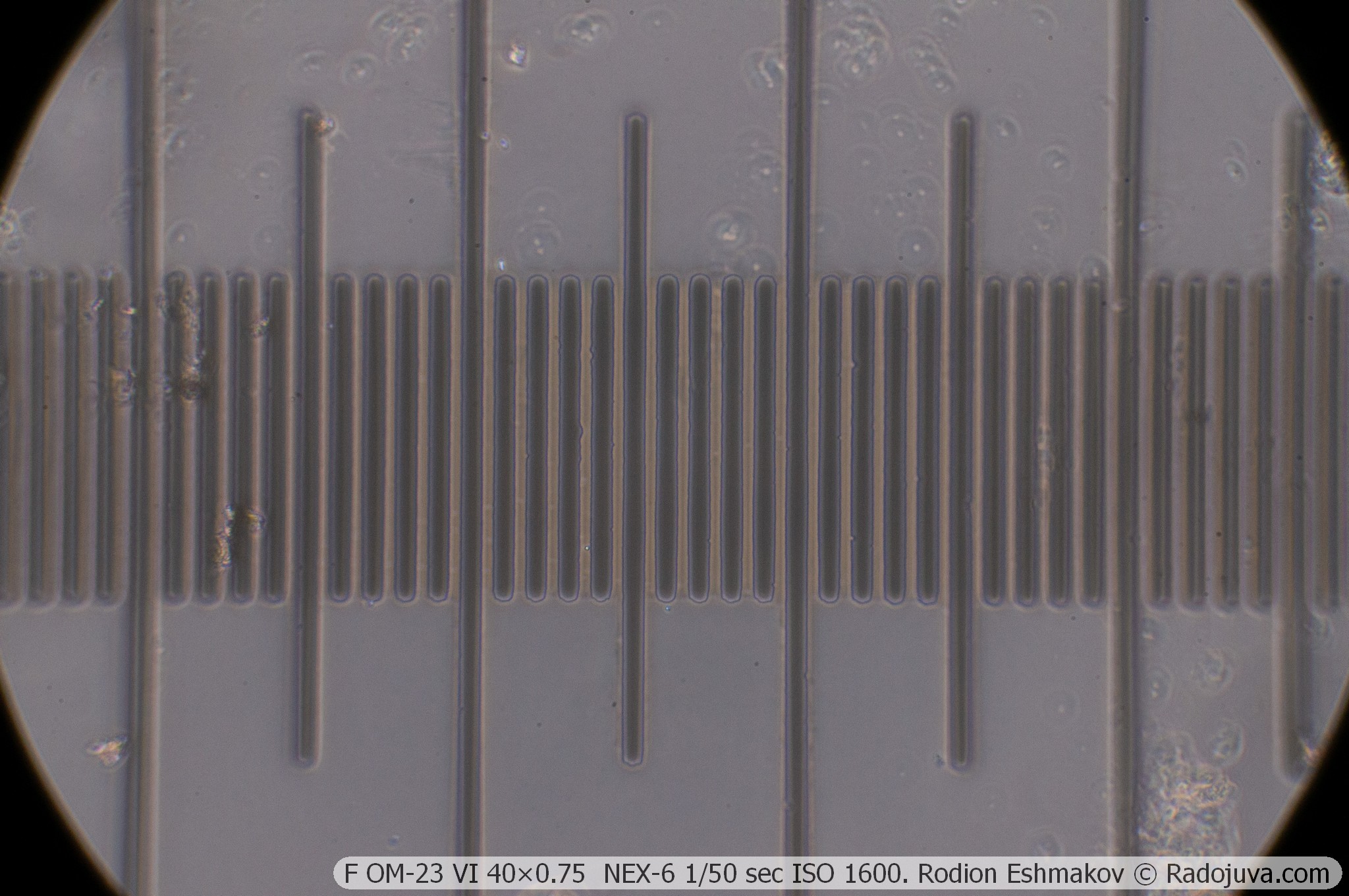
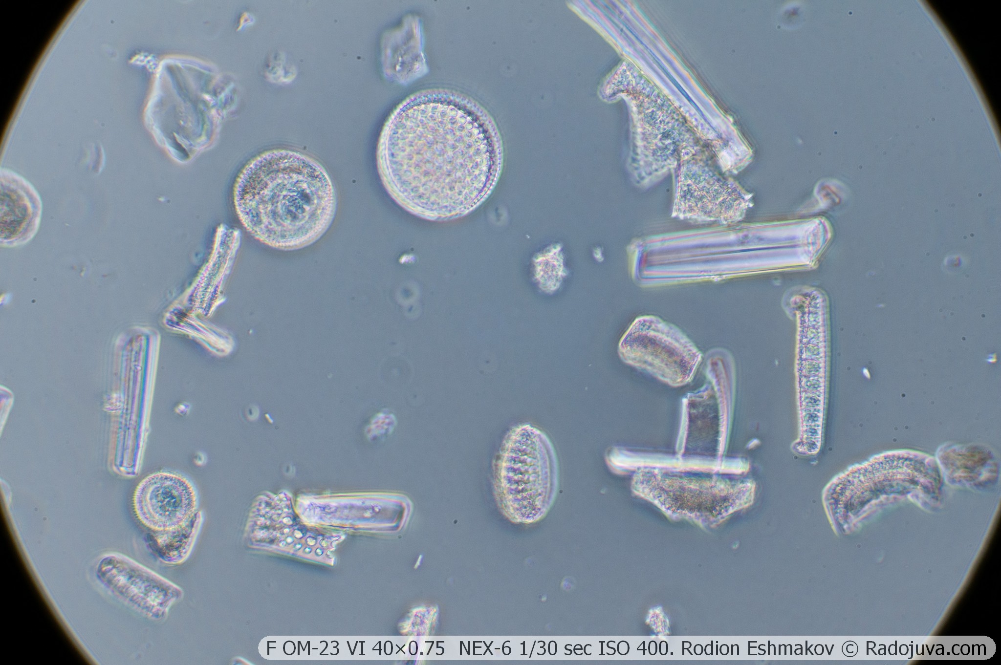
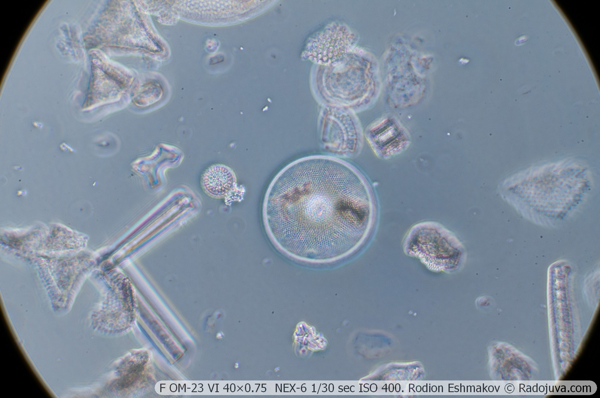
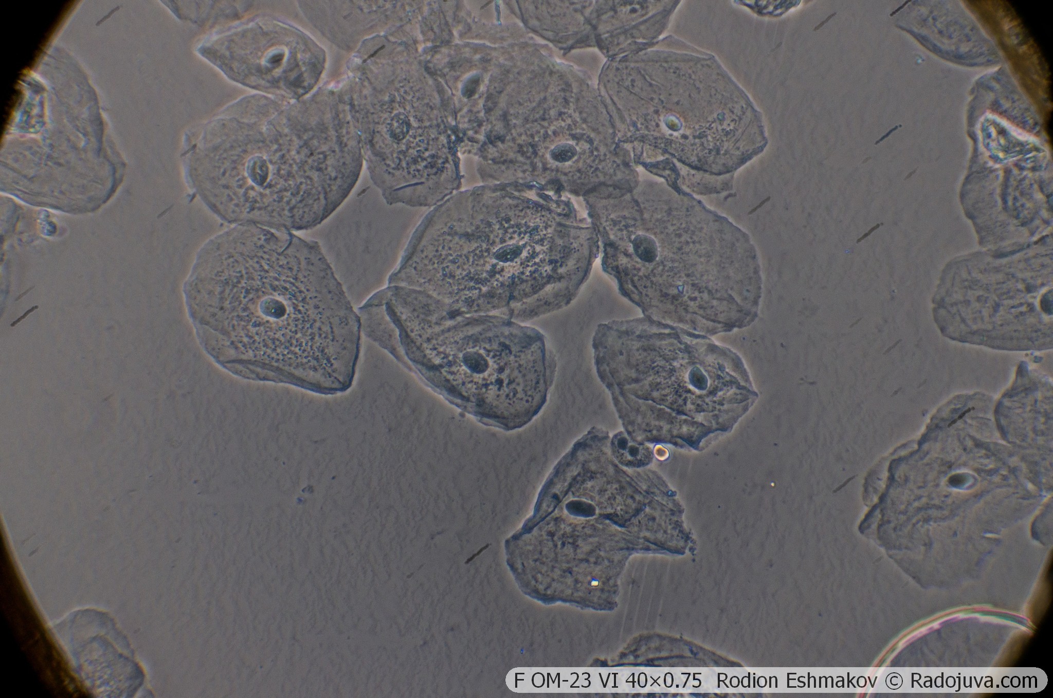
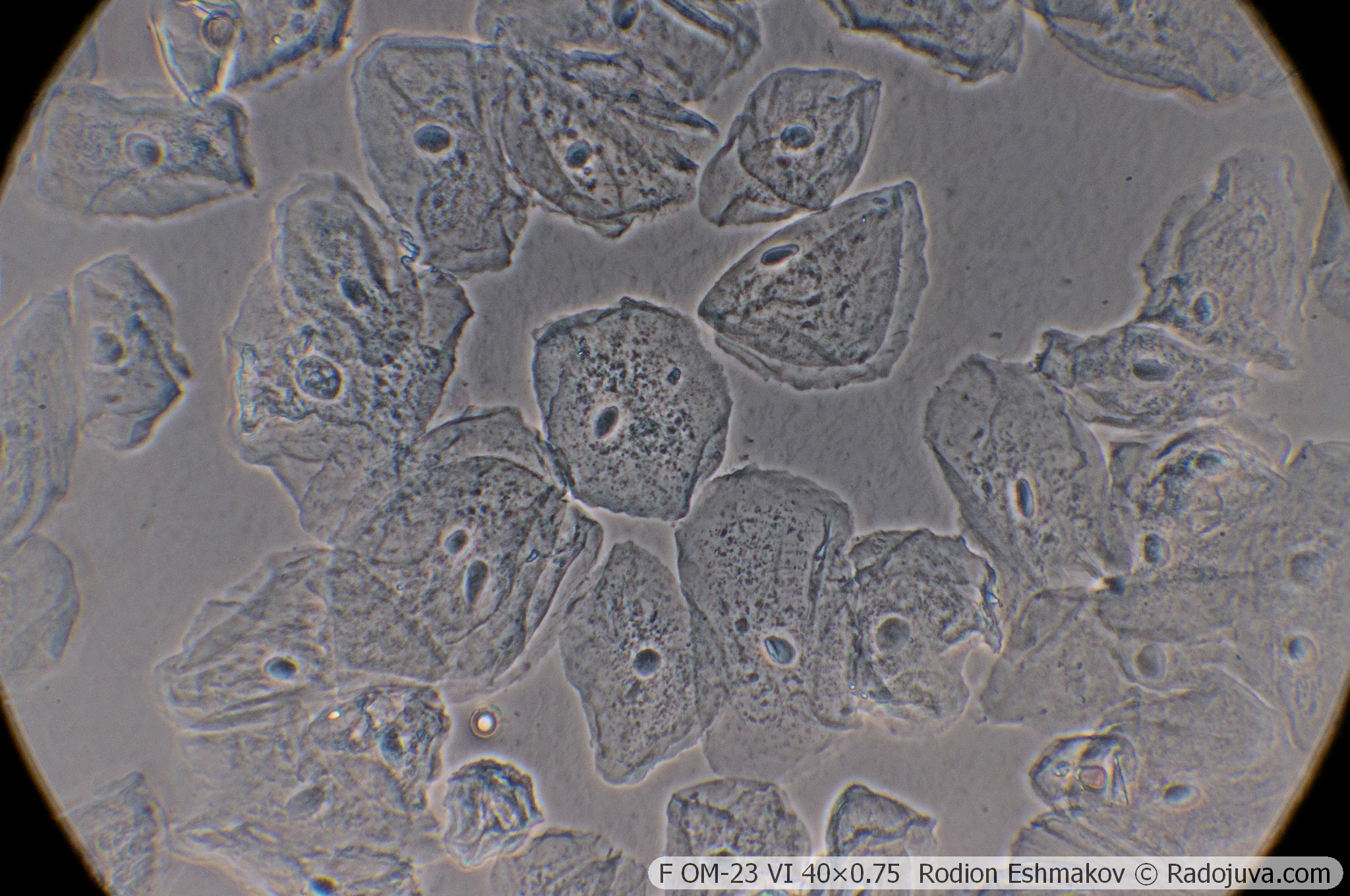
Below are examples of photos with stacking.
Lenses for the KF-1: 70×1.25 F VI OM-25
Optical design – 10 lenses in 5 groups, 3 fluorite (CaF2) lenses, 1 alum (KAl(SO4)2·12H2O) lens, movable lens group for spherical aberration correction; high-aperture apochromat-aplanat;
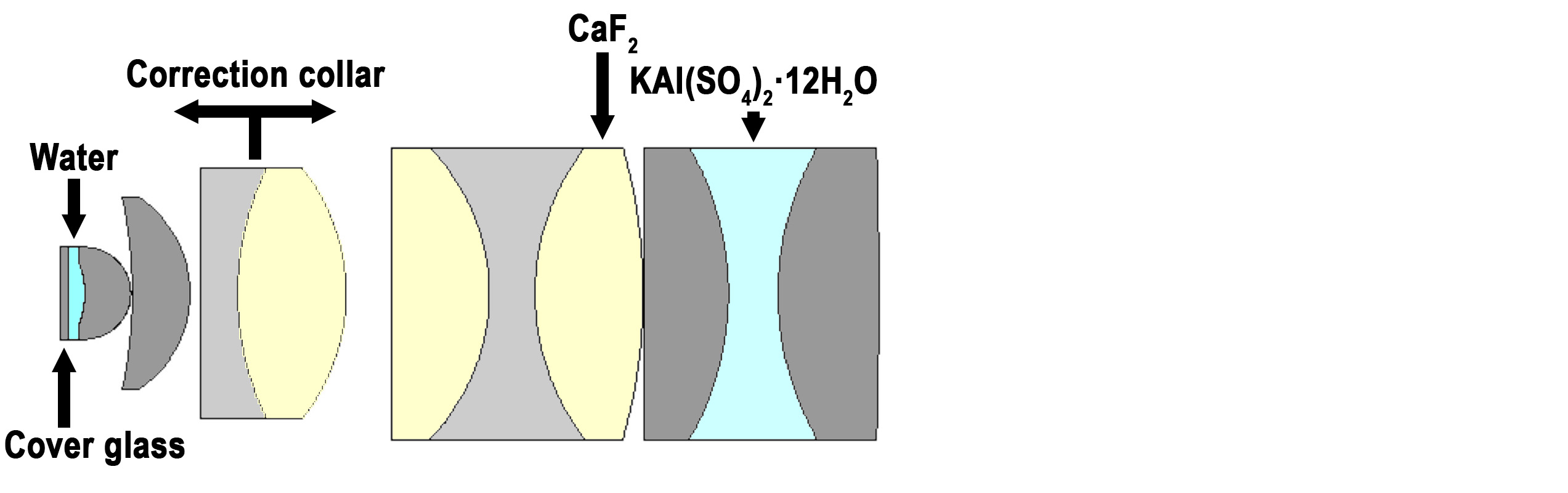
Drawing of the optical diagram of the F APO VI OM-25 70×1.25.
Correction type – apochromat;
Tube distance – 160 mm;
Parfocal distance – 32.7 mm;
Magnification: 70×;
Numerical aperture – 1.25 (sometimes marked 1.23, with KF-1 0.8, phase ring 0.37);
Immersion required – yes (water);
Working distance – 0.14-0.04 mm;
Cover glass thickness: 0.10-0.23 mm (adjustable using a correction collar);
Mounting type – RMS standard (4/5” × 1/36” thread);
Lateral chromatic aberration – corrected with a compensating eyepiece (such as LOMO K5×);
Features - microscopic lens, does not have an iris diaphragm and a focusing mechanism.
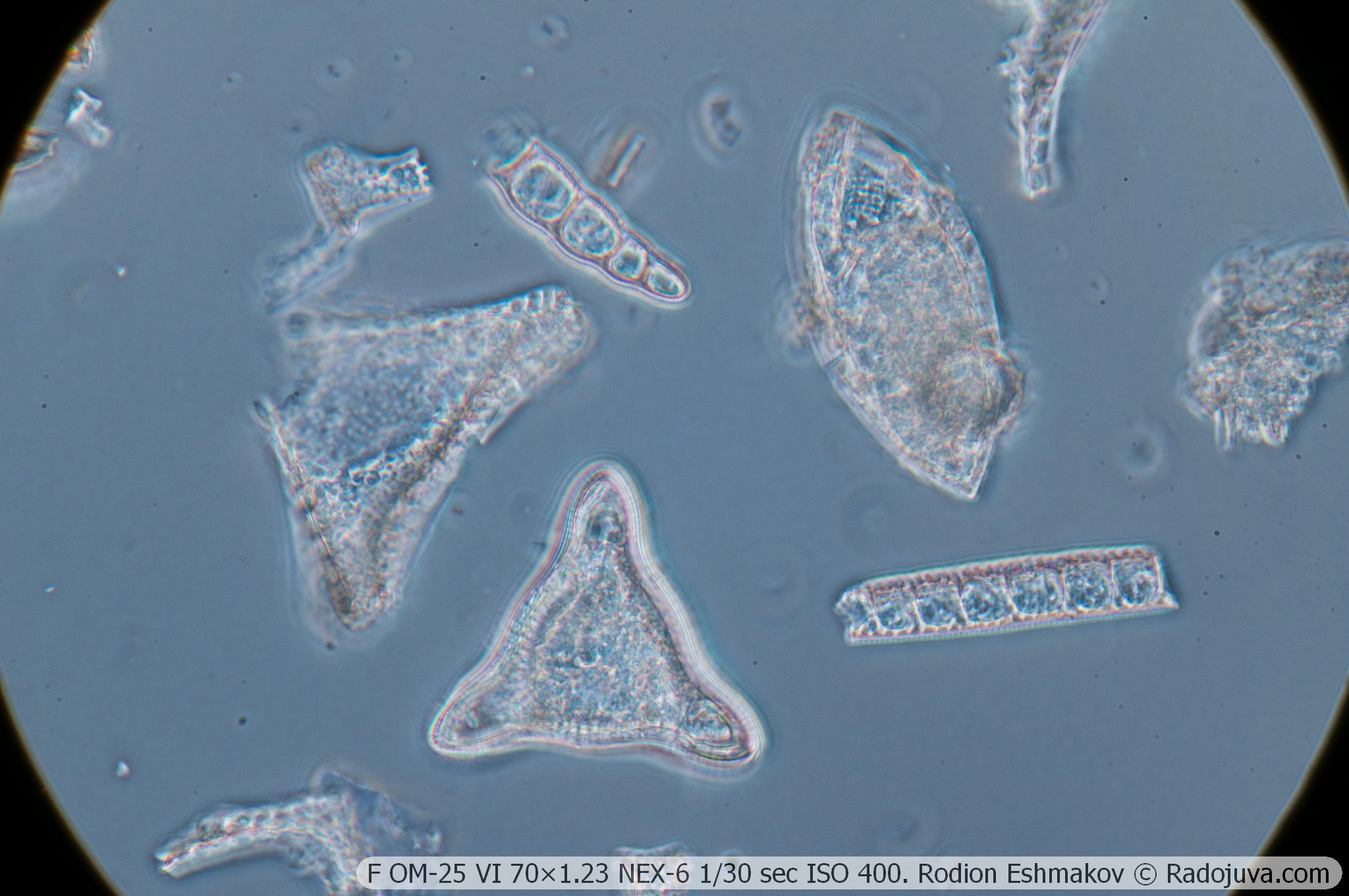
This lens features a complex optical design using unusual materials. Oddly enough, its original design was most likely developed by Ernst Abbe in the late 19th century. Three lenses in the F VI APO 70×1.25 OM-25 are made of single-crystal fluorite, and one lens is made of potassium alum. Alum is a very exotic material characterized by very low chemical and thermal stability compared to optical glass. It is highly soluble in water and gradually degrades upon exposure to air, especially dry air, and when heated above 100°C. Alum is even softer and more brittle than fluorite. The use of such a difficult material is due to the lack of special flints with the required parameters, without which effective correction of chromatic aberration is extremely difficult. The alum lens is embedded in a triple bond between two lenses made of simple optical glass, which also serves as a protective layer. The lens is very sensitive to storage conditions—changes in temperature and humidity can cause degradation of the bonds.
The complex optical design of this old apochromat is also quite primitive. A lens with this design cannot correct for field aberrations and lateral chromatic aberration. Even with a compensating eyepiece like the K-5× ML-2, pronounced astigmatism significantly degrades image quality off-center.
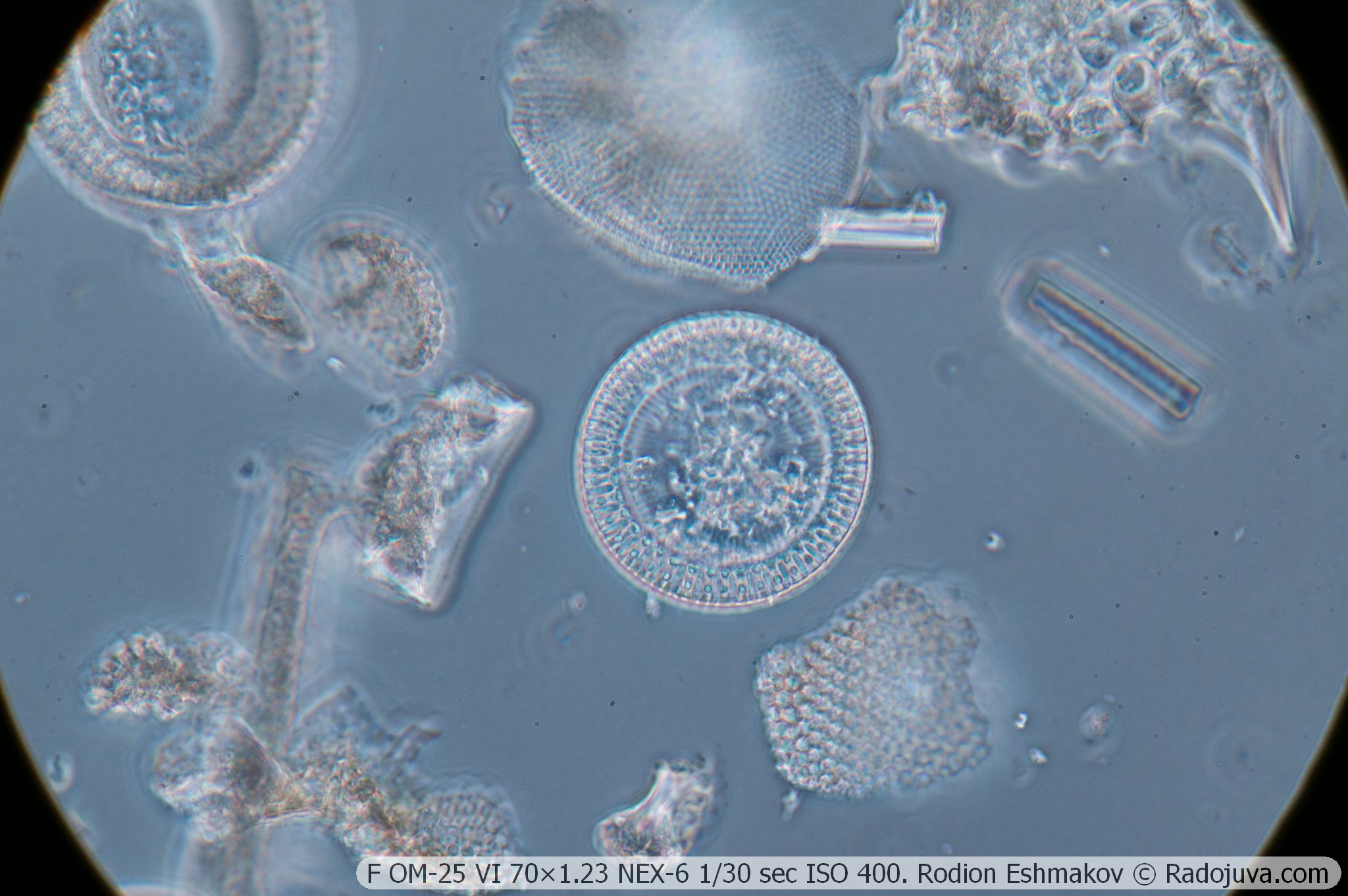
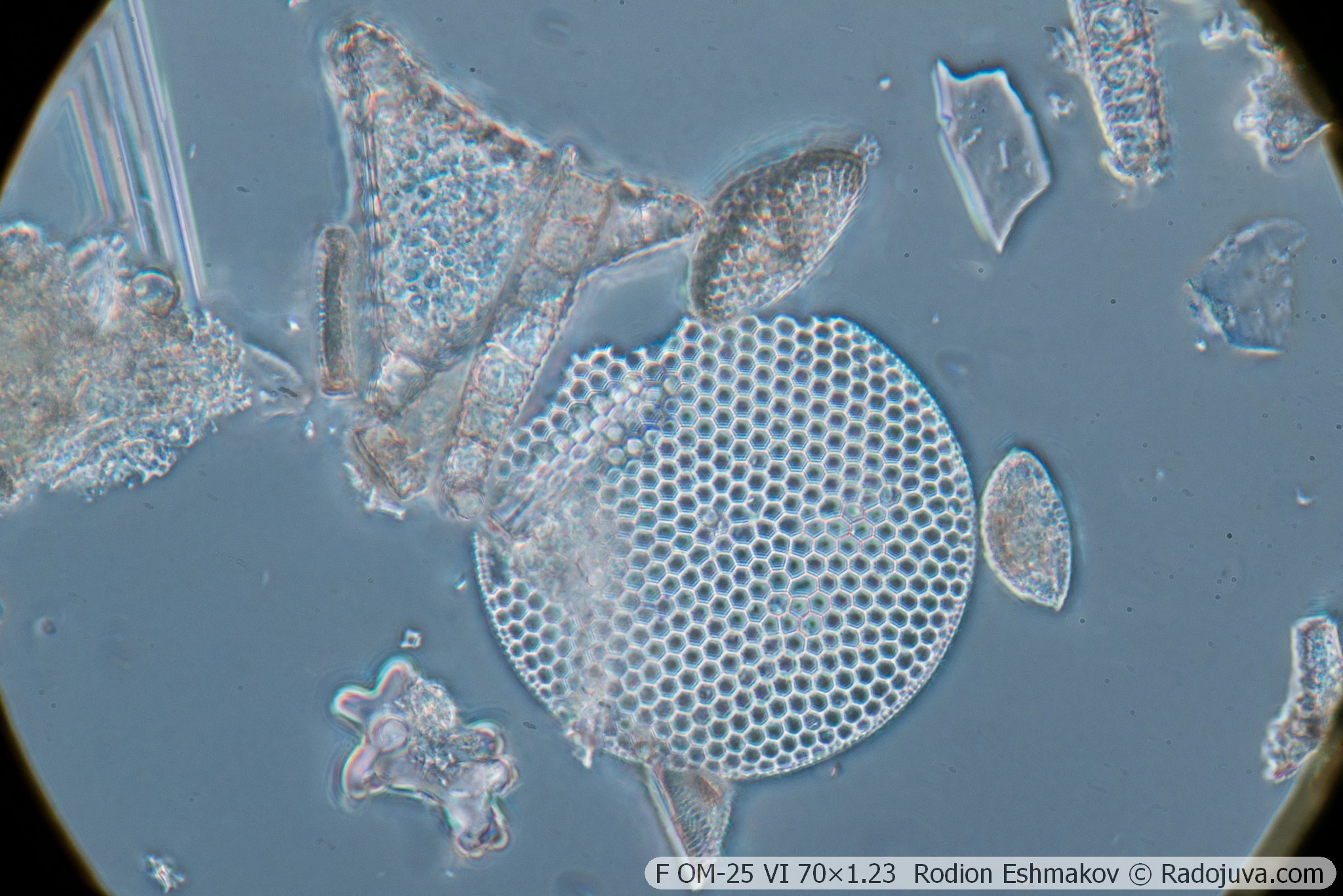
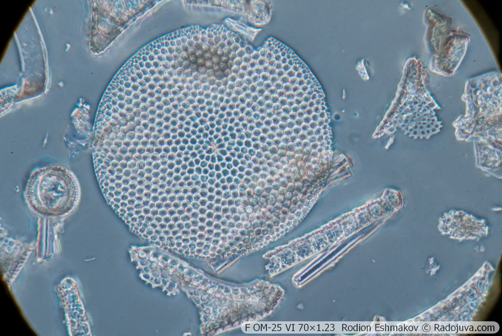
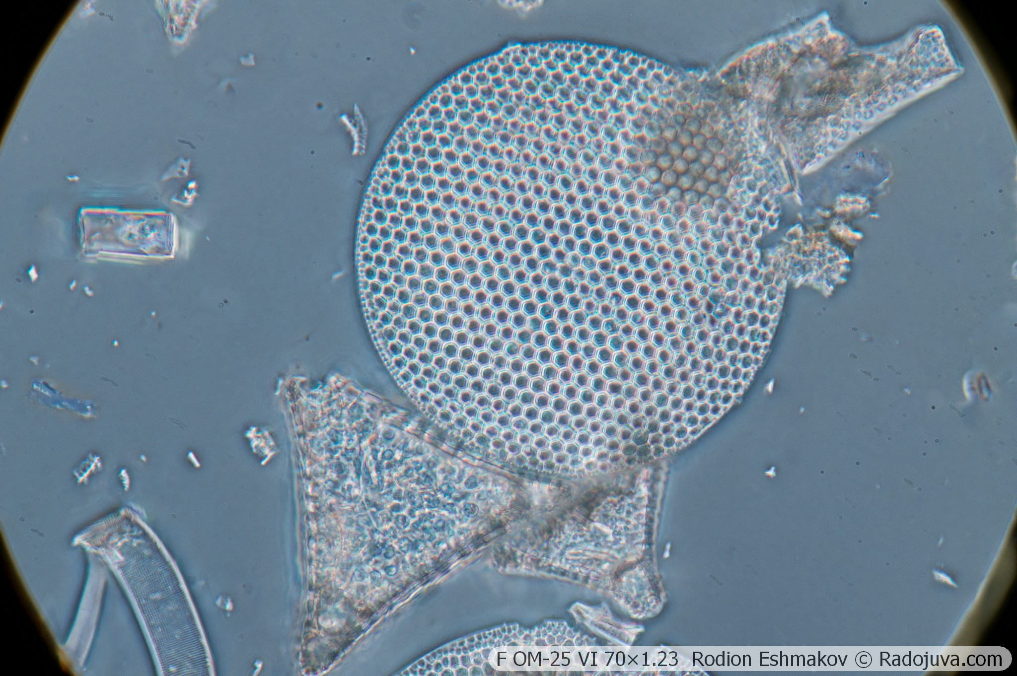
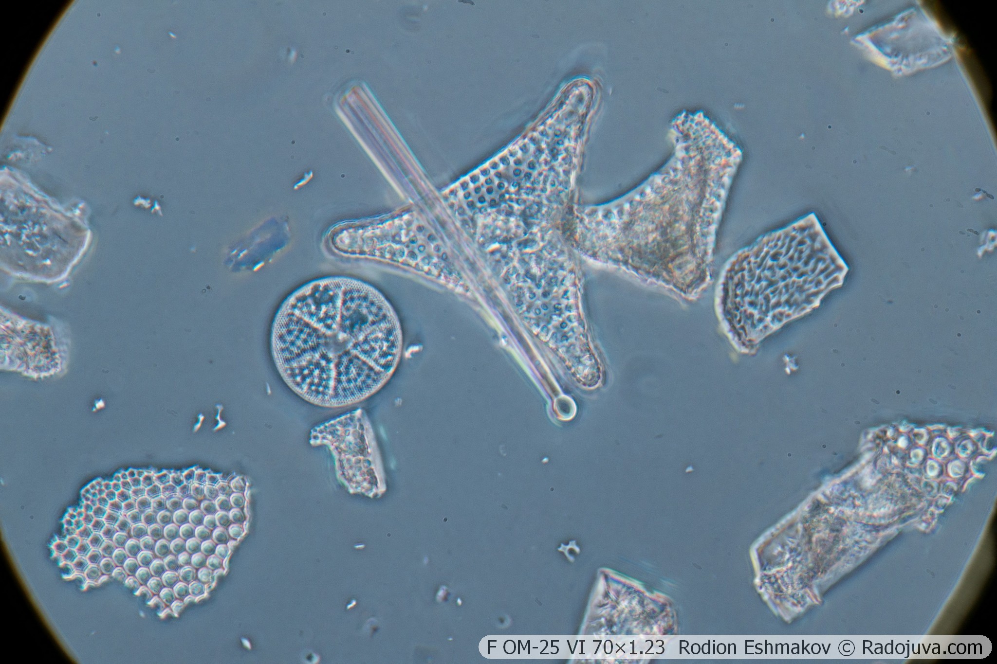
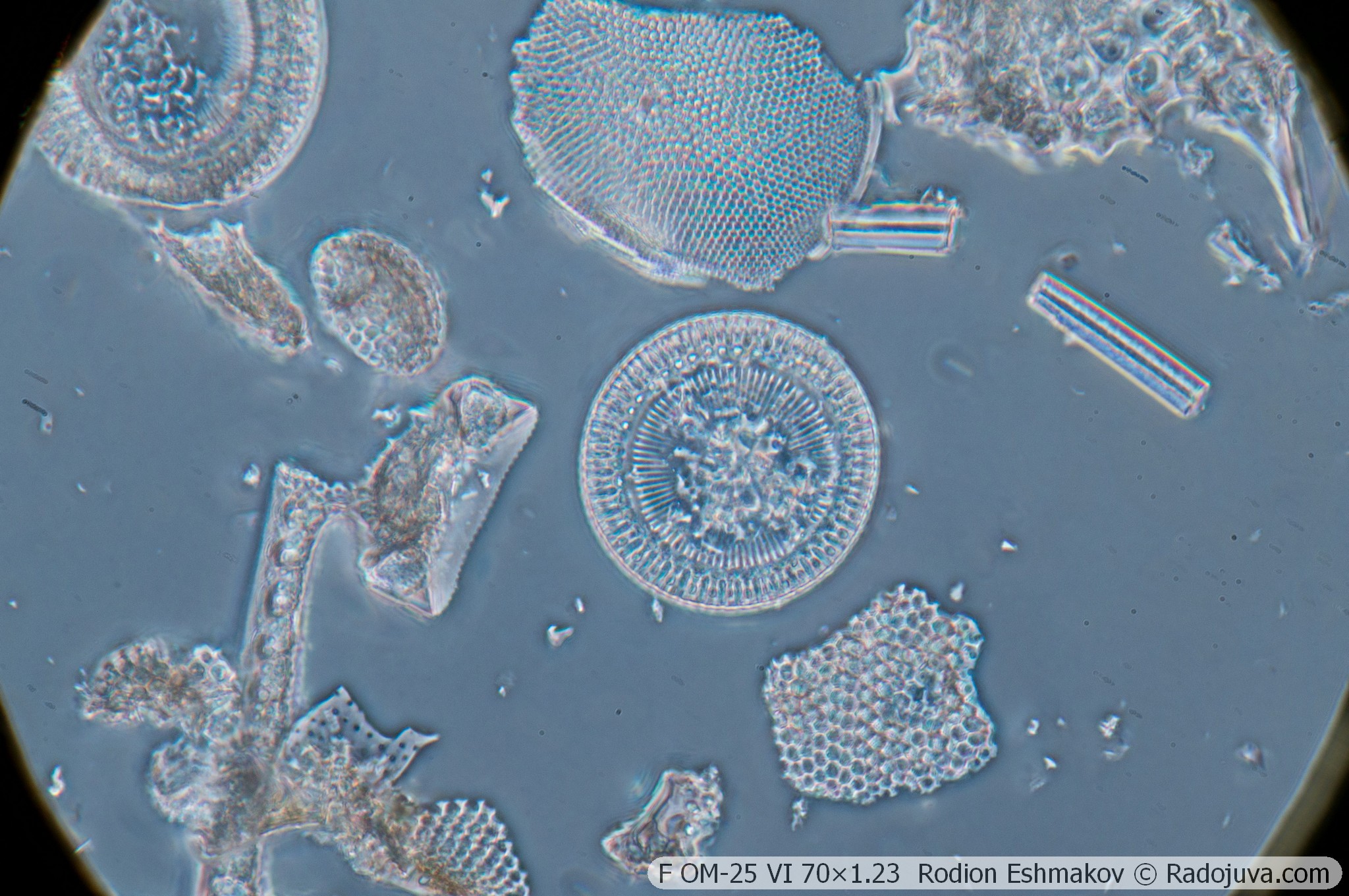
Nevertheless, based on its combined parameters, this lens is the most interesting for use with the KF-1 condenser. Compared to other 40-90× achromatic objectives, the 70×1.23 apochromat offers significantly better correction of aberrations in the center of the field, and its secondary spectrum is approximately 5-10 times smaller than that of achromatic objectives. Combined with its lower magnification compared to the F OM-41 MI, this makes the F OM-25 70×1.25 more suitable for photography.
There are, however, complications associated with using water immersion instead of oil immersion. For example, due to the difference in the refractive index of glass and water, the objective lens is quite sensitive to deviations in the cover slip thickness from the calculated 0.17 mm. Therefore, the F OM-25 VI 70×1.25 has a unique element—the so-called correction collar. This ring, when rotated, moves the two-lens assembly relative to the other elements of the optical system, allowing for correction of spherical aberration and compensation for deviations in cover slip thickness from the calculated value. The objective collar is marked in millimeters from 0.10 to 0.23. On my objective, it rotates very stiffly due to hardened lubricant—a common problem with these lenses. However, when using phase contrast, the collar may not be necessary due to the low working numerical aperture.
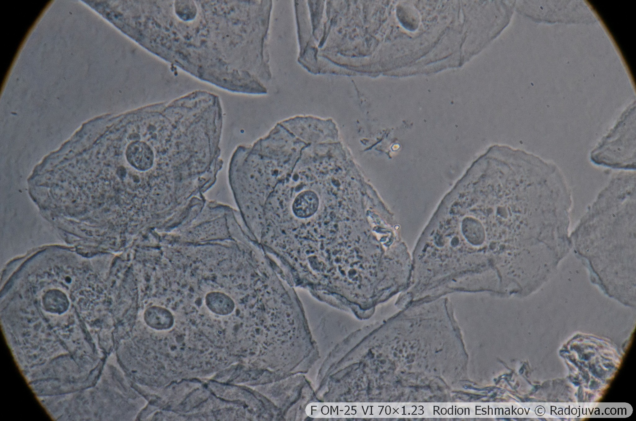
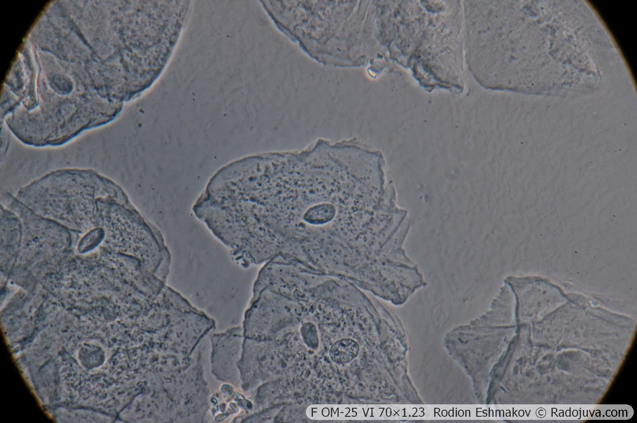
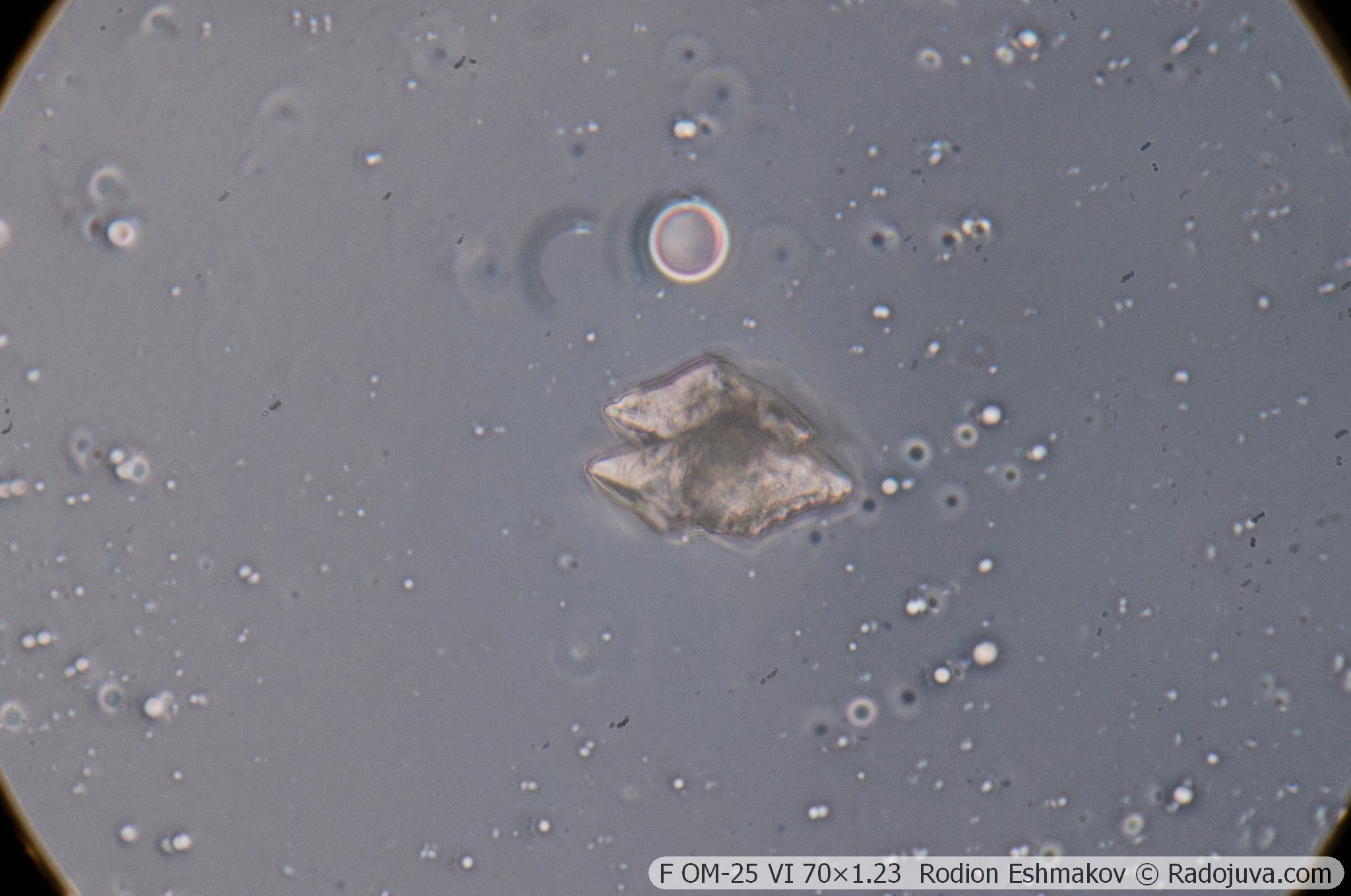
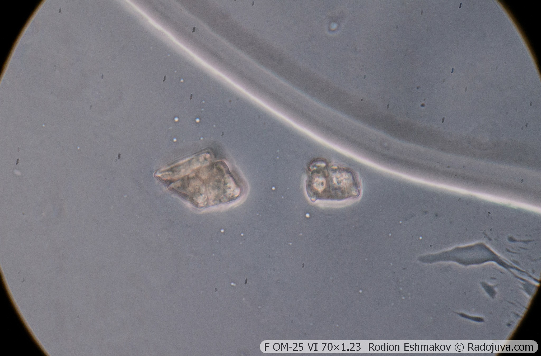
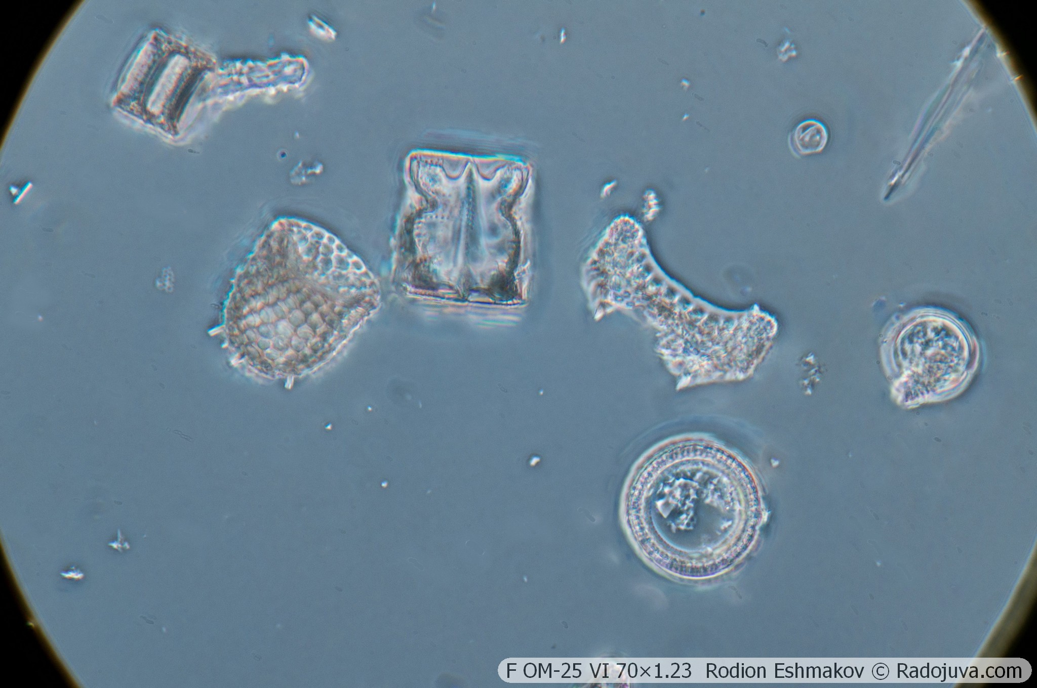
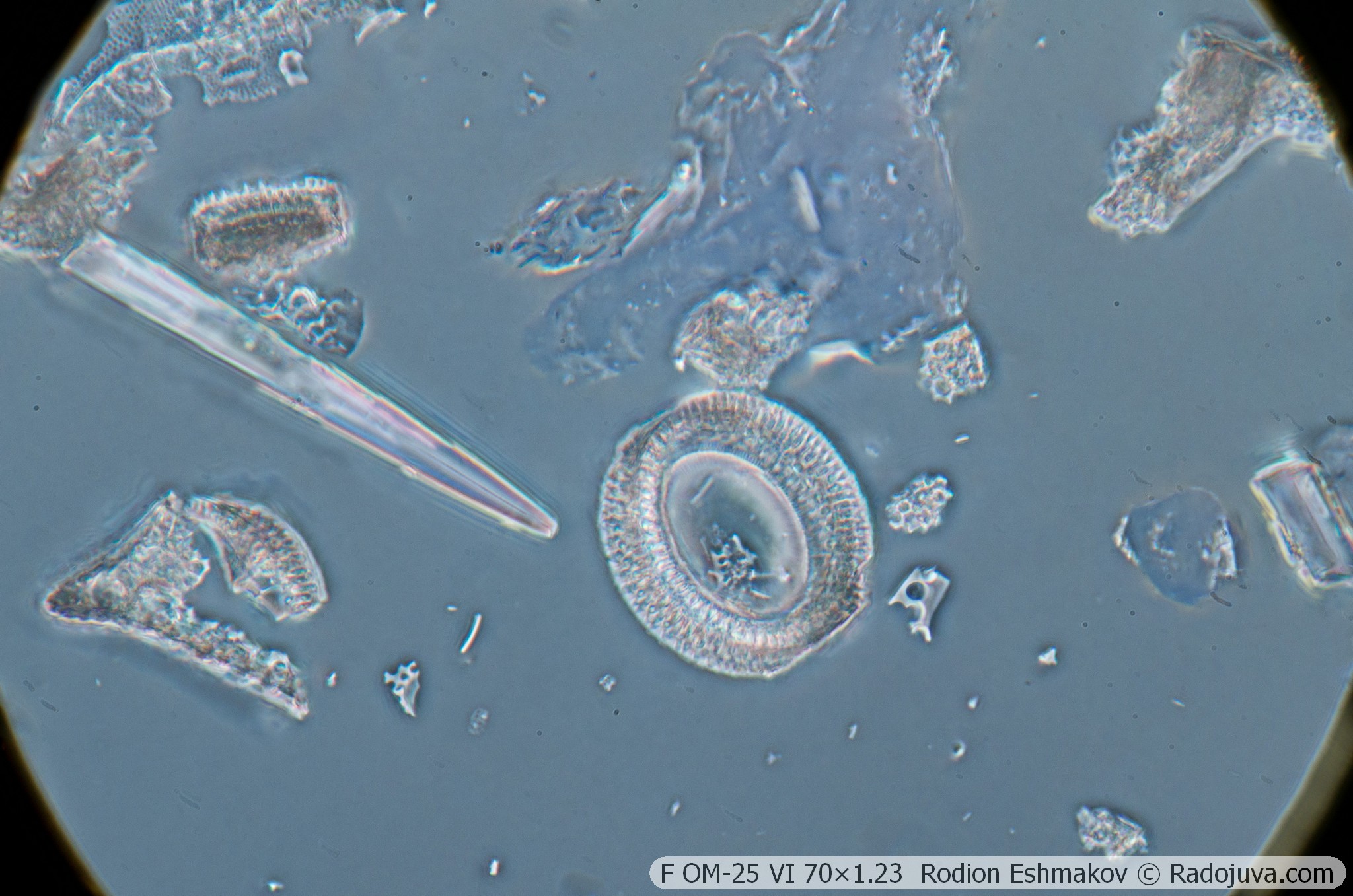
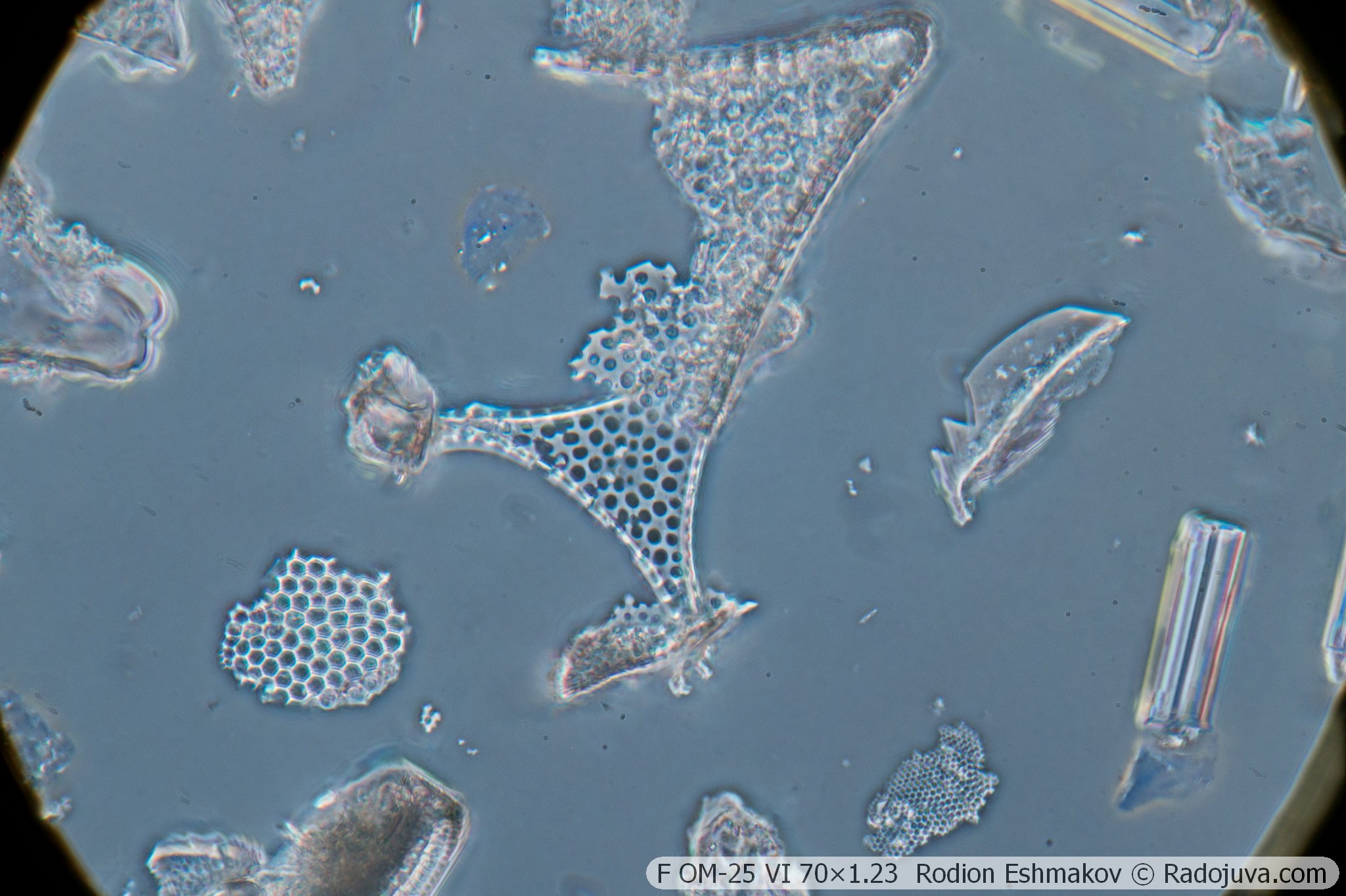
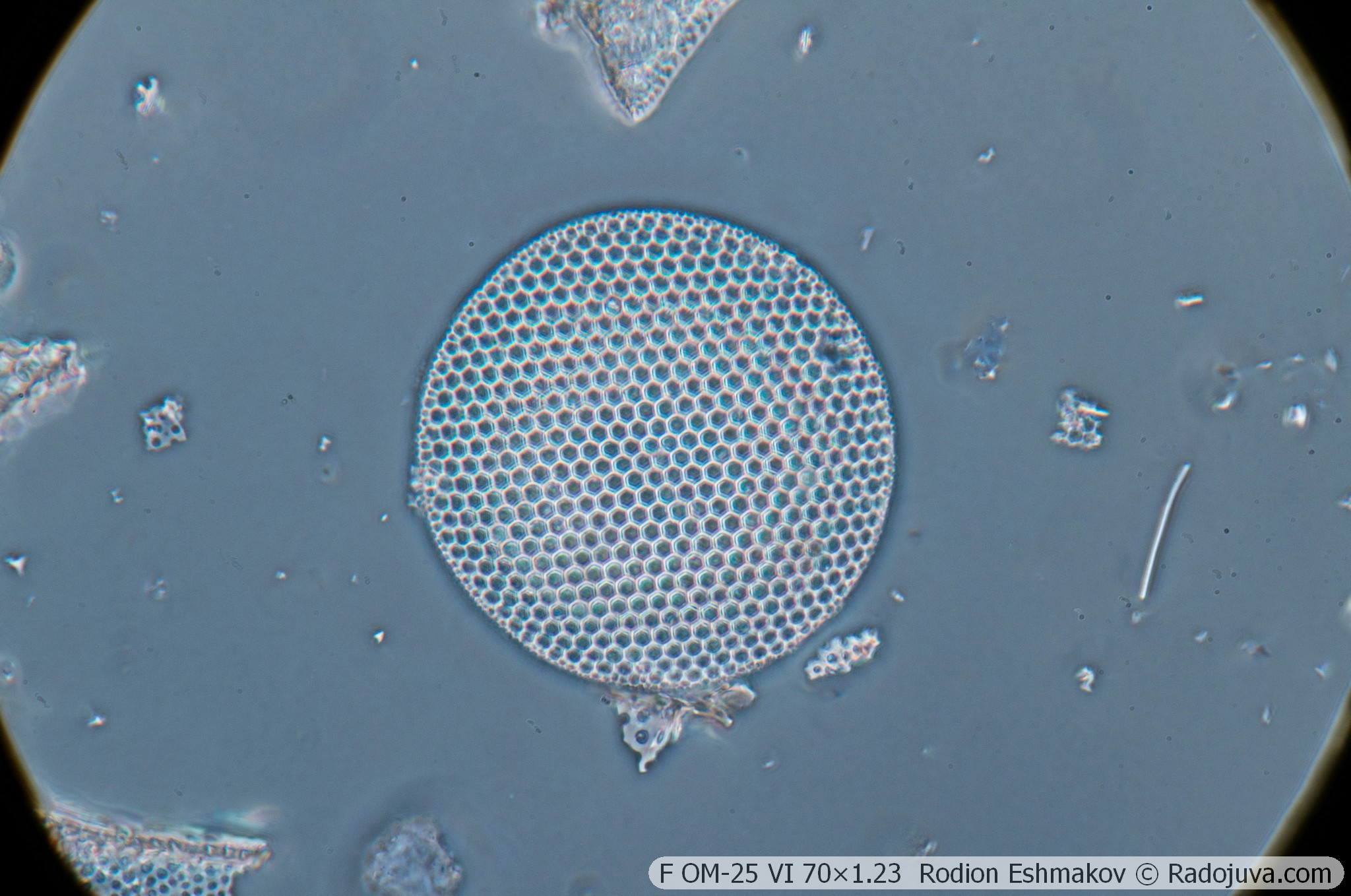
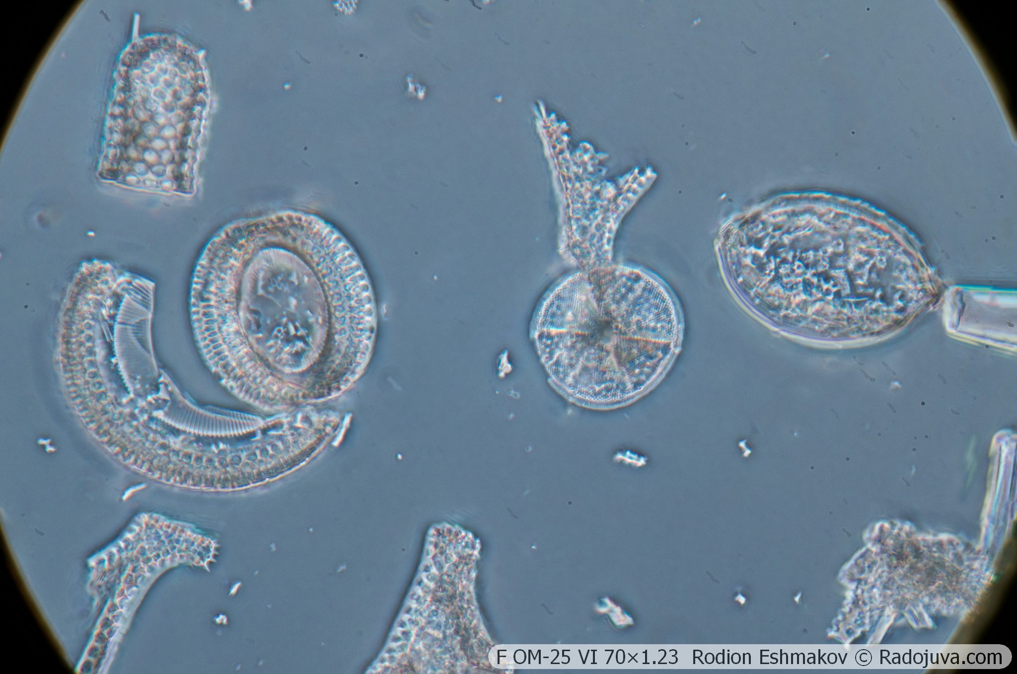
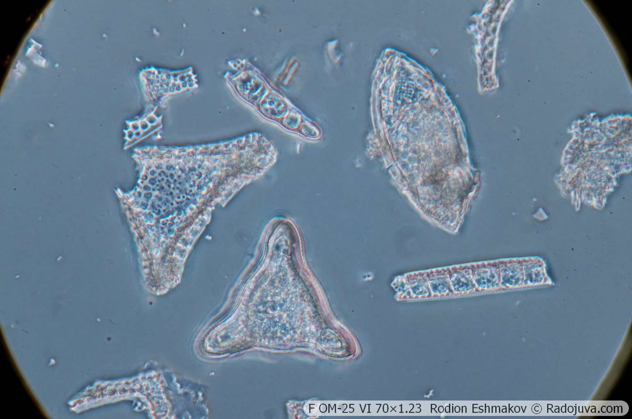
Below are examples of photos taken on a Sony NEX-6 and a F OM-25 VI 70×1.25 lens using a relay from a K5× ML-2 eyepiece and a WitrueHD 75/2.8 10 Mpix lens.
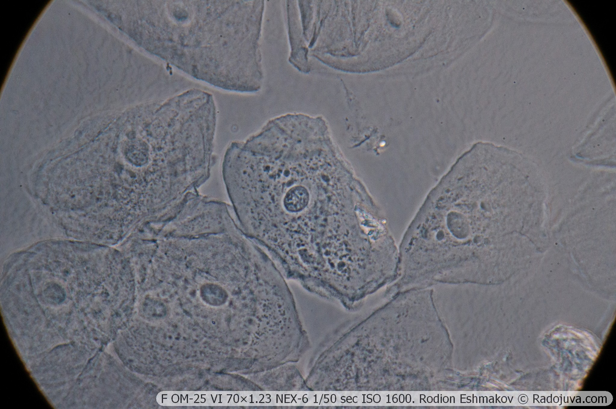
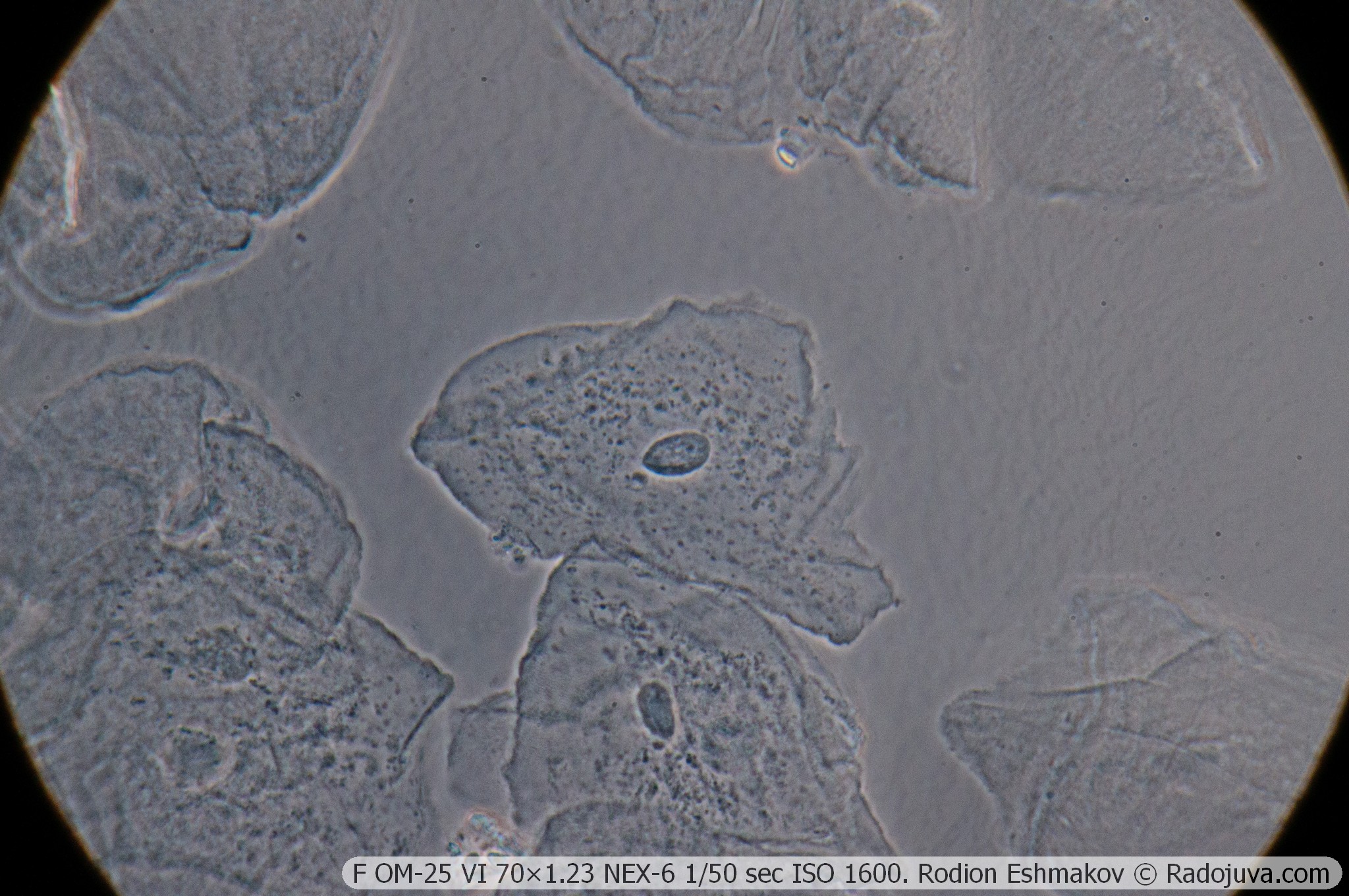
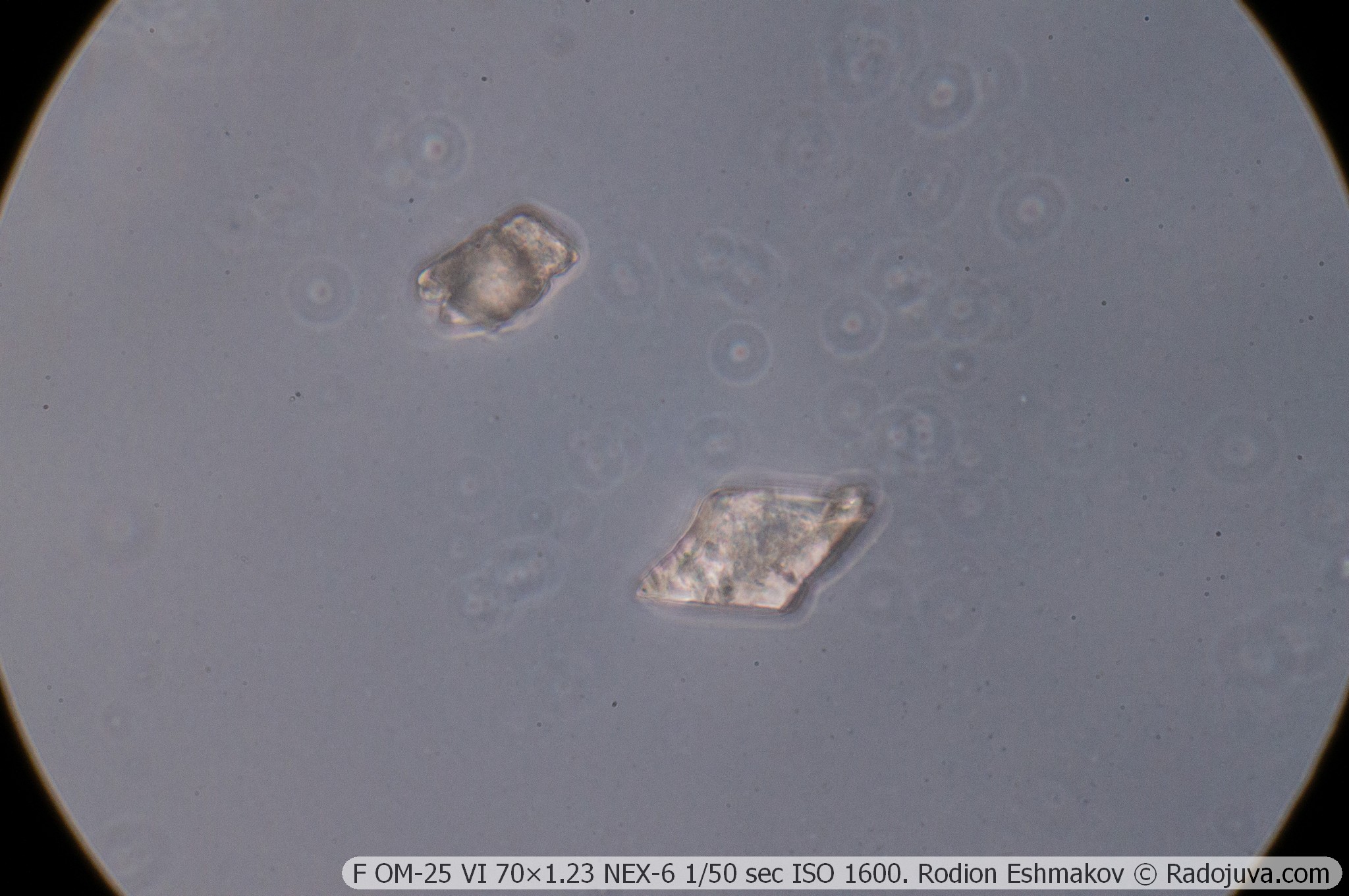
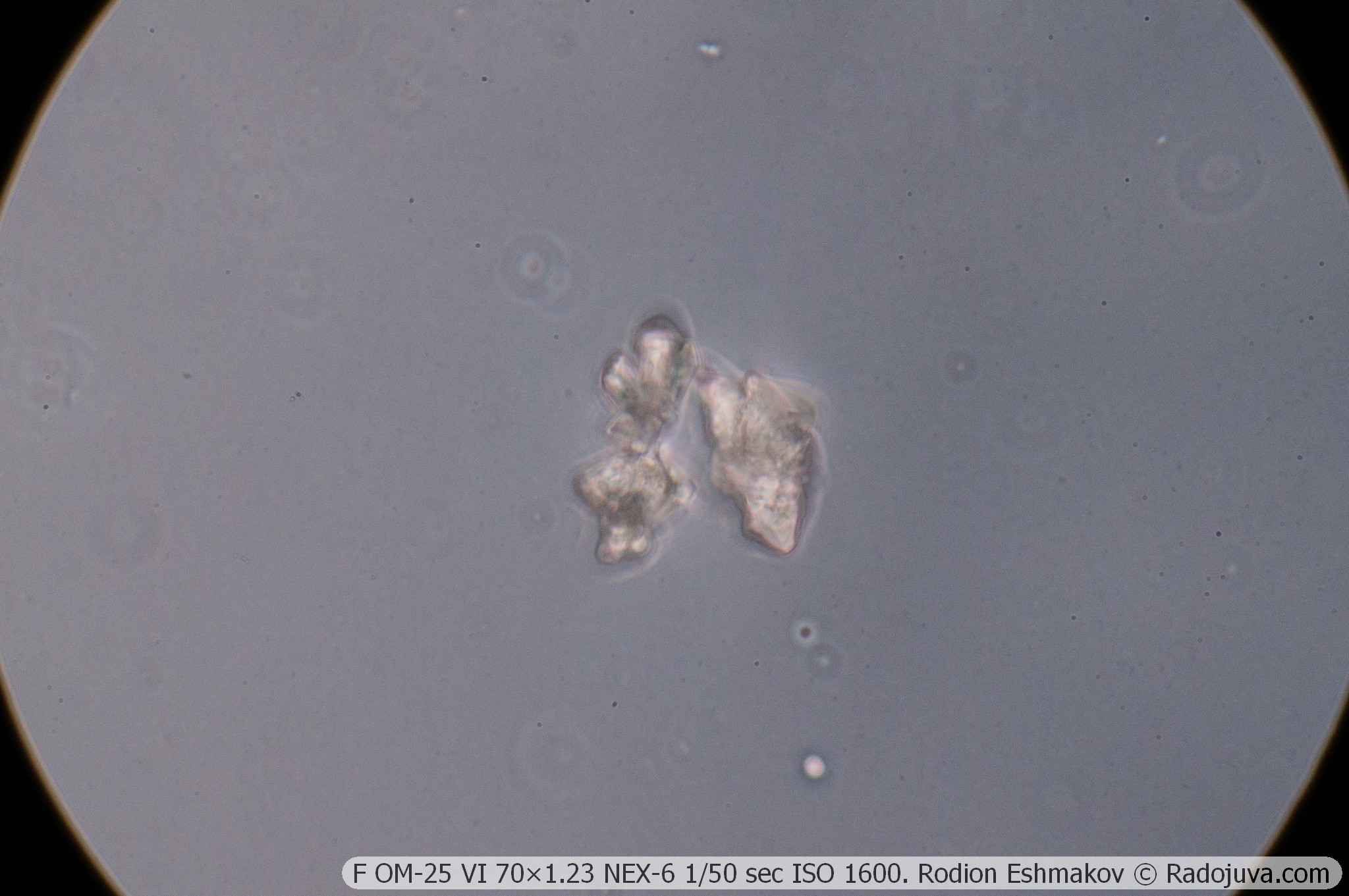
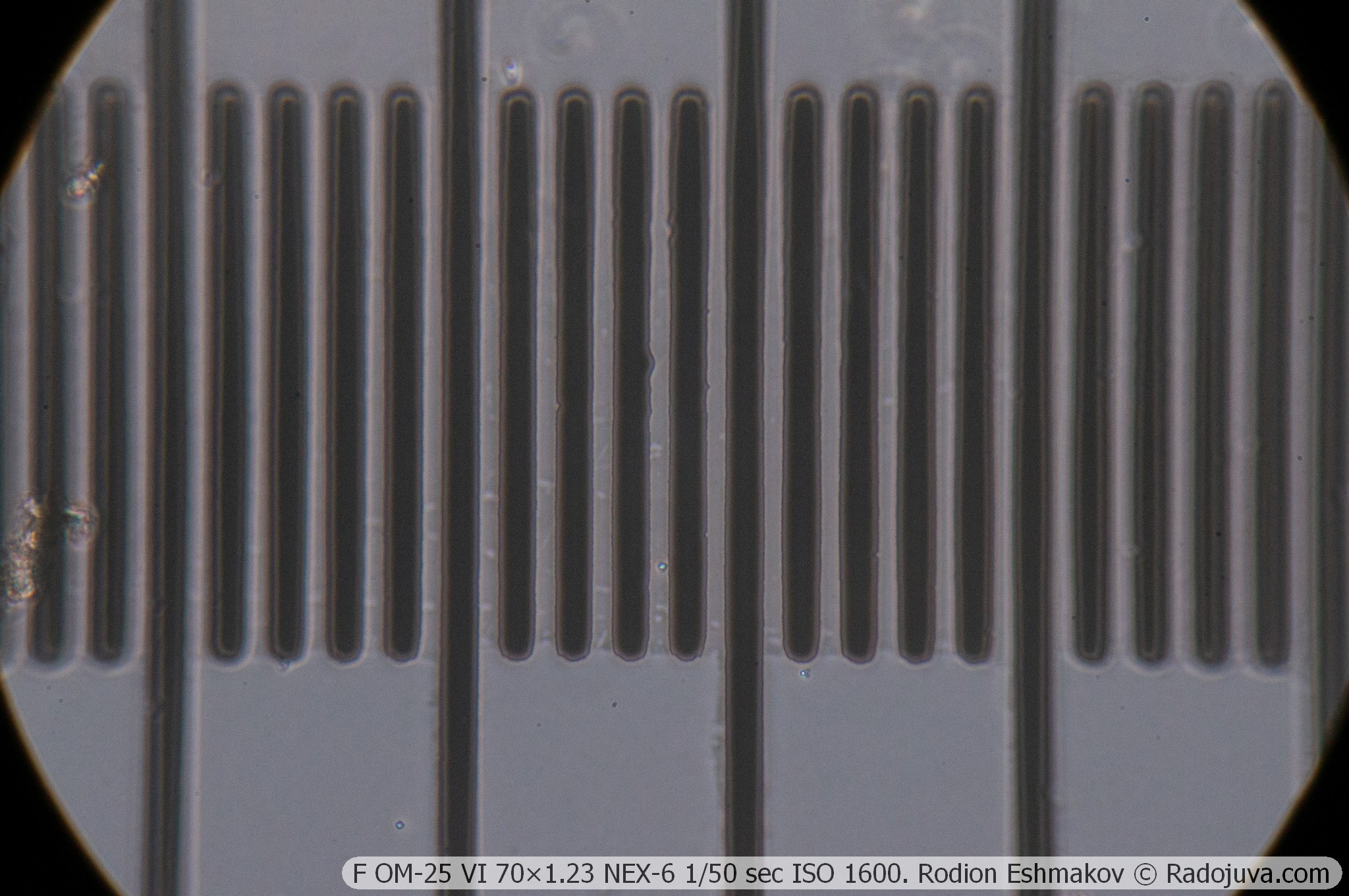
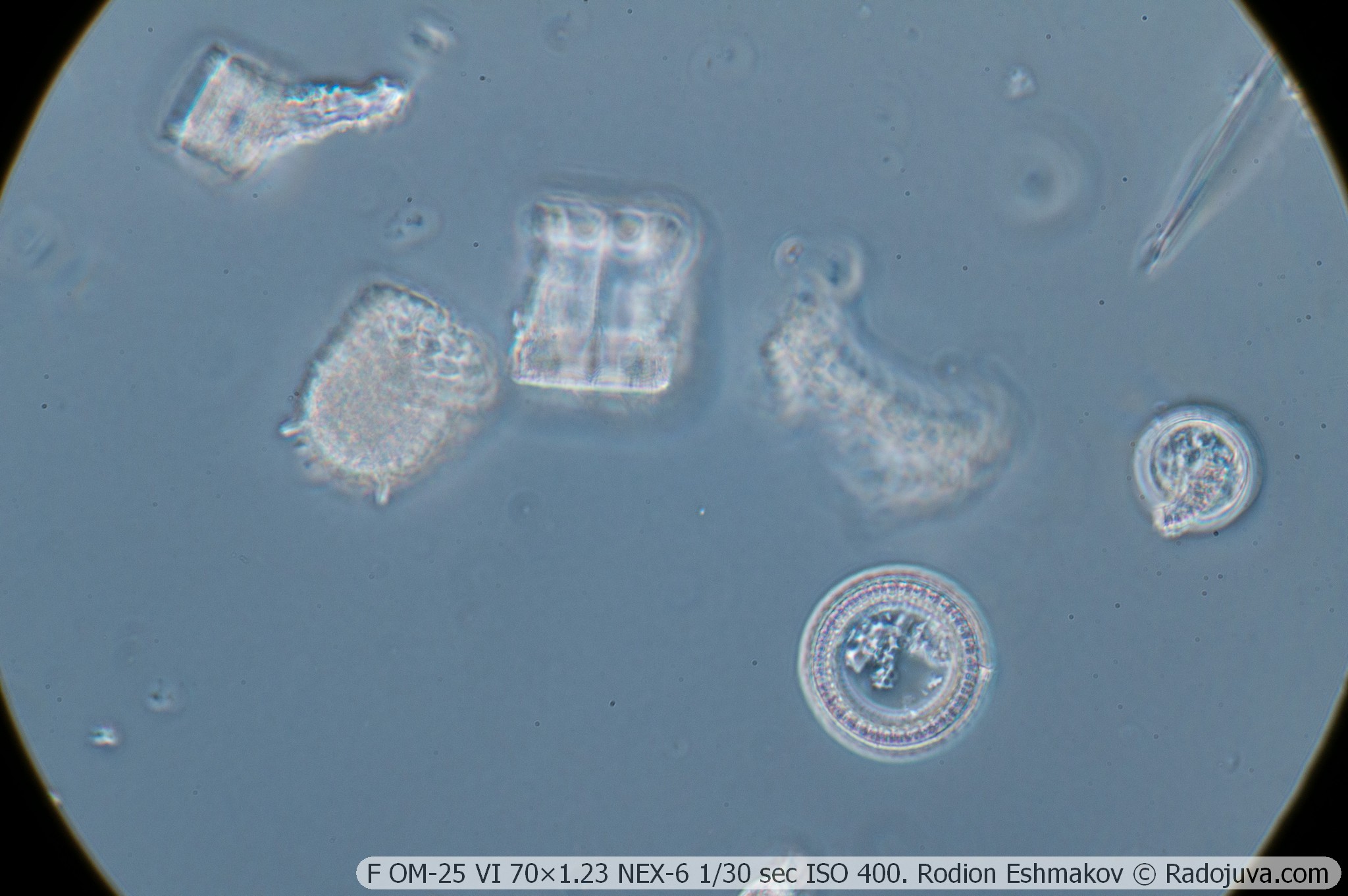
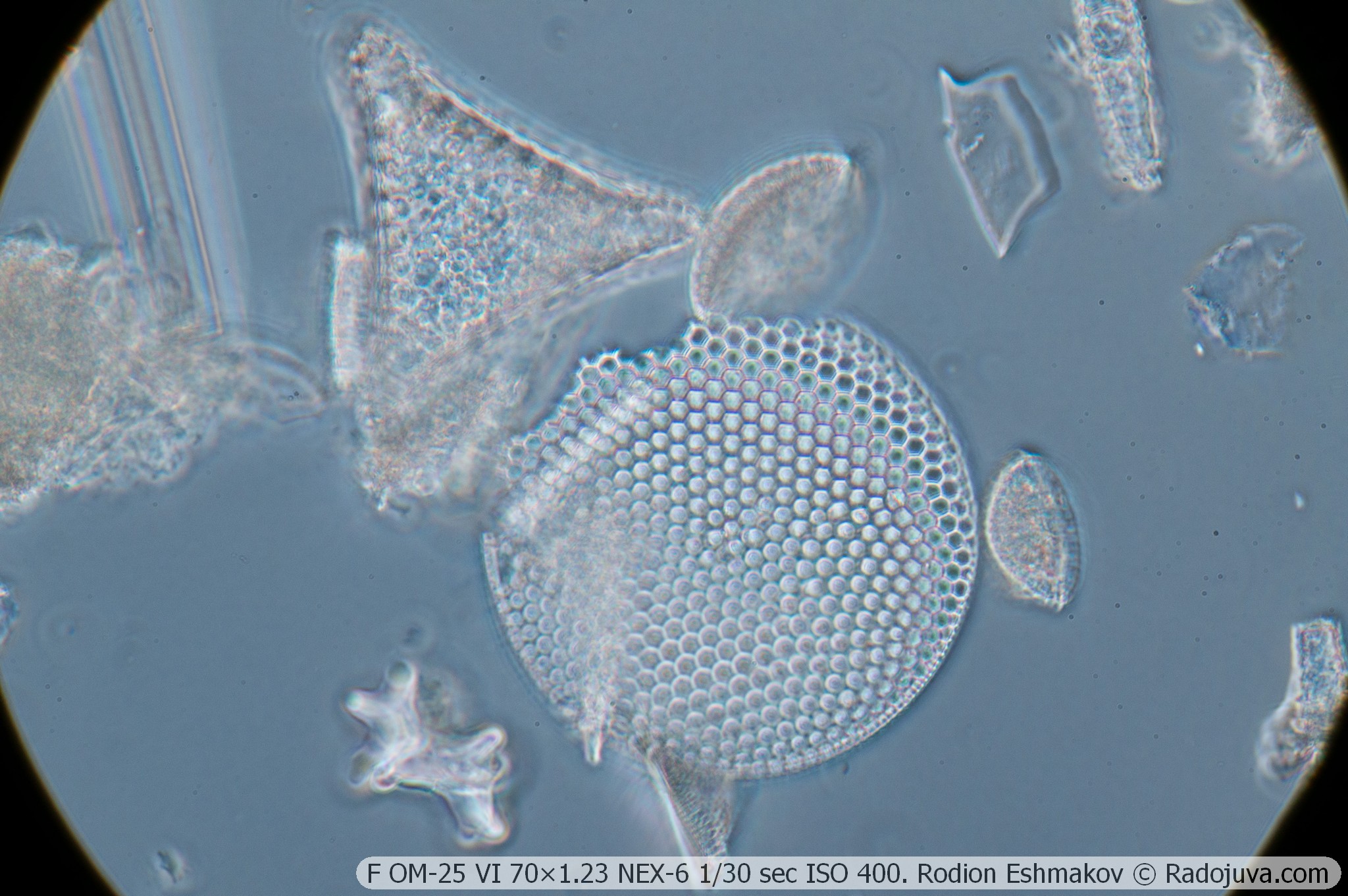
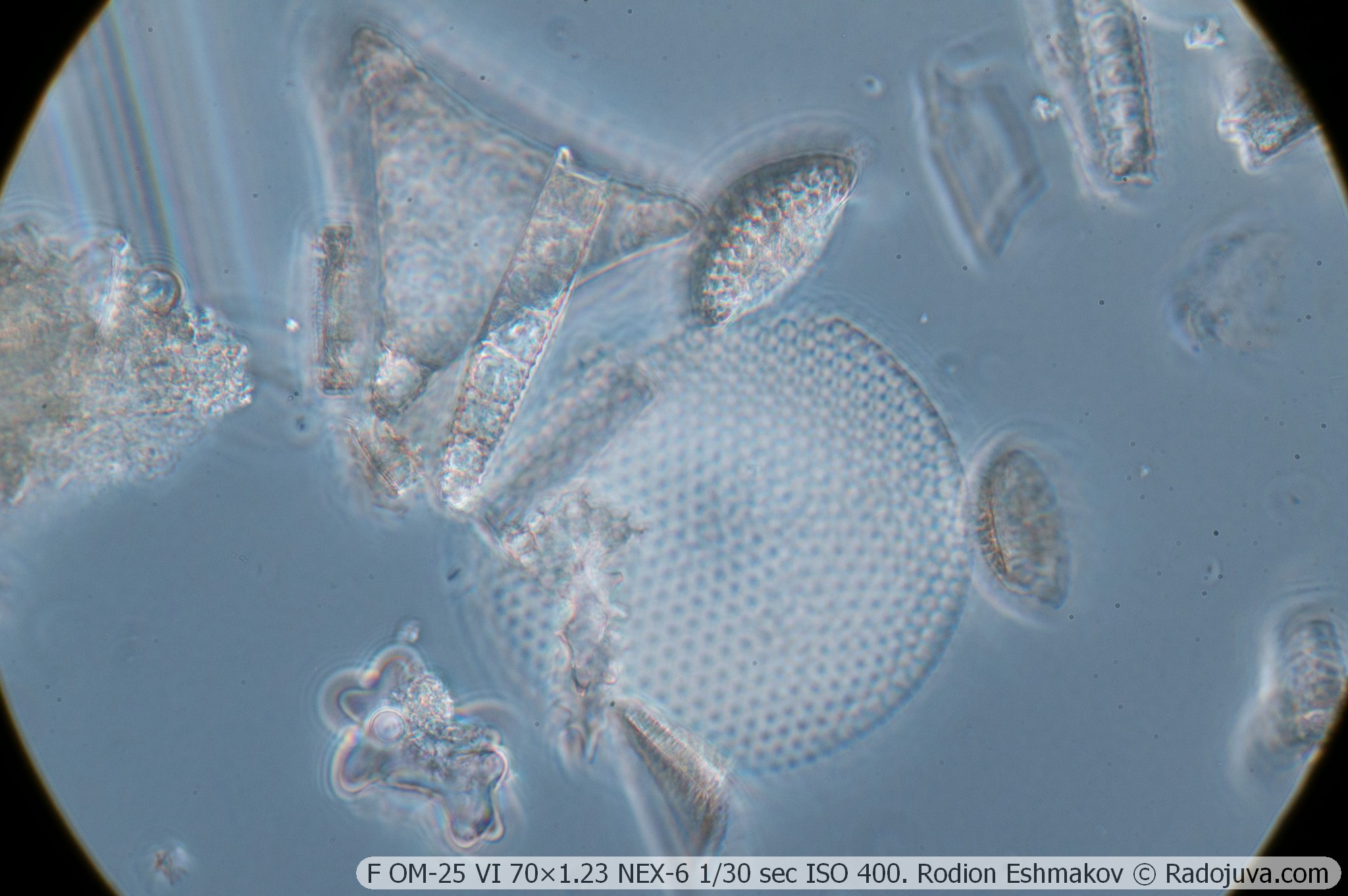
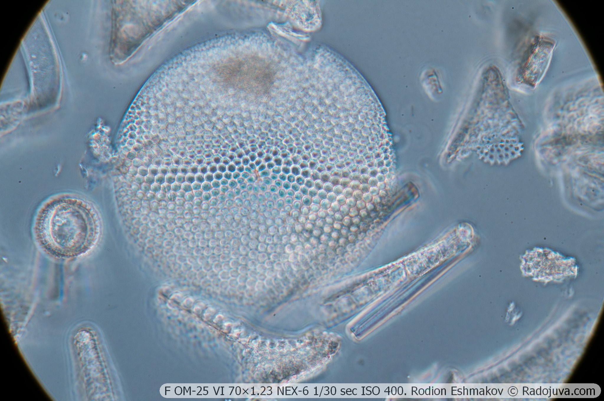
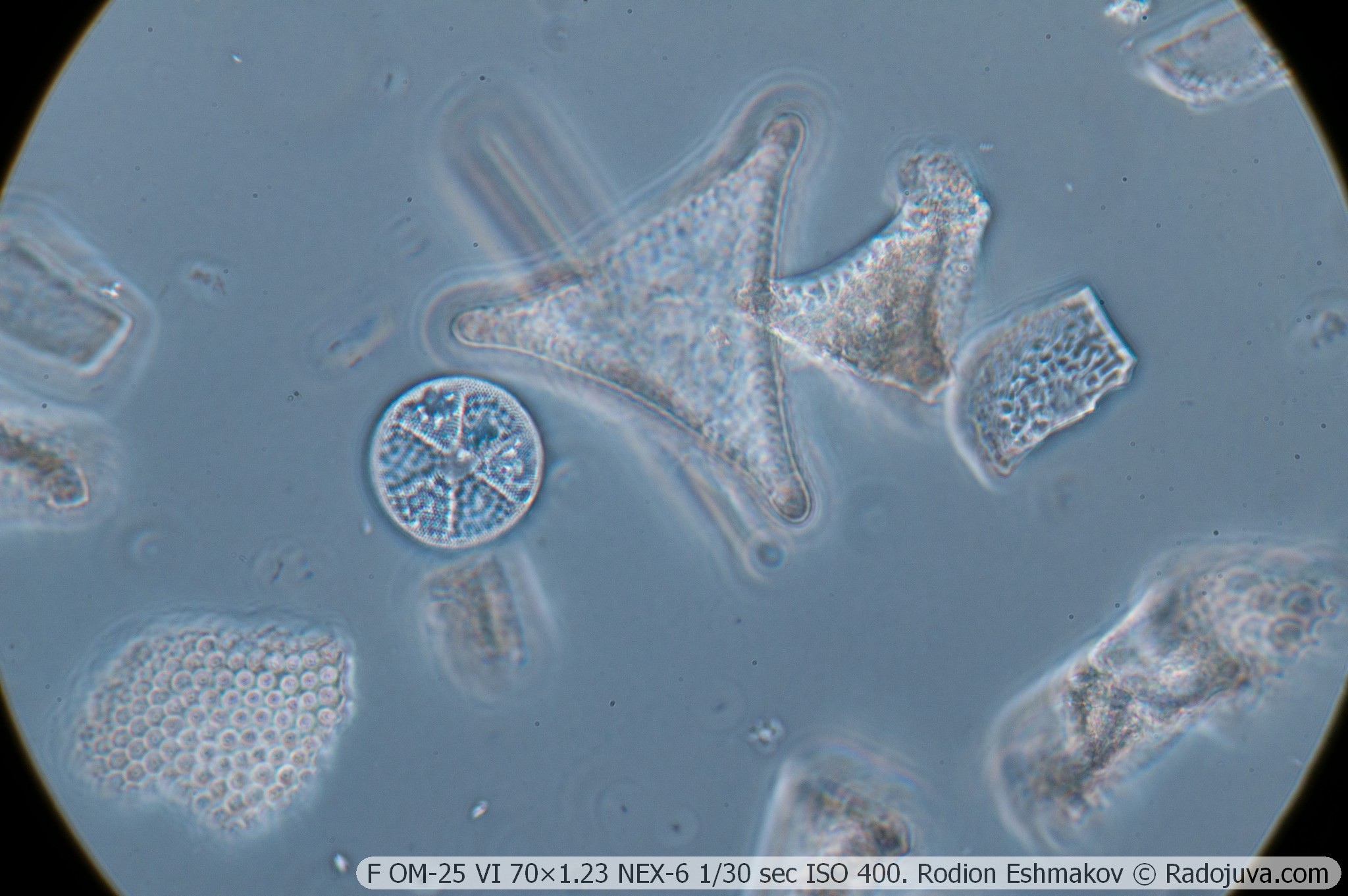
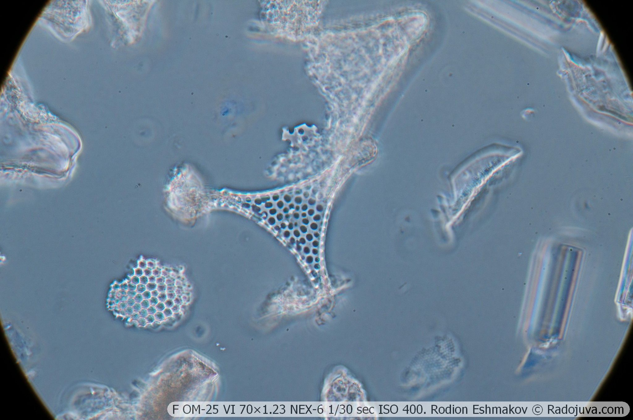
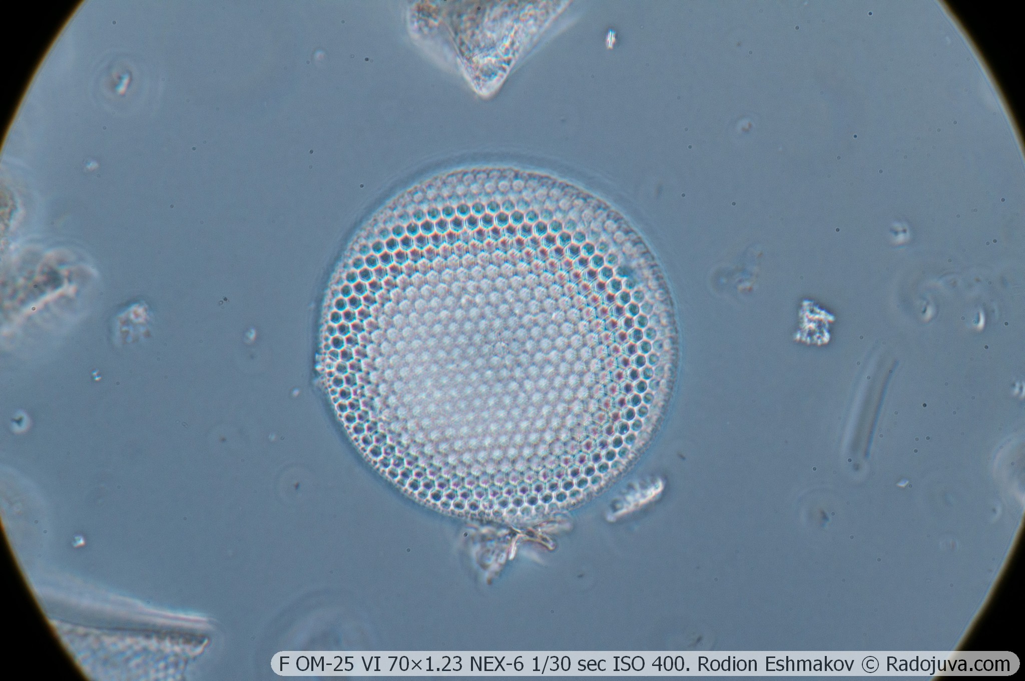
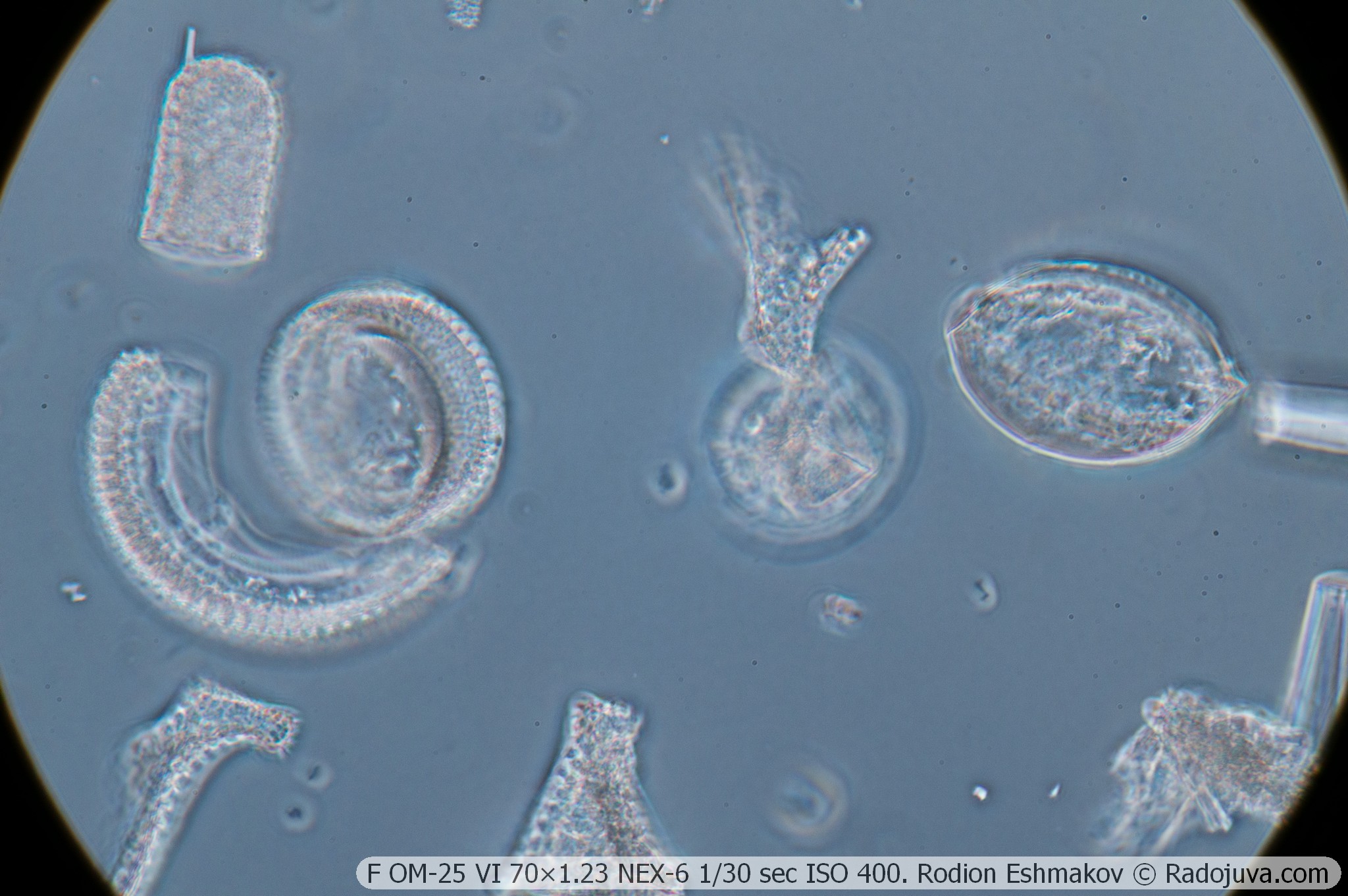
Below are examples of photos with stacking.
Conclusions
The KF-1 phase contrast device is today not only a museum exhibit but also one of the most accessible options for introducing phase contrast microscopy. The method itself significantly expands the range of objects and phenomena accessible for observation, making it of great interest.
The advantage of the KF-1 condenser is its compatibility with many Soviet and German microscopes. It is well suited for brightfield work and allows the use of darkfield illumination for objectives with small apertures. Objectives for the KF-1 are readily available on the used market, and are compatible with both the newer, more advanced objectives from the KF-4M condenser (10-40×) and the unique F APO VI 70×1.25 apochromatic water immersion objective. The KF-1's empty slots allow for modifications to the condenser for use with other objectives, such as the Carl Zeiss Jena PhV or modern Chinese ones.
The disadvantages of the KF-1 kit include the need for constant adjustment of the aperture position when changing lenses, the mediocre optical quality of the condenser lens block and, most importantly, the generally low optical quality of the kit lenses, especially uncorrected lateral chromatic aberration and low working apertures.
More micro-optics reviews here.